. 2017 Nov 15;37(46):11085-11100.
doi: 10.1523/JNEUROSCI.1294-17.2017. Epub 2017 Oct 13.

Dysregulation of the Mitochondrial Unfolded Protein Response Induces Non-Apoptotic Dopaminergic Neurodegeneration in C. elegans Models of Parkinson's Disease

Affiliations

Dysregulation of the Mitochondrial Unfolded Protein Response Induces Non-Apoptotic Dopaminergic Neurodegeneration in C. elegans Models of Parkinson's Disease

Bryan A Martinez et al. J Neurosci. .

Abstract

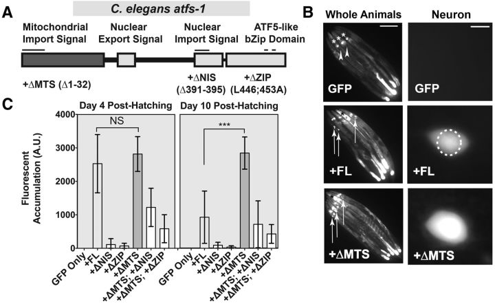
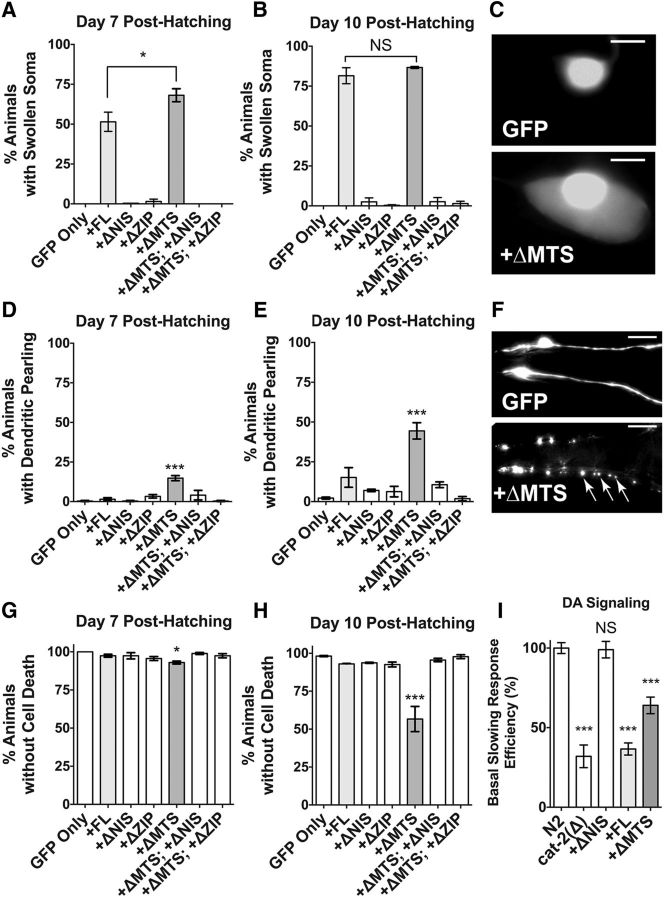
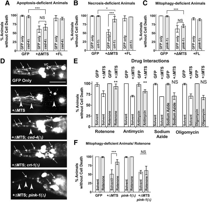
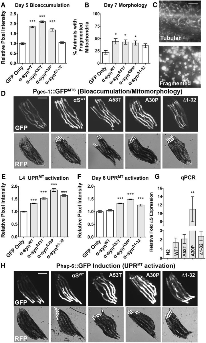
Due to environmental insult or innate genetic deficiency, protein folding environments of the mitochondrial matrix are prone to dysregulation, prompting the activation of a specific organellar stress-response mechanism, the mitochondrial unfolded protein response (UPRMT). In Caenorhabditis elegans, mitochondrial damage leads to nuclear translocation of the ATFS-1 transcription factor to activate the UPRMT After short-term acute stress has been mitigated, the UPRMT is eventually suppressed to restore homeostasis to C. elegans hermaphrodites. In contrast, and reflective of the more chronic nature of progressive neurodegenerative disorders such as Parkinson's disease (PD), here, we report the consequences of prolonged, cell-autonomous activation of the UPRMT in C. elegans dopaminergic neurons. We reveal that neuronal function and integrity decline rapidly with age, culminating in activity-dependent, non-apoptotic cell death. In a PD-like context wherein transgenic nematodes express the Lewy body constituent protein α-synuclein (αS), we not only find that this protein and its PD-associated disease variants have the capacity to induce the UPRMT, but also that coexpression of αS and ATFS-1-associated dysregulation of the UPRMT synergistically potentiate dopaminergic neurotoxicity. This genetic interaction is in parallel to mitophagic pathways dependent on the C. elegans PINK1 homolog, which is necessary for cellular resistance to chronic malfunction of the UPRMT Given the increasingly recognized role of mitochondrial quality control in neurodegenerative diseases, these studies illustrate, for the first time, an insidious aspect of mitochondrial signaling in which the UPRMT pathway, under disease-associated, context-specific dysregulation, exacerbates disruption of dopaminergic neurons in vivo, resulting in the neurodegeneration characteristic of PD.SIGNIFICANCE STATEMENT Disruptions or alterations in the activation of pathways that regulate mitochondrial quality control have been linked to neurodegenerative diseases due in part to the central role of mitochondria in metabolism, ROS regulation, and proteostasis. The extent to which these pathways, including the mitochondrial unfolded protein response (UPRMT) and mitophagy, are active may predict severity and progression of these disorders, as well as sensitivity to compounding stressors. Furthermore, therapeutic strategies that aim to induce these pathways may benefit from increased study into cellular responses that arise from long-term or ectopic stimulation, especially in neuronal compartments. By demonstrating the detrimental consequences of prolonged cellular activation of the UPRMT, we provide evidence that this pathway is not a universally beneficial mechanism because dysregulation has neurotoxic consequences.

Keywords: C. elegans; alpha-synuclein; dopaminergic; mitochondria; neurodegeneration; stress.


Figures

Figure 1.
Figure 1.
ATFS-1 and constructs expressing modified ATFS-1 structural variants are stable and exhibit variable activities in C. elegans dopaminergic neurons. A, The ATFS-1 domain structure possesses a MTS, NIS, nuclear export signal, and a conserved bZIP domain (ZIP). Into this cDNA backbone, we introduced three mutations: ΔMTS (Δ1–32), ΔNIS (Δ391–395), and ΔZIP (L446, 453A). Furthermore, ΔMTS and either ΔNIS or ΔZIP were paired together to engineer two mutations in the same backbone. B, Animals bearing the cDNA constructs under control of a dopaminergic promoter (Pdat-1) were crossed to worm strain SJ4100 (Phsp-6:: GFP) to determine whether the varied ATFS-1 constructs are stable and to what degree they confer transcriptional activity to the neuronal compartment. Whole animal images show five representative juxtaposed animals per transgenic construct. Arrowheads indicate the absence of dopaminergic GFP accumulation. Asterisks indicate background pharyngeal GFP expression that was not scored in these assays. Arrows indicate the presence of GFP accumulation in anterior DA neurons. Scale bar, 100 μm. Neuron images provide higher-magnification views of individual images from each strain used in the quantitation of fluorescent accumulation. The dashed circle indicates the fluorescent scoring region. Scale bar, 5 μm. C, Fluorescent intensity of GFP accumulation was measured in an anatomically consistent region comprising only the anterior dopaminergic compartments at days 4 and 10 posthatching. Data are shown as the average of 20 animals, inclusive of background fluorescence subtraction of one representative stable transgenic line, with error bars indicating SD. Statistics were performed using a one-way ANOVA with a Tukey's post hoc test. NS p > 0.05, ***p < 0.001.
Figure 2.
Figure 2.
ATFS-1 overexpression induces dopaminergic neurodegenerative phenotypes and cell death in an age-dependent manner. Animals expressing ATFS-1 variant constructs under the Pdat-1 promoter were crossed into animals that express GFP in their dopaminergic neurons (strain BY250; Pdat-1::GFP) as a means to monitor cellular health when ATFS-1 variants are overexpressed. Four neurodegenerative phenotypes were observed: Soma swelling (AC), dendritic pearling (DF), neuron cell death (G,H), and dopaminergic signaling reduction (I). A, B, Animals were assayed at days 7 and 10 posthatching for dopaminergic soma swelling phenotypes characterized by enlargement of the cytosolic space surrounding the nucleus of the dopaminergic neuron. Data are shown as the average of three separate transgenic lines with three replicates scored per line per construct. Error bars indicate the SEM of the average of each transgenic line. Statistics were performed using a one-way ANOVA with a Tukey's post hoc test. NS p > 0.05, *p < 0.05. C, Representative images of swollen dopaminergic neuron soma. GFP refers to BY250 (Pdat-1::GFP). Scale bar, 5 μm. D, E, Animals were assayed at days 7 and 10 posthatching for irregular dendritic swelling phenotypes (pearling) characterized by enlargement of the cytosolic space of the dendritic sensillary processes of the CEP dopaminergic neurons. Data are shown as the average of three separate transgenic lines with three replicates per line per construct. Error bars indicate the SEM of the average of each transgenic line. Statistics were performed using a one-way ANOVA with a Tukey's post hoc test. ***p < 0.001. F, Representative images of dendritic pearling in the anterior dopaminergic CEP neurons. GFP-only refers to BY250 (Pdat-1::GFP). Arrows represent irregular GFP puncta. Scale bar, 10 μm. G, H, Animals were assayed at days 7 and 10 posthatching for dopaminergic cell death, characterized by the absence of any of six anterior dopaminergic neurons in part or in whole. Data are shown as the average of three separate transgenic lines with three replicates per line per construct. Error bars indicate the SEM of the average of each transgenic line. Statistics were performed using a one-way ANOVA with a Tukey's post hoc test. *p < 0.05, ***p < 0.001. I, Animals were assayed at day 7 posthatching for basal slowing response activity by measurement of the peristaltic speed on food and off food, expressed as a ratio, and normalized to the ratio quantified for N2 control worms. Data are shown as the average of two transgenic lines using n = 10 animals per each of four replicates per construct. Error bars indicate the SEM of the average. Statistics were performed using a one-way ANOVA with a Tukey's post hoc test. NS, p > 0.05, ***p < 0.001.
Figure 3.
Figure 3.
ATFS-1-induced dopaminergic cell death is necrotic and is attenuated by rotenone though pink-1. AC, C. elegans dopaminergic neurons expressing functional ATFS-1 structural variant constructs were crossed independently to three mutations that may affect cell death and analyzed at 10 d posthatching. Data are shown as the average of three separate transgenic lines with one to two replicates per line per construct analyzed. Error bars indicate the SD of the average of all replicates. Statistics were performed using a two-way ANOVA with a Tukey's post hoc test. NS p > 0.05, *p < 0.05, ***p < 0.001. A, ATFS-1ΔMTS and ATFS-1FL variants were crossed to a mutation of ced-4(n1162). B, ATFS-1ΔMTS and ATFS-1FL variants were crossed to a mutation of crt-1(jh101). C, ATFS-1ΔMTS and ATFS-1FL variants were crossed to a mutation of pink-1(tm1779). D, Representative images of the six anterior dopaminergic neurons scored in the analyses displayed in 3A-C. Arrows represent intact sensillary dopaminergic process of four CEP neurons and the other pair of two ADE neurons. Alterations in this count are considered positive for cell death phenotypes (as depicted with arrowheads). Asterisks in some of these images represent the ventral deirid commissure (VC); this is not scored in our assays. Scale bar, 30 μm. The selected images depicted are slightly overexposed to facilitate visualization of neuronal processes. E, Analysis of dopaminergic neurons from transgenic C. elegans expressing either Pdat-1::GFP alone or with the gain-of-function ATFS-1ΔMTS exposed to drugs that modulate mitochondrial electron transport chain function. Concentrations used were as follows: 1 μm rotenone, 1 μm antimycin, 100 μm sodium azide, 2 μm oligomycin. Sodium azide was dissolved in water; all remaining chemicals were dissolved in ethanol (final concentration 2%). Data are shown as the average of 30 worms per replicate per construct using three separate transgenic lines with one to two replicates per line per construct. Error bars indicate the SD of the average of all replicates. Statistics were performed using a two-way ANOVA with a Tukey's post hoc test. NS p > 0.05, **p < 0.01. F, ATFS-1ΔMTS variants were crossed to a mutation of pink-1(tm1779) and exposed to 1 μm rotenone then assayed for dopaminergic cell death phenotypes as in D and E. Error bars indicate the SD of the average of all replicates. Statistics were performed using a two-way ANOVA with a Tukey's post hoc test. NS p > 0.05, ***p < 0.001.
Figure 4.
Figure 4.
αS variants induce bioaccumulation of mitochondria, mitochondrial fragmentation, and mitochondrial stress. A, D, Bioaccumulation of mitochondria was assayed using the SJ4143 (Pges-1::GFP) animals crossed to intestinally expressing αS variants and assayed at day 5 posthatching for GFP intensity differences by imaging animals at a consistent exposure time and measuring with a standardized 80 × 80 pixel region box (∼30 × 30 μm) at the anterior-most region of the intestine. Data are shown as the average of 90 animals per line with two to three transgenic lines per construct analyzed. Values were standardized relative to the GFP-only control. Error bars indicate SEM and statistics were performed using a one-way ANOVA with a Dunnett's post hoc analysis. ***p < 0.001. B, Strains from A were also analyzed at day 7 posthatching for mitochondrial fragmentation, characterized by circularization of the mitochondrial tubular networks that exemplify normal mitochondrial morphology. Data are represented as an average of three sets of 30 animals per line and two to three transgenic lines were analyzed per construct. Error bars indicate SEM and statistical analysis was performed using a one-way ANOVA with a Dunnett's post hoc test. *p < 0.05. C, Representative images of mitochondrial morphology that can be either tubular or fragmented, as visualized using SJ4143 (Pges-1::GFPMTS) transgenic integrated line. Scale bar, 20 μm. D, Bioaccumulation of mitochondria is displayed with images of five representative, juxtaposed Pges-1::GFPMTS animals per transgenic construct. Two filters were used: GFP indicates accumulation of mitochondrial-associated GFP and RFP shows light-field worm images with a red fluorescent marker in the pharynx that indicates the animals carry a transgenic αS transgenic array (Pmyo-2::mCherry). Scale bar, 300 μm. E, F, SJ4100 (Phsp-6::GFP) animals were crossed to intestinally expressing αS variants and assayed at larval stage 4 (L4) or 6 d posthatching adults to examine UPRMT mitostress by via GFP intensity differences. Animals were imaged at consistent exposure levels and anatomical location at the anterior region of the intestine using an 80 × 80 pixel region box (∼30 × 30 μm). Data are shown as the average of 90 animals per line and two to three transgenic lines were examined per construct. Values were standardized relative to the GFP-only control. Error bars indicate SEM and statistics were performed using a one-way ANOVA with a Dunnett's post hoc. ***p < 0.001. G, Each of three transgenic lines was probed by standard qRT-PCR methods in duplicate biological samples per line. Relative fold expression of Pges-1::αSWT (as well as the other αS variants shown in the graph) was estimated using three reference genes. Data are shown as the average relative expression values of all transgenic lines in arbitrary units. N2 had no αS expression. Not shown, Phsp-6::GFP and Pges-1::GFPMTS show similar results to N2. Average expression was measured statistically using a one-way ANOVA with a Dunnett's post hoc test among transgenic lines compared with αSWT. Data are shown as average ± SEM. **p < 0.01; NS p > 0.05. H, Representative images of the data graphed in F. UPRMT mitostress is displayed as images of five representative juxtaposed animals per transgenic construct. Animals were imaged at consistent anatomical location at the anterior region of the intestine. Two filters are shown: GFP indicates mitostress (Phsp-6::GFP) and RFP shows light-field worm images with a red fluorescent marker in the pharynx indicating that the animals carry a transgenic αS transgenic array (Pmyo-2::mCherry). Scale bar, 300 μm.
Figure 5.
Figure 5.
ATFS-1 overactivity potentiates αS toxicity synergistically and independently of mitophagic downregulation. A, Loss-of-function (LoF: gk3094) and gain-of-function (GoF: et15) atfs-1 mutants were crossed into BY250 (Pdat-1::GFP) to monitor cell death phenotypes at two age points. Data are shown as the average ± SD of three to four replicates with 30 animals per replicate. Statistics were performed using a one-way ANOVA with a Tukey's post hoc test. NS p > 0.05. B, LoF and GoF atfs-1 mutants were crossed into UA44 (Pdat-1::GFP; αSWT) to monitor cell death phenotypes at two points of aging. Data are shown as the average ± SD of 3–4 replicates, with 30 animals/replicate. Statistics were performed using a one-way ANOVA with a Tukey's post hoc test. *p < 0.05, **p < 0.01, ***p < 0.001. C, Representative anterior images of worm dopaminergic neurons. αSWT-expressing animals generally exhibit cell death (arrowheads), but the full anterior dopaminergic neuron count (6 neurons; visualized with arrows) is retained in this representative image of an αSWT; atfs-1 ΔLoF animal. Scale bar, 30 μm. D, LoF and GoF atfs-1 mutants were crossed into UA49 (Punc-54::GFP:αSWT) to monitor αS aggregation, which was assessed by aggregate size and number using an arbitrary value scale. Data are shown as the average ± SD of three to four replicates of 30 animals. Statistics were performed using a one-way ANOVA with a Tukey's post hoc test. ***p < 0.001. E, Representative Punc-54::GFP::αSWT muscle cells used in the analysis described in D. Arrows indicate GFP:: αSWT puncta. F, G, Dopaminergic cell death analysis in animals expressing Pdat-1:: αSWT and Pdat-1::GFP. The assay was performed at days 7 and 10 posthatching. Data are shown as the average of three separate transgenic lines with three to four replicates per line per construct. Error bars indicate the SEM of the average of each transgenic line. Statistics were performed using a one-way ANOVA with a Tukey's post hoc test. **p < 0.01, ***p < 0.001. H, Animals were assayed at day 5 for cell death in the presence of αSWT in the dopaminergic neurons in two strains competent for neuronal RNAi (UA196 and UA309) targeting the listed gene products. Data are shown as the average of 30 worms per replicate comprising three separate transgenic lines with two replicates per line per construct. Error bars indicate the SD of the average of each transgenic line. Statistics were performed using a two-way ANOVA series with a Tukey's post hoc test. NS p > 0.05 *p < 0.01 **p < 0.01, ***p < 0.001. I, J, ATFS-1FL and ATFS-1ΔNIS variants were crossed to a mutation of either pink-1(tm1779) or lrk-1(tm1898). Data are shown as the average of 30 worms per replicate comprising three separate transgenic lines with two replicates per line per construct. Error bars indicate the SD of the average of each transgenic line. Statistics were performed using a two-way ANOVA with a Tukey's post hoc test. **p < 0.01, ***p < 0.001.

Similar articles

Cited by

References

    1. Baba M, Nakajo S, Tu PH, Tomita T, Nakaya K, Lee V, Trojanowski JQ, Iwatsubo T (1998) Aggregation of a-synuclein in Lewy bodies of sporadic Parkinson's disease and dementia with Lewy bodies. Am J Pathol 152:879–884. - PMC - PubMed
    1. Baker BM, Haynes CM (2011) Mitochondrial protein quality control during biogenesis and aging. Trends Biochem Sci 36:254–261. 10.1016/j.tibs.2011.01.004 - DOI - PubMed
    1. Baker MJ, Tatsuta T, Langer T (2011) Quality control of mitochondrial proteostasis. Cold Spring Harb Perspect Biol 3: pii: a007559. 10.1101/cshperspect.a007559 - DOI - PMC - PubMed
    1. Benedetti C, Haynes CM, Yang Y, Harding HP, Ron D (2006) Ubiquitin-like protein 5 positively regulates chaperone gene expression in the mitochondrial unfolded protein response. Genetics 174:229–239. 10.1534/genetics.106.061580 - DOI - PMC - PubMed
    1. Bennett CF, Vander Wende H, Simko M, Klum S, Barfield S, Choi H, Pineda VV, Kaeberlein M (2014) Activation of the mitochondrial unfolded protein response does not predict longevity in Caenorhabditis elegans. Nat Commun 5:3483. 10.1038/ncomms4483 - DOI - PMC - PubMed

Publication types

MeSH terms

Substances

LinkOut - more resources