Variation and Distribution of L-A Helper Totiviruses in Saccharomyces sensu stricto Yeasts Producing Different Killer Toxins
- PMID: 29019944
- PMCID: PMC5666360
- DOI: 10.3390/toxins9100313
Variation and Distribution of L-A Helper Totiviruses in Saccharomyces sensu stricto Yeasts Producing Different Killer Toxins
Abstract
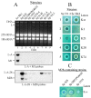
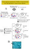
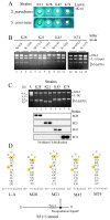
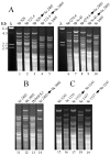
Yeasts within the Saccharomyces sensu stricto cluster can produce different killer toxins. Each toxin is encoded by a medium size (1.5-2.4 Kb) M dsRNA virus, maintained by a larger helper virus generally called L-A (4.6 Kb). Different types of L-A are found associated to specific Ms: L-A in K1 strains and L-A-2 in K2 strains. Here, we extend the analysis of L-A helper viruses to yeasts other than S. cerevisiae, namely S. paradoxus, S. uvarum and S. kudriavzevii. Our sequencing data from nine new L-A variants confirm the specific association of each toxin-producing M and its helper virus, suggesting co-evolution. Their nucleotide sequences vary from 10% to 30% and the variation seems to depend on the geographical location of the hosts, suggesting cross-species transmission between species in the same habitat. Finally, we transferred by genetic methods different killer viruses from S. paradoxus into S. cerevisiae or viruses from S. cerevisiae into S. uvarum or S. kudriavzevii. In the foster hosts, we observed no impairment for their stable transmission and maintenance, indicating that the requirements for virus amplification in these species are essentially the same. We also characterized new killer toxins from S. paradoxus and constructed "superkiller" strains expressing them.
Keywords: L-A totivirus; Saccharomyces sensu stricto; double-stranded RNA virus; yeast killer toxins.
Conflict of interest statement
The authors declare no conflict of interest. The founding sponsors had no role in the design of the study; in the collection, analyses, or interpretation of data; in the writing of the manuscript, and in the decision to publish the results.
Figures


Similar articles
-
Relationships and Evolution of Double-Stranded RNA Totiviruses of Yeasts Inferred from Analysis of L-A-2 and L-BC Variants in Wine Yeast Strain Populations.Appl Environ Microbiol. 2017 Feb 1;83(4):e02991-16. doi: 10.1128/AEM.02991-16. Print 2017 Feb 15. Appl Environ Microbiol. 2017. PMID: 27940540 Free PMC article.
-
L-A-lus, a new variant of the L-A totivirus found in wine yeasts with Klus killer toxin-encoding Mlus double-stranded RNA: possible role of killer toxin-encoding satellite RNAs in the evolution of their helper viruses.Appl Environ Microbiol. 2013 Aug;79(15):4661-74. doi: 10.1128/AEM.00500-13. Epub 2013 May 31. Appl Environ Microbiol. 2013. PMID: 23728812 Free PMC article.
-
A population study of killer viruses reveals different evolutionary histories of two closely related Saccharomyces sensu stricto yeasts.Mol Ecol. 2015 Aug;24(16):4312-22. doi: 10.1111/mec.13310. Epub 2015 Jul 30. Mol Ecol. 2015. PMID: 26179470
-
Viral induced yeast apoptosis.Biochim Biophys Acta. 2008 Jul;1783(7):1413-7. doi: 10.1016/j.bbamcr.2008.01.017. Epub 2008 Feb 7. Biochim Biophys Acta. 2008. PMID: 18291112 Review.
-
Yeast Killer Toxin K28: Biology and Unique Strategy of Host Cell Intoxication and Killing.Toxins (Basel). 2017 Oct 20;9(10):333. doi: 10.3390/toxins9100333. Toxins (Basel). 2017. PMID: 29053588 Free PMC article. Review.
Cited by
-
The Effect of Trichoderma harzianum Hypovirus 1 (ThHV1) and Its Defective RNA ThHV1-S on the Antifungal Activity and Metabolome of Trichoderma koningiopsis T-51.J Fungi (Basel). 2023 Jan 28;9(2):175. doi: 10.3390/jof9020175. J Fungi (Basel). 2023. PMID: 36836290 Free PMC article.
-
Identification and Molecular Characterization of Novel Mycoviruses in Saccharomyces and Non-Saccharomyces Yeasts of Oenological Interest.Viruses. 2021 Dec 29;14(1):52. doi: 10.3390/v14010052. Viruses. 2021. PMID: 35062256 Free PMC article.
-
Expression of the K74 Killer Toxin from Saccharomyces paradoxus Is Modulated by the Toxin-Encoding M74 Double-Stranded RNA 5' Untranslated Terminal Region.Appl Environ Microbiol. 2022 Apr 26;88(8):e0203021. doi: 10.1128/aem.02030-21. Epub 2022 Apr 7. Appl Environ Microbiol. 2022. PMID: 35389250 Free PMC article.
-
RNA viruses, M satellites, chromosomal killer genes, and killer/nonkiller phenotypes in the 100-genomes S. cerevisiae strains.G3 (Bethesda). 2023 Sep 30;13(10):jkad167. doi: 10.1093/g3journal/jkad167. G3 (Bethesda). 2023. PMID: 37497616 Free PMC article.
-
The Species-Specific Acquisition and Diversification of a K1-like Family of Killer Toxins in Budding Yeasts of the Saccharomycotina.PLoS Genet. 2021 Feb 4;17(2):e1009341. doi: 10.1371/journal.pgen.1009341. eCollection 2021 Feb. PLoS Genet. 2021. PMID: 33539346 Free PMC article.
References
-
- Wickner R.B., Ghabrial S.A., Nibert M.L., Patterson J.L., Wang C.C. Family Totiviridae. In: King A.M.Q., Adams M.J., Carstens E.B., Lefkowits E.J., editors. Virus Taxonomy: Classification and Nomenclature of Viruses: Ninth Report of the International Committee on Taxonomy of Viruses. Elsevier Academic Press; Tokyo, Japan: 2011. pp. 639–650.
-
- Icho T., Wickner R.B. The double-stranded RNA genome of yeast virus L-A encodes its own putative RNA polymerase by fusing two open reading frames. J. Biol. Chem. 1989;264:6716–6723. - PubMed
Publication types
MeSH terms
Substances
LinkOut - more resources
Full Text Sources
Other Literature Sources

