. 2017 Oct 7;9(10):292.
doi: 10.3390/v9100292.

Polyprotein Processing as a Determinant for in Vitro Activity of Semliki Forest Virus Replicase

Affiliations

Polyprotein Processing as a Determinant for in Vitro Activity of Semliki Forest Virus Replicase

Maija K Pietilä et al. Viruses. .

Abstract

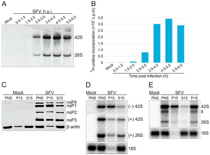
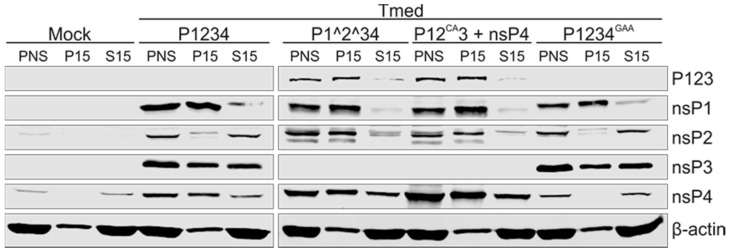
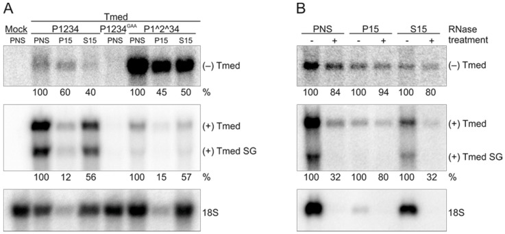
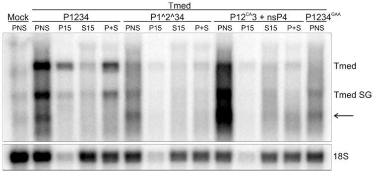
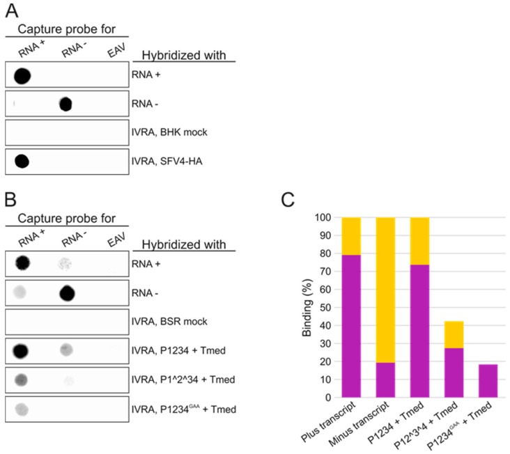
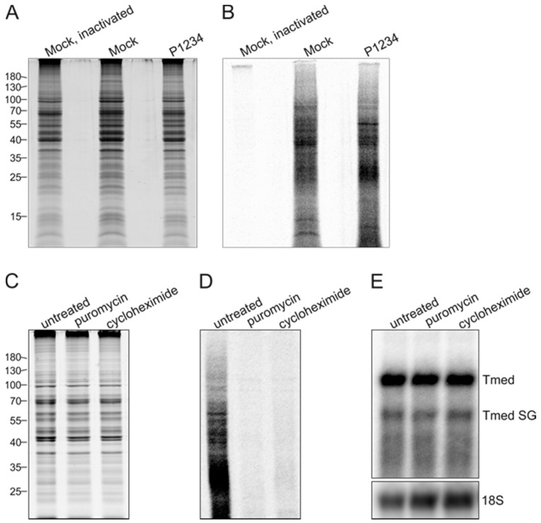
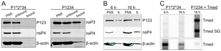
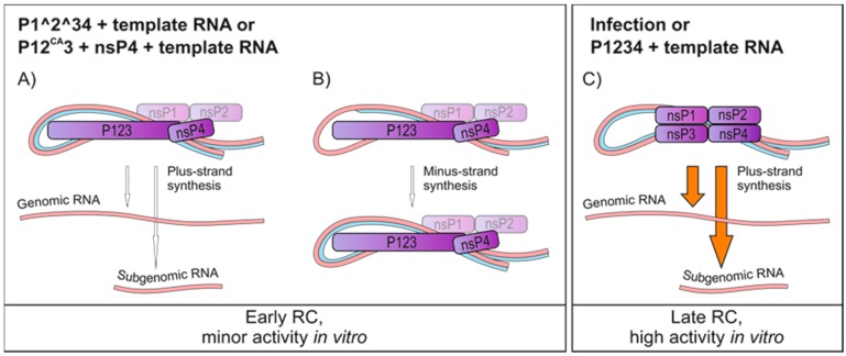
Semliki Forest virus (SFV) is an arthropod-borne alphavirus that induces membrane invaginations (spherules) in host cells. These harbor the viral replication complexes (RC) that synthesize viral RNA. Alphaviruses have four replicase or nonstructural proteins (nsPs), nsP1-4, expressed as polyprotein P1234. An early RC, which synthesizes minus-strand RNA, is formed by the polyprotein P123 and the polymerase nsP4. Further proteolytic cleavage results in a late RC consisting of nsP1-4 and synthesizing plus strands. Here, we show that only the late RCs are highly active in RNA synthesis in vitro. Furthermore, we demonstrate that active RCs can be isolated from both virus-infected cells and cells transfected with the wild-type replicase in combination with a plasmid expressing a template RNA. When an uncleavable polyprotein P123 and polymerase nsP4 were expressed together with a template, high levels of minus-strand RNA were produced in cells, but RCs isolated from these cells were hardly active in vitro. Furthermore, we observed that the uncleavable polyprotein P123 and polymerase nsP4, which have previously been shown to form spherules even in the absence of the template, did not replicate an exogenous template. Consequently, we hypothesize that the replicase proteins were sequestered in spherules and were no longer able to recruit a template.

Keywords: RNA synthesis; Semliki Forest virus; alphavirus; in vitro replication; nonstructural protein; polymerase; replication complex.


Conflict of interest statement

The authors declare no conflict of interest.

Figures

Figure 1
Figure 1
Kinetics of Semliki Forest virus (SFV) RNA synthesis and isolation of active replication complexes for in vitro analysis. (A) SFV-infected baby hamster kidney (BHK) cells were pulse-labeled with 3H-uridine for 1 h during the time periods indicated. After labeling, RNA was isolated, analysed by denaturing agarose gel electrophoresis, and visualized by fluorography. The positions of genomic and subgenomic RNA are indicated; (B) Liquid scintillation counts of the same RNA as in A; (C) Expression of the replicase proteins and their distribution between the P15 and S15 fractions as studied by Western blotting with specific antibodies. Distribution of the cytosolic marker β-actin is also shown. Post-nuclear supernatant (PNS) was prepared at 4 h post infection (p.i.), and equivalent amounts of PNS, P15, and S15 were analysed from mock- and SFV-infected cells. Data are a representative example of two independent experiments; (D) Distribution of SFV minus- and plus-strand RNA determined by in-gel hybridization with a probe specific to minus- or plus-strand RNA. The probe against plus-strand RNA detects both genomic and subgenomic RNA. As a control, a probe against ribosomal 18S RNA was used; (E) RNA synthesizing activity of various subcellular fractions analysed in vitro by determining incorporation of 32P-CTP into RNA. After an in vitro replication assay (IVRA), RNA was analysed in a denaturing agarose gel. P+S indicates a mixture of equal volumes of P15 and S15 from SFV-infected cells. 18S rRNA was detected by in-gel hybridization of the same gel. Representative experiments are shown in (D) and (E).
Figure 2
Figure 2
Distribution of nsPs expressed in transfected cells. BSR cells were transfected with the replicase plasmid(s) indicated at the top and Tmed template plasmid. At 16 h post transfection, PNS was prepared and fractionated into P15 and S15, and nsPs were detected by Western blotting with specific antibodies. The polyprotein P123 was recognized using anti-nsP3 antibody. β-actin was used as a cytosolic marker. Equivalent amounts of PNS, P15, and S15 were loaded. Two independent experiments were performed and one representative result is shown here.
Figure 3
Figure 3
Distribution and stability of endogenous minus- and plus-strand RNA in P15 and S15 fractions prepared from transfected BSR cells. (A) PNS was prepared at 16 h post transfection, and total RNA was isolated from equivalent amounts of PNS, P15, and S15 followed by in-gel hybridization with probes that detect either minus- or plus-strand RNA. Subgenomic (SG) RNA is indicated by Tmed SG. Numbers below the lanes indicate the percentage of 32P-label detected in the genomic RNA compared to PNS. For the plus-strand RNA, the genomic Tmed was quantified. As a control, a probe against ribosomal 18S RNA was used; (B) PNS, P15, and S15 from cells co-transfected with the P1^2^34 and Tmed constructs were treated with RNase A/T1 under low salt conditions, and RNA was detected as in A. Numbers below the lanes indicate the percentage of 32P-signal in genomic RNA compared to the untreated sample. Representative experiments are shown.
Figure 4
Figure 4
In vitro activity of replication complexes formed by expressing different SFV replicase constructs and a template in the trans-replication system. BSR cells were transfected and PNS, P15, and S15 were prepared as in Figure 3. Replication activity was determined by measuring the incorporation of 32P-CTP into viral RNA after separation in denaturing agarose gels. The genomic and subgenomic RNA are indicated by Tmed and Tmed SG, respectively. P+S indicates a reaction containing equal volumes of P15 and S15 fractions, and each reaction contained an equivalent amount of PNS, P15, or S15. The arrow indicates an unspecific band. Ribosomal 18S RNA was detected by in-gel hybridization of the same gel. Data are representative of at least two independent experiments.
Figure 5
Figure 5
Determination of the polarity of in vitro synthesized RNA. (A) RNA probes with the sequence of the 3′ end of the SFV genome, recognizing minus-strand RNA (RNA−), or its complementary sequence, recognizing plus-strand RNA (RNA+), were immobilized on membranes. As a negative control, an unrelated transcript (equine arteritis virus (EAV)) was also immobilized. The membranes were then hybridized with 32P-labeled SFV transcripts of positive or negative polarity as well as 32P-labeled IVRA products. IVRA reactions with PNS samples from mock and SFV-infected cells were included; (B) RNA probes with the plus sequence of Tmed, recognizing minus-strand RNA (RNA−), or its complementary sequence, recognizing plus-strand RNA (RNA+), were immobilized on membranes as well as EAV transcript followed by hybridization with 32P-labeled Tmed plus and minus transcripts as well as 32P-labeled IVRA products. PNS samples from both mock and transfected cells were included; (C) binding of Tmed plus and minus transcripts as well as IVRA products to the capture probes was quantified from two independent experiments. The binding of the IVRA products made by replication complexes (RCs) from P1^2^34 or P1234GAA + Tmed-transfected cells were normalized to the binding of the IVRA products made by RCs from P1234 + Tmed-transfected cells. Magenta indicates binding to the capture probe for the plus-strand RNA and orange to the capture probe for the minus-strand RNA.
Figure 6
Figure 6
In vitro RNA-synthesizing activity is not dependent on translation. BSR cells were either mock-transfected or co-transfected with the P1234 and Tmed constructs, and PNS was prepared at 16 h post transfection. (A) Protein profiles of PNS samples in an SDS-polyacrylamide gel stained with Coomassie blue. One of the mock PNS samples was heat inactivated, and then all samples were incubated with 35S-l-methionine and 35S-l-cysteine. Numbers on the left indicate the molecular masses (kDa) of marker proteins; (B) a Phosphor Imager screen was exposed to the same gel as in A to detect 35S-labeled proteins; (C) PNS samples from the cells transfected with P1234 + Tmed were treated with translation inhibitors, puromycin or cycloheximide, and then incubated with 35S-methionine and 35S-l-cysteine. Protein profiles in an SDS-polyacrylamide gel stained with Coomassie blue are shown; (D) A Phosphor Imager screen was exposed to the same gel as in C to detect 35S-labeled proteins; (E) P1234 + Tmed PNS was treated with translation inhibitors as in C followed by an IVRA. 18S rRNA was detected by in-gel hybridization of the same gel. The results of one of two independent experiments is shown.
Figure 7
Figure 7
Exogenous template RNA is not replicated in vitro. (A) BSR cells were transfected with the polyprotein construct P1^2^34 or P1234 and cells were collected at 16 h post transfection followed by homogenization and HA-affinity capture. The HA tag was in nsP3. After PNS was incubated with HA-specific agarose beads, they were washed and boiled with Laemmli sample buffer. The presence of polyprotein P123, nsP3, nsP4, and β-actin in PNS as well as in unbound and bound fractions was studied by Western blotting. P123 and nsP3 were detected using an antibody against the HA tag; (B) BSR cells were transfected with the polyprotein construct P1^2^34, cells were collected at 6 or 16 h post transfection, and membranes were purified from PNS using flotation centrifugation. A Western blot analysis shows the presence of P123, nsP4, and β-actin in the PNS and membrane fractions (fr.); (C) IVRA was performed with the flotation fractions by adding 1 µg of Tmed in vitro transcript. As a control, replication activity from the endogenous template is shown for PNS prepared from BSR cells co-transfected with the P1234 and Tmed constructs. The lower panel shows the presence of exogenous Tmed transcript in the membrane fractions after the IVRA, detected by in-gel hybridization from the same gel using a probe specific to the plus-stranded Tmed.
Figure 8
Figure 8
Schematic representation of the possible activities of various RCs that can be produced with the trans-replication system. (A,B) When cells are transfected with the partially uncleavable replicase and template constructs, PNS shows very little activity in vitro indicated by the white arrows. A minor amount of plus-strand RNA is made, and it is also possible that RCs finish initiated minus strands. These complexes represent early RCs; (C) when cells are infected or transfected with the wild-type replicase and template constructs, the prepared PNS is highly active in vitro and synthesizes RNA of positive polarity, both genomic and subgenomic, indicated by the orange arrows. These complexes are late RCs.

Similar articles

Cited by

References

    1. King A.M.Q., Adams M.J., Carstens E.B., Lefkowitz E.J. Virus Taxonomy: Ninth Report of the International Commitee on Taxonmy of Viruses. Elsevier Academic Press; London, UK: 2012. pp. 10–14.
    1. Koonin E.V., Dolja V.V., Krupovic M. Origins and evolution of viruses of eukaryotes: The ultimate modularity. Virology. 2015;479:2–25. doi: 10.1016/j.virol.2015.02.039. - DOI - PMC - PubMed
    1. Amraoui F., Failloux A.B. Chikungunya: An unexpected emergence in Europe. Curr. Opin. Virol. 2016;21:146–150. doi: 10.1016/j.coviro.2016.09.014. - DOI - PubMed
    1. Tsetsarkin K.A., Chen R., Weaver S.C. Interspecies transmission and chikungunya virus emergence. Curr. Opin. Virol. 2016;16:143–150. doi: 10.1016/j.coviro.2016.02.007. - DOI - PMC - PubMed
    1. Strauss J.H., Strauss E.G. The alphaviruses: Gene expression, replication, and evolution. Microbiol. Rev. 1994;58:491–562. - PMC - PubMed

MeSH terms

LinkOut - more resources