. 2017 Nov 30;91(24):e01658-17.
doi: 10.1128/JVI.01658-17. Print 2017 Dec 15.

Induction of DNA Damages upon Marek's Disease Virus Infection: Implication in Viral Replication and Pathogenesis

Affiliations

Induction of DNA Damages upon Marek's Disease Virus Infection: Implication in Viral Replication and Pathogenesis

Djihad Bencherit et al. J Virol. .

Abstract

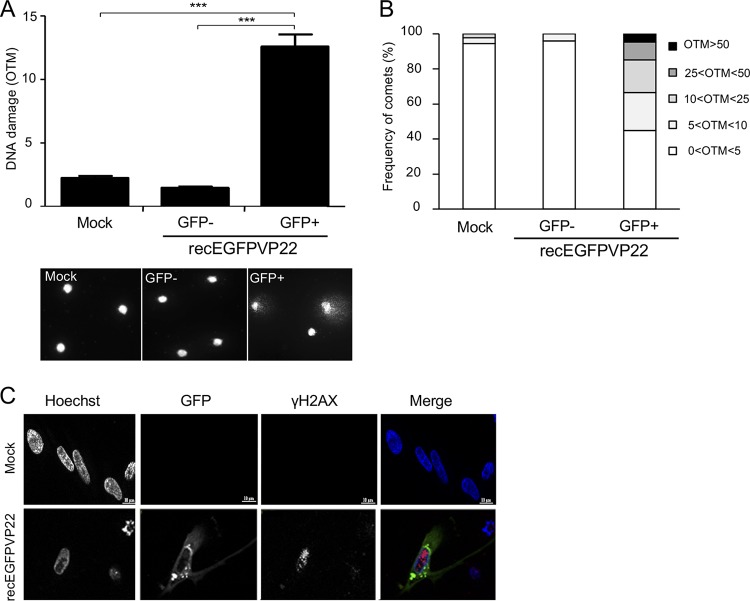
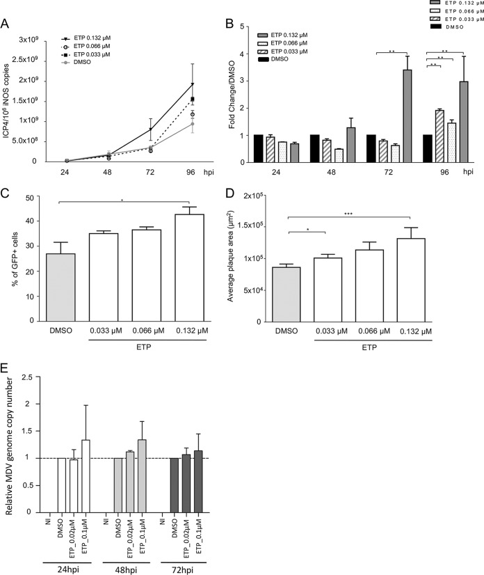
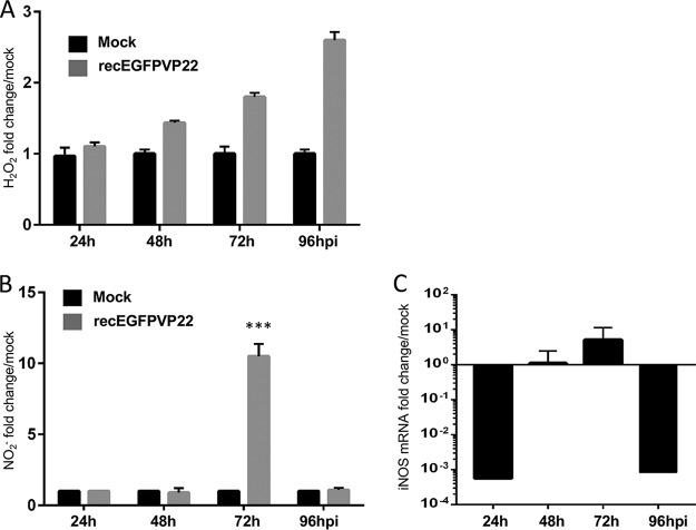
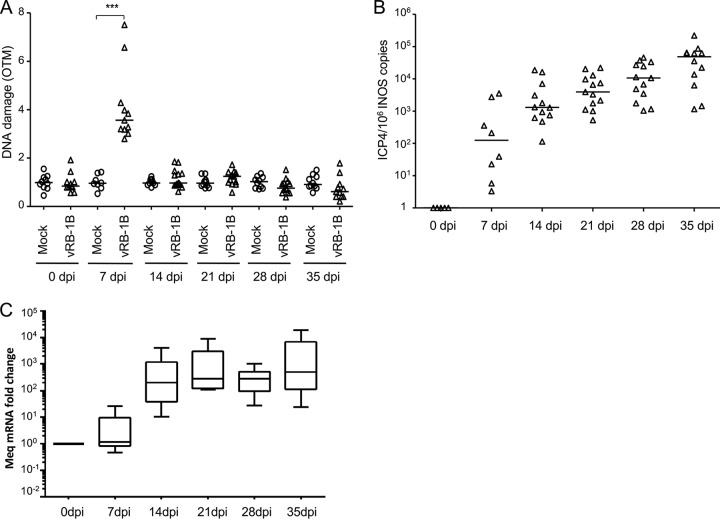
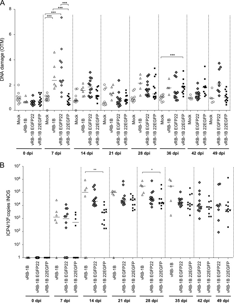
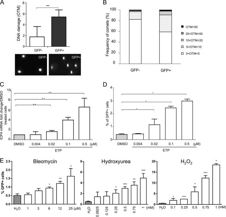
Marek's disease virus (MDV) is a highly contagious alphaherpesvirus that infects chickens and causes a deadly neoplastic disease. We previously demonstrated that MDV infection arrests cells in S phase and that the tegument protein VP22 plays a major role in this process. In addition, expression of VP22 induces double-strand breaks (DSBs) in the cellular DNA, suggesting that DNA damage and the associated cellular response might be favorable for the MDV life cycle. Here, we addressed the role of DNA damage in MDV replication and pathogenesis. We demonstrated that MDV induces DSBs during lytic infection in vitro and in the peripheral blood mononuclear cells of infected animals. Intriguingly, we did not observe DNA damage in latently infected MDV-induced lymphoblastoid cells, while MDV reactivation resulted in the onset of DNA lesions, suggesting that DNA damage and/or the resulting DNA damage response might be required for efficient MDV replication and reactivation. In addition, reactivation was significantly enhanced by the induction of DNA damage using a number of chemicals. Finally, we used recombinant viruses to show that VP22 is required for the induction of DNA damage in vivo and that this likely contributes to viral oncogenesis.IMPORTANCE Marek's disease virus is an oncogenic alphaherpesvirus that causes fatal T-cell lymphomas in chickens. MDV causes substantial losses in the poultry industry and is also used in small-animal models for virus-induced tumor formation. DNA damage not only is implicated in tumor development but also aids in the life cycle of several viruses; however, its role in MDV replication, latency, and reactivation remains elusive. Here, we demonstrate that MDV induces DNA lesions during lytic replication in vitro and in vivo DNA damage was not observed in latently infected cells; however, it was reinitiated during reactivation. Reactivation was significantly enhanced by the induction of DNA damage. Recombinant viruses that lacked the ability to induce DNA damage were defective in their ability to induce tumors, suggesting that DNA damage might also contribute to cellular transformation processes leading to MDV lymphomagenesis.

Keywords: DNA damage; Marek's disease virus; VP22; cell cycle; herpesvirus; oncogenesis; viral replication.


Figures

FIG 1
FIG 1
Induction of DNA damage in cells lytically infected with MDV. CESCs were infected with 104 PFU of recEGFPVP22. (A) Analysis of DNA damage in mock- or recEGFPVP22-infected CESCs. At 4 dpi, EGFP-positive and -negative cells were sorted by flow cytometry, and DNA damage in 2 × 10⁵ cells was analyzed by alkaline comet assays. Two slides per comet assay were prepared for each condition and analyzed using CometScore software. Results are presented as the mean OTM score ± SD (***, P < 0,001) (top), and representative photographs of comets are shown (bottom). (B) Frequency distribution of the comets with respect to their OTM values. (C) Expression and localization of γ-H2AX in CESCs infected with recEGFPVP22. At 4 dpi, mock- and recEGFPVP22-infected CESCs were subjected to immunofluorescence using a mouse anti-γ-H2AX monoclonal antibody and an Alexa Fluor 594-conjugated secondary antibody (red). Nuclei were stained with Hoechst 33342 (blue), and infected cells expressing EGFP-tagged VP22 were directly visualized by fluorescence microscopy (green).
FIG 2
FIG 2
DNA damage induction enhances MDV replication. (A to D) CESCs were infected with recEGFPVP22 and treated at 6 hpi with etoposide (ETP), bleomycin, hydroxyurea (HU), and H2O2 at the indicated concentrations or with DMSO or H2O (as negative controls). (A and B) At 24, 48, 72, and 96 hpi, DNA was extracted from cells treated with ETP and MDV replication was assessed using qPCR. For each group, the number of MDV genome copies (corresponding to the ICP4 copy number) was normalized to 106 cells (estimated by the iNOS copy number). (A) Representative growth curve from a total of 3 independent experiments. Means ± SDs from triplicate qPCRs are indicated. (B) Fold change in the number of MDV copies in ETP-treated cells relative to the value for DMSO-treated cells. **, P < 0.05. (C) Number of cells lytically infected with MDV upon ETP treatment. The percentage of viable GFP-positive infected cells was determined at 96 hpi by fluorescence-activated cell sorting. Viable cells were detected using the viability dye eFluor 780. Means ± SDs are represented as bars. *, P < 0.05. (D) Effect of ETP on MDV plaque size. Images of fluorescent MDV plaques were taken and plaque sizes were measured at 48 hpi. Means ± SDs are presented as histograms. *, P < 0.05; ***, P < 0.001. (E) Impact of ETP-induced DNA damage on MDV replication in RECC-CU91 T cells. RECC-CU91 cells were infected with strain RB-1B_TK-GFP and treated with 0.02 and 0.1 μM ETP or with DMSO (as a negative control). At 24, 48, and 72 hpi, the MDV genome copy number was quantified by qPCR, and the data are shown as the mean fold change ± SD relative to the value for DMSO-treated cells. NI, noninfected.
FIG 3
FIG 3
MDV replication induces production of ROS and NO. CESCs were mock infected or infected with recEGFPVP22. (A) ROS accumulation in the supernatants of mock- and recEGFPVP22-infected cells. At 24, 48, 72, and 96 hpi, the supernatants of mock- and recEGFPVP22-infected cells were collected, and the level of H2O2 accumulation was quantified using an ROS-Glo kit (Promega). Results were normalized to the RLU values obtained from mock-infected cells and are expressed as means ± SDs. (B) NO production in the supernatants of mock- and recEGFPVP22-infected cells. At the indicated time points, the supernatants of mock- and recEGFPVP22-infected cells were collected, and nitrite accumulation was quantified using the Griess reaction. Results are presented as the mean fold change in the level of NO2 in the supernatants of infected cells relative to that in the supernatants of mock-infected cells ± SDs. ***, P < 0.001. (C) Expression of inducible nitric oxide synthase (iNOS) in MDV-infected cells. Total mRNA was isolated from mock- and MDV infected CESCs at the indicated time points, and qRT-PCRs were performed with iNOS-specific primers. Results were normalized to the level of GAPDH expression and are expressed as the mean fold change in iNOS mRNA expression compared to that in mock-infected cells ± SD.
FIG 4
FIG 4
Induction of DNA damage in PBMCs of chickens infected with MDV. Specific-pathogen-free (SPF) susceptible White Leghorn chicks (B13/B13 haplotype) were inoculated intramuscularly with 1,000 PFU of the very virulent MDV strain vRB-1B. DNA damage onset in PBMCs from 10 noninfected chickens (circles) and 13 birds infected with vRB-1B (triangles) was assessed. Blood was collected from all birds at the indicated time points. (A) Analysis of DNA damage in PBMCs isolated from mock- and vRB-1B-infected chickens by alkaline comet assays. Two slides per comet assay were prepared for each animal at each time point. A minimum of 50 comets were observed and further analyzed on each replicate slide using CometScore software. Results are presented as a dot plot, with each dot representing an animal and the mean OTM for each group being indicated as a bar. ***, P < 0.001. (B) Viral load estimated after extraction of DNA from whole blood and quantification of MDV genome copies using qPCR. For each group, the number of ICP4 copies in the MDV genome was normalized to 106 copies of the cellular genome, estimated by the detection of iNOS copies. The median copy numbers are indicated as bars. (C) Meq mRNA expression upon MDV infection. Total RNA was extracted from PBMCs isolated from the blood of birds infected with vRB-1B. Quantitative RT-PCRs were performed in order to detect the expression of Meq mRNA. The level of gene expression was normalized to that of GAPDH expression, and fold changes are presented as box plots (minimum and maximum).
FIG 5
FIG 5
Role of VP22 and DNA damage in MDV-mediated oncogenicity in chickens. SPF White Leghorn chicks were inoculated with 1,500 PFU of vRB-1B, vRB-1B EGFP22, or vRB-1B 22EGFP. Blood was collected from all birds at the indicated time points, and PBMCs were isolated. (A) DNA damage was quantified from 2 × 10⁵ PBMCs using the alkaline comet assay. Results are presented as dot plots, with each dot representing an animal. For each group, the median OTM is indicated as a bar, ***, P < 0.001. (B) The MDV viral load was evaluated by qPCR on DNA extracted from PBMCs. The number of MDV copies per 106 cells is presented as a dot plot, with each dot representing an animal. For each group, the median is indicated as a bar. *, P < 0.05; **, P < 0.005.
FIG 6
FIG 6
DNA damage during MDV reactivation. 3867K cells undergoing MDV lytic replication were sorted by cytometry on the basis of the expression of the UL47 gene tagged with EGFP. (A) DNA damage analysis in lytically infected (GFP-positive) and latently infected (GFP-negative) cells. The alkaline comet assay was performed on EGFP-positive and -negative sorted cells. Results are presented as the mean OTM ± SD (***, P < 0.001) (top), and representative comet images are shown (bottom). (B) Frequency distribution of the comets with respect to their OTM values. (C to E) Effect of DNA-damaging pharmacological agents on MDV reactivation. 3867K cells were treated with etoposide (ETP), bleomycin, hydroxyurea (HU), or H2O2 at the indicated concentrations for 48 h. DMSO and H2O were added to the culture media as negative controls. (C) MDV replication was evaluated by quantifying the expression of mRNA for the immediate early gene ICP4 by qRT-PCR. The level of ICP4 expression was normalized to the level of expression of GAPDH, and results are presented as means ± SDs. **, P < 0.005. (D and E) Number of 3867K cells in which MDV was reactivated. The percentage of viable GFP-positive cells (expressing the EGFP-tagged UL47 protein) was determined by cytometry 48 h posttreatment. Viable cells were labeled using the viability dye eFluor 780. Means ± SDs are represented as bars. *, P < 0.05. Results are representative of those from 3 independent experiments realized in triplicate.

Similar articles

Cited by

References

    1. Jha HC, Banerjee S, Robertson ES. 2016. The role of gammaherpesviruses in cancer pathogenesis. Pathogens 5:E18. doi:10.3390/pathogens5010018. - DOI - PMC - PubMed
    1. Delecluse HJ, Hammerschmidt W. 1993. Status of Marek's disease virus in established lymphoma cell lines: herpesvirus integration is common. J Virol 67:82–92. - PMC - PubMed
    1. Kaufer BB, Jarosinski KW, Osterrieder N. 2011. Herpesvirus telomeric repeats facilitate genomic integration into host telomeres and mobilization of viral DNA during reactivation. J Exp Med 208:605–615. doi:10.1084/jem.20101402. - DOI - PMC - PubMed
    1. Arbuckle JH, Medveczky MM, Luka J, Hadley SH, Luegmayr A, Ablashi D, Lund TC, Tolar J, De Meirleir K, Montoya JG, Komaroff AL, Ambros PF, Medveczky PG. 2010. The latent human herpesvirus-6A genome specifically integrates in telomeres of human chromosomes in vivo and in vitro. Proc Natl Acad Sci U S A 107:5563–5568. doi:10.1073/pnas.0913586107. - DOI - PMC - PubMed
    1. Wallaschek N, Sanyal A, Pirzer F, Gravel A, Mori Y, Flamand L, Kaufer BB. 2016. The telomeric repeats of human herpesvirus 6A (HHV-6A) are required for efficient virus integration. PLoS Pathog 12:e1005666. doi:10.1371/journal.ppat.1005666. - DOI - PMC - PubMed

MeSH terms

LinkOut - more resources