. 2017 Jul 22;8(35):58903-58917.
doi: 10.18632/oncotarget.19440. eCollection 2017 Aug 29.

Oroxylin A inhibits colitis by inactivating NLRP3 inflammasome

Affiliations

Oroxylin A inhibits colitis by inactivating NLRP3 inflammasome

Wei Zhou et al. Oncotarget. .

Abstract

NLRP3 inflammasome is a novel therapeutic target for inflammatory bowel disease (IBD). The aim of this study was to investigate the anti-inflammatory effect of a bioactive flavonoid-oroxylin A on the treatment of dextran sulfate sodium (DSS)-induced murine colitis via targeting NLRP3 inflammasome. In this study, we found that oroxylin A attenuated experimental colitis in mice, including loss of body weights, shortening of the colon lengths and infiltration of inflammatory cells. The production of IL-1β, IL-6 and TNF-α in colon was also markedly reduced by oroxylin A. Moreover, oroxylin A significantly decreased the expression of NLRP3 in intestinal mucosal tissue. In addition, NLRP3-/- mice were observably protected from DSS-induced acute colitis, and oroxylin A treatment had no effects on attenuating inflammation in NLRP3-/- mice. Further study found that the activation of NLRP3 inflammasome was dose-dependently inhibited by oroxylin A in both THP-Ms and BMDMs, followed by decrease in the cleavage of caspase-1 and secretion of IL-1β. This inhibitory effect of oroxylin A was due to restraint of the NLRP3 protein expression and the inflammasome formation in macrophages. Furthermore, the reduction of NLRP3 protein expression by oroxylin A was dependent on the inhibition of NF-κB p65 expression and nuclear translocation. Besides, oroxylin A directly suppressed the ASC speck formation and the inflammasome assembly which in turn restrained the activation of NLRP3 inflammasome. Our findings demonstrated that oroxylin A inhibited NLRP3 inflammasome activation and could potentially be used for the treatment of IBD.

Keywords: DSS-induced colitis; NF-κB; NLRP3 inflammasome; inflammatory bowel disease; oroxylin A.


Conflict of interest statement

CONFLICTS OF INTEREST There is no conflicts of interest.

Figures
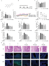
Figure 1
Figure 1. Dietary administration of oroxylin A attenuated DSS-induced chronic colitis
(A) Molecular structure of oroxylin A (C16H12O5. MW: 284.27). (B) The experimental protocol for the treatment with oroxylin A in chronic colitis model. (C, D) The lengths of colons and spleen indexes of each group were measured (n = 10). (E) The changes of body weight in each group during the disease process (n = 10). (F) Histopathological scores of each group were determined (n = 6). (G) MPO and (H) iNOS activities in the colonic tissues were detected (n = 6). (I) The secretion of IL-1β in serum was determined by ELISA. (JL) The protein levels of IL-1β, TNF-α and IL-6 in colonic tissue were determined. (M) Serial sections of colon tissues were stained with HE. Sections of colonic tissue were immunostained for DAPI (blue) and CD11b-FITC (green) or F4/80-FITC (green) (scale bar, 100 μm). Data were shown as means ± SD (n = 3). Statistical analysis was performed using one-way ANOVA coupled with a post hoc test. Significant differences were indicated as *P < 0.05 vs. control group (#P < 0.05 vs. DSS group); **P < 0.01 vs. control group (##P < 0.01 vs. DSS group).
Figure 2
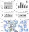
Figure 2. Oroxylin A suppressed the activation of NLRP3 inflammasome in DSS-induced chronic colitis
(A) The protein levels of cleaved-caspase-1 (caspase-1 p10), cleaved-IL-1β and NLRP3 in colonic tissues were detected by western blot. (B) The mRNA levels of NLRP3, IL-1β and IL-6 in colonic homogenate were determined by real-time RT-PCR. (C) The NF-κB p65, IκBα phosphorylation in colonic tissues were detected by western blot. (D) The relative expressions of cytoplastic protein and nuclear protein NF-κB p65 in colonic tissues were assessed by western blot. (E) Immunohistochemistry of IL-1β, NLRP3 and p65 in colonic tissues of each group was measured. Brown colored is positive (scale bar, 100 μm). Data were shown as means ± SD (n = 3). Statistical analysis was performed using one-way ANOVA coupled with a post hoc test. Significant differences were indicated as * P < 0.05 vs. control group (#P < 0.05 vs. DSS group); ** P < 0.01 vs. control group (##P < 0.01 vs. DSS group).
Figure 3
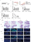
Figure 3. Oroxylin A ameliorated the symptoms of DSS-induced acute colitis via NLRP3 inflammasome
(A) The experimental protocol for the treatment with oroxylin A in acute colitis model. (B) The changes of body weight in each group during the disease process. Disease activity index (DAI) of each group was evaluated. (C, D) The lengths of colons and spleen indexes of each group were measured (n = 6). (E) MPO and (F) iNOS activities in the colonic tissues were detected (n = 6). (G) Histopathological scores of each group were determined (n = 6). (H) Serial sections of colon tissues were stained with HE. Sections of colonic tissue were immunostained for DAPI (blue) and CD11b-FITC (green) or F4/80-FITC (green) (scale bar, 100 μm). Data were shown as means ± SD (n = 3). Statistical analysis was performed using one-way ANOVA coupled with a post hoc test. Significant differences were indicated as *P < 0.05 vs. control group (#P < 0.05 vs. DSS group); **P < 0.01 vs. control group (##P < 0.01 vs. DSS group).
Figure 4
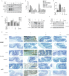
Figure 4. Oroxylin A down-regulated the activation of NLRP3 inflammasome in DSS-induced acute colitis
(A) The protein levels of cleaved-caspase-1 (caspase-1 p10), cleaved-IL-1β and NLRP3 in colonic tissues were detected by western blot. (B) The serum IL-1β, colonic IL-1β and (C) colonic IL-6 were determined by ELISA. (D) The mRNA levels of NLRP3, IL-1β, IL-6 and TNF-α in colonic tissue were determined by real-time RT-PCR. (E) The NF-κB p65 and IκBα phosphorylation in colonic tissues were detected by western blot. (F) The relative expressions of cytoplastic protein NF-κB p65 and nuclear protein NF-κB p65 in colonic tissues were assessed by western blot. (G) Immunohistochemistry of NLRP3, IL-1β and p65 in colonic tissues of each group was measured. Brown colored is positive (scale bar, 100 μm). Data were shown as means ± SD (n = 3). Statistical analysis was performed using one-way ANOVA coupled with a post hoc test. Significant differences were indicated as *P < 0.05 vs. control group (#P < 0.05 vs. DSS group); **P < 0.01 vs. control group (##P < 0.01 vs. DSS group).
Figure 5
Figure 5. Oroxylin A inhibited NLRP3 inflammasome activation in THP-Ms and BMDMs
(A) THP-Ms were stimulated with LPS (100 μM) and ATP (5 mM), then treated with indicated concentrations of oroxylin A for 12 h. The expressions of NLRP3, pro-caspase 1 and cleaved-caspase-1 (caspase-1 p10) proteins were assessed by western blot. (B) The secretion of IL-1β in supernatant was determined by ELISA. (C) The mRNA levels of NLRP3 and IL-1β were detected by real-time RT-PCR. (D) THP-Ms were treated with oroxylin A for 12 h at indicated doses, then protein levels of NLRP3, pro-caspase 1 and cleaved-caspase-1 (caspase-1 p10) were assessed by western blot. (E) BMDMs were stimulated with LPS (100 μM) and ATP (5 mM), then treated with oroxylin A for 12 h. The expressions of NLRP3, pro-caspase-1 and cleaved-caspase-1 (caspase-1 p10) proteins were assessed by western blot. (F) The amount of IL-1β in supernatant was determined by ELISA. (G) The expressions of NLRP3 and IL-1β mRNA in BMDMs were detected by real-time RT-PCR. (H) BMDMs were treated with oroxylin A for 12 h at indicated doses, the protein levels of NLRP3 and pro-caspase 1 were assessed by western blot. (I) The protein expressions of NF-κB p65, p-IκB and IκB were measured by western blot in THP-Ms. (J) The relative expressions of cytoplastic protein NF-κB p65 and nuclear protein NF-κB p65 in THP-Ms were assessed by western blot. (K) The mRNA expressions of IL-6 and TNF-α in THP-Ms were detected by real-time RT-PCR. (L) Immunofluorescence was performed to analyze NF-κB p65 nuclear translocation (scale bar, 20 μm). Data were shown as means ± SD (n = 3). Statistical analysis was performed using one-way ANOVA coupled with a post hoc test. Significant differences were indicated as *P < 0.05 vs. control group (#P < 0.05 vs. DSS group); **P < 0.01 vs. control group (##P < 0.01 vs. DSS group).
Figure 6
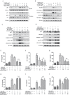
Figure 6. Oroxylin A blocked NLRP3 inflammasome activation through inhibiting the NF-κB signaling
(A) After pretreatment with bay11-7082 (20 μM), THP-Ms were stimulated with oroxylin A at indicated doses for 12 h. The expressions of cytoplastic protein p65, p-IκB, IκB and nuclear protein NF-κB p65 in THP-Ms were assessed by western blot. (B) The expressions of NLRP3, cleaved-caspase-1 (caspase-1 p10) and cleaved-IL-1β proteins were assessed by western blot. (C) BMDMs were transfected with NF-κB p65 siRNA for 24 h. Transfected BMDMs were stimulated with LPS and ATP for the indicated time. Protein levels of NLRP3, cleaved-caspase-1 (caspase-1 p10) and cleaved-IL-1β were determined by western Blot. (D) BMDMs were transfected with NF-κB p65 overexpression plasmid for 24 h. Transfected BMDMs were stimulated with LPS and ATP for the indicated time. Protein levels of NLRP3, cleaved-caspase-1 (caspase-1 p10) and cleaved-IL-1β were determined by western Blot. (EG) BMDMs were transfected with NF-κB p65 siRNA for 24 h. Transfected BMDMs were stimulated with LPS and ATP for the indicated time. IL-1β, IL-6 and TNF-α in supernatant were detected by ELISA. (HJ) BMDMs were transfected with NF-κB p65 overexpression plasmid for 24 h. Transfected BMDMs were stimulated with LPS and ATP for the indicated time. IL-1β, IL-6 and TNF-α in supernatant were detected by ELISA. Data were shown as means ± SD (n = 3). Statistical analysis was performed using one-way ANOVA coupled with a post hoc test. Significant differences were indicated as *P < 0.05 vs. control group (#P < 0.05 vs. DSS group); **P < 0.01 vs. control group (##P < 0.01 vs. DSS group).
Figure 7
Figure 7. Oroxylin A blocked ASC speck formation and inflammasome assembly
(A) Oroxylin A-treated THP-Ms were pretreated with cycloheximide for 1 h, and then stimulated with LPS and ATP. Cleaved-caspase-1 (caspase-1 p10), cleaved-IL-1β and NLRP3 were detected by western blot. (B) Oroxylin A-treated THP-Ms were stimulated with LPS together with ATP for 1 h. Cleaved-caspase-1 (caspase-1 p10), cleaved-IL-1β and NLRP3 were detected by western blot. (C) Immunoblotting of ASC was performed in crosslinked pellets and in cell lysates. (D) Immunostaining of endogenous ASC was performed (scale bar, 20 μm). The results are representative of three independent experiments.

Similar articles

Cited by

References

    1. Ponder A, Long MD. A clinical review of recent findings in the epidemiology of inflammatory bowel disease. Clin Epidemiol. 2013;5:237–247. - PMC - PubMed
    1. Podolsky DK. Inflammatory bowel disease. N Engl J Med. 2002;347:417–429. - PubMed
    1. Tontini GE, Vecchi M, Pastorelli L, Neurath MF, Neumann H. Differential diagnosis in inflammatory bowel disease colitis: state of the art and future perspectives. World J Gastroenterol. 2015;21:21–46. - PMC - PubMed
    1. Zaki MH, Lamkanfi M, Kanneganti TD. The Nlrp3 inflammasome: contributions to intestinal homeostasis. Trends Immunol. 2011;32:171–179. - PMC - PubMed
    1. Biasi F, Leonarduzzi G, Oteiza PI, Poli G. Inflammatory bowel disease: mechanisms, redox considerations, and therapeutic targets. Antioxid Redox Signal. 2013;19:1711–1747. - PMC - PubMed

LinkOut - more resources