. 2017 Sep 19;20(12):2921-2934.
doi: 10.1016/j.celrep.2017.08.078.

Human Tissue-Resident Memory T Cells Are Defined by Core Transcriptional and Functional Signatures in Lymphoid and Mucosal Sites

Affiliations

Human Tissue-Resident Memory T Cells Are Defined by Core Transcriptional and Functional Signatures in Lymphoid and Mucosal Sites

Brahma V Kumar et al. Cell Rep. .

Abstract

Tissue-resident memory T cells (TRMs) in mice mediate optimal protective immunity to infection and vaccination, while in humans, the existence and properties of TRMs remain unclear. Here, we use a unique human tissue resource to determine whether human tissue memory T cells constitute a distinct subset in diverse mucosal and lymphoid tissues. We identify a core transcriptional profile within the CD69+ subset of memory CD4+ and CD8+ T cells in lung and spleen that is distinct from that of CD69- TEM cells in tissues and circulation and defines human TRMs based on homology to the transcriptional profile of mouse CD8+ TRMs. Human TRMs in diverse sites exhibit increased expression of adhesion and inhibitory molecules, produce both pro-inflammatory and regulatory cytokines, and have reduced turnover compared with circulating TEM, suggesting unique adaptations for in situ immunity. Together, our results provide a unifying signature for human TRM and a blueprint for designing tissue-targeted immunotherapies.

Keywords: RNA-seq; human immunology; memory T cells; mucosal immunity.


Figures

Figure 1
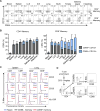
Figure 1. CD69+ memory T cells are prevalent in tissues and do not show features of activation
(A) Expression of CD69 and CD103 by CD4+ (top) and CD8+ (lower) memory T cells (CCR7-CD45RA-) within each indicated site from one individual (donor 332) representative of 6 donors. LLN: Lung lymph node, ILN: inguinal lymph node, MLN: mesenteric lymph node. (B) Frequency of CD69+CD103+ (grey) and CD69+CD103+ (blue) CD4+ (left) and CD8+ memory T cells in each tissue compiled from 16–22 donors. (C) Activation profile of T cells from human tissues. Flow cytometry plots show expression of CD25, CD38, and HLA-DR by naïve T cells (CCR7+ CD45RA+, blue line), and CD69 (black) and CD69+ (red) subsets of memory T cells (CCR7CD45RA). Data are representative of 3 donors. (D) Sorting strategy for isolation of CD4+ and CD8+ CD45RACCR7CD69+ and CD45RACCR7CD69 T cells for RNA sequencing is shown from spleen.
Figure 2
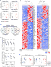
Figure 2. CD69 expression defines a transcriptionally distinct memory subset in humans with features of tissue residency
Whole transcriptome profiling by RNA sequencing was performed on CD69 and CD69+ subsets of CD4+ and CD8+ memory T cells from spleen and lungs of 3 donors (Donors 226, 233, 250; see methods). (A) Principle Component Analysis (PCA) of paired CD69+ and CD69 samples from spleen and lung and for CD4+ and CD8+ subsets, based on the global transcriptome (~20000 genes). (B) Diagram shows the number of significant differentially expressed genes (FDR≤0.05 and log2 fold-change ≥1) between CD69 and CD69+ samples within each tissue for CD4+ and CD8+ T cells showing overlap between tissues. (C) Heat map showing normalized expression levels of the overlap genes identified in (B) for CD4+ (77 genes) and CD8+ (133 genes) CD69 vs. CD69+ subsets from spleen (S) and lung (L). (D) Transcriptional downregulation of S1PR1 and KLF2 in all CD69+ vs. CD69 subsets. Normalized expression levels of S1PR1 (top) and KLF2 (bottom) transcripts in CD69 and CD69+ samples from spleen (S) and lung (L) of each donor are shown. Individual donors are indicated by distinct symbols, and lines connect samples from identical donors within a tissue. **** FDR≤10−5, *** FDR≤10−3. (E) PCA of CD69+ (red) and CD69 (black: tissue, blue: blood) memory subsets based on the genes in (C). S=spleen, L=lung, B=blood. See also Figure S1 and Table S6.
Figure 3
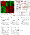
Figure 3. A core gene signature defines tissue CD69+ memory T cells distinct from circulating CD69− cells in tissues and blood
(A) Heatmap shows normalized expression of genes with significant differential expression between CD69+ and CD69 memory T cells for all subsets (CD4+, CD8+) and tissues (spleen, lung). (B) Network analysis of the core gene set in (A) showing known and predicted interactions (activating, inhibitory) between proteins encoded by the core genes that are upregulated (red) or downregulated (green) by TRM compared with TEM with key pathways indicated in the shaded boxes. Relationships were determined using IPA software, String Protein database, GeneCards, and Pubmed literature searches. (C-G) Normalized mRNA expression levels of ITGA1 (C), ITGAE (D), CXCR6 (E), CX3CR1 (F), and PDCD1 (G) by CD4+ and CD8+ CD69+ and CD69 memory subsets in blood (B), spleen (S) and lung (L) of each individual donor.*FDR≤0.05, ** FDR≤10−2, ***FDR≤10−3, ****FDR≤10−5. Each donor is represented by a unique shape as indicated. See also Figures S2 and S3.
Figure 4
Figure 4. Comparison of the human and mouse TRM transcriptome
(A) PCA was performed using RNA-Seq data presented here (black symbols) compared to mouse herpes simplex virus (HSV)-specific CD8+ TRM from skin and CD8+TEM from spleen (“mouse HSV”, yellow) and LCMV-specific CD8+TRM from intestine and CD8+TEM from spleen (“Mouse LCMV”, red) (Mackay et al. 2016). Left: PCA comparing whole transcriptomes of each dataset comprising 15571 common genes between human and mouse. Right: PCA comparing the core human gene signature (Fig. 3) to the mouse datasets. (B) Gene set enrichment analysis (GSEA) comparing our human CD8+ (left) and CD4+ (left) gene sets to published microarray data of CD103+ brain TRM vs. spleen TEM (top row), gut TRM vs. spleen TEM (middle row), and lung TRM vs. spleen TEM (middle row) (Wakim et al. 2012; Mackay et al. 2013). In each plot, the x-axis shows the genes ranked with absolute value of log fold change between TRM vs. TEM and y-axis shows running enrichment score (ES) comparing the ranked list of genes with indicated p values. (C) Comparison of Hobit gene expression in mouse and human datasets. Violin plots show Z score of gene expression levels from mouse TRM (from Mackay et al. 2016) and human CD69+ memory T cells (this study). Red dots represent Hobit, blue dots represent the housekeeping gene GAPDH, green dots represent CD69, and the white dot represents median gene expression.
Figure 5
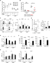
Figure 5. TCR clonal analysis, turnover, and function of CD69+ and CD69 cells
(A) CD8+ T cells have reduced TCR repertoire diversity compared with CD4+ cells. CDR3 sequences were inferred from RNA-Seq data using TRUST (See methods). Graph shows the number of unique CDR3 calls (clonotypes) in each sample per 103 reads mapped to the TCR region. (B) Increased tissue overlap of TCR clones within CD8+ CD69 compared to CD69+ memory T cells. Graph shows percentage of overlapping clones between lung and spleen samples from each donor, calculated by dividing the total number of overlapping clones by the total number of unique clones present in both tissues. See also Figure S4. (C) Reduced proliferative turnover by CD69+ memory T cells. Left: Representative flow cytometry plots of intracellular Ki67 expression from spleen and lungs of one individual donor. Right: Ki67 expression compiled from 10 donors depicted as mean frequency KI67+±SEM. (D) Increased CD57 by CD69 compared to CD69+ cells. Left: CD57 expression by CD69 and CD69+ memory T cell subsets from spleen and lung of one representative donor. Right: CD57 expression compiled from 11 donors displayed as mean percent positive ±SEM. * p≤0.05, ** p≤0.01. (E-G) Distinct functional profile of CD69+ cells. CD4+ and CD8+ CD69 and CD69+ memory T cells isolated from spleens and lungs were stimulated with PMA/Ionomycin and cytokine production was assessed by intracellular cytokine staining (ICS) (for IL-2, IFN-γ, IL-17), or were stimulated with anti-CD3/CD28 beads (for IL-10) and IL-10 levels in the supernatant were assessed by BD cytokine bead array. (E) Graph shows mean frequency of CD69 and CD69+ cells producing IL-2. (F) Graph shows mean ±SEM IL-10 production in pg/ml. (G) Graph shows mean frequency of CD69 and CD69+ cells producing IFN-γ (left) and IL-17A (right) ±SEM. n=6 donors spleen, 10 donors lung for IL-2, IFN-γ, IL-17, n=3 donors for IL-10. *p<0.05, **p<0.01, ***p<0.001. Unstimulated cells had minimal cytokine production (<5%).
Figure 6
Figure 6. Lineage and tissue-specific transcription and phenotypic profiles in human CD69+ memory T cells
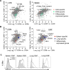
(A) Analysis of lineage-specific gene expression in CD69+ and CD69 memory T cells. Scatter plots display log2 fold change of CD4+CD69+ vs. CD69 on the x axis and CD8+ subsets on the y axis from lung (left) and spleen (right). Grey dots represent genes with significant differential expression in any paired CD69+ vs. CD69 sample. Orange dots (“CD4 specific”) represent genes with significant differential expression in CD4+ CD69+ vs. CD69 but not in CD8+ samples. Green dots (“CD8 specific”) represent genes with significant differential expression in CD8+CD69+ vs. CD69 but not in CD4+ samples. (B) Analysis of tissue-specific genes in CD69+ and CD69 memory T cells. Scatter plots display log2 fold change of lung CD69+ vs. CD69 samples on the x axis and spleen samples on the y axis for CD4+ (left) and CD8+ (right) T cells using the same strategy as in (A) with red dots denoting “spleen specific” and blue dots denoting “lung specific” transcripts in the paired analysis. (C) CD101 expression in human tissues. Representative plots show CD101 expression in CD69+ (black outline) and CD69 (shaded) cells from one individual donor. Data are representative of 15 donors (see Figure S5).
Figure 7
Figure 7. TRM are a phenotypically distinct subset across multiple tissues
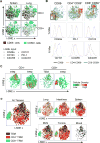
Simultaneous expression of CD49a, CD103, CD101, CXCR6, CX3CR1, PD-1, and CD69 was visualized using t-SNE analysis. (A) CD69+ and CD69− memory T cells are phenotypically distinct in spleen and lung. Plots show CD69+ memory T cells (color coded green) and CD69 memory T cells (color coded black) from spleen and lungs of an individual donor (Donor 321) representative of 5 donors. (B) Defining the phenotype of TRM and TEM clusters. Regions with high cellular density were manually gated within TEM (CD69), CD4+ TRM (CD69+), and CD8+ TRM (CD69+) fractions (top row). Histograms show expression levels of CD49a, CD103, CD101, CXCR6, CX3CR1, and PD-1 within gated regions (bottom row). (C-D) The core TRM phenotype is observed across multiple tissues. Phenotype analysis as in (A) was performed using lung, intestine, spleen, mesenteric lymph node (MLN), tonsils, and blood samples from one representative donor (Donor 332). (C) Plots show CD4+ (left) and CD8+ (right) TRM and TEM subsets from all tissues with cell number density color coded. (D) Plots shows cells from all tissues (left large plot) or each individual site (Right smaller plots) color coded by cell type (CD4+ TRM, red; CD8+ TRM, green; TEM, black) of one donor representative of 4 donors. See also Figure S6.

Similar articles

Cited by

References

    1. Anderson KG, Mayer-Barber K, Sung H, Beura L, James BR, Taylor JJ, Qunaj L, Griffith TS, Vezys V, Barber DL, et al. Intravascular staining for discrimination of vascular and tissue leukocytes. Nat Protoc. 2014;9:209–222. - PMC - PubMed
    1. Barber DL, Wherry EJ, Masopust D, Zhu B, Allison JP, Sharpe AH, Freeman GJ, Ahmed R. Restoring function in exhausted CD8 T cells during chronic viral infection. Nature. 2006;439:682–687. - PubMed
    1. Bergsbaken T, Bevan MJ. Proinflammatory microenvironments within the intestine regulate the differentiation of tissue-resident CD8(+) T cells responding to infection. Nat Immunol. 2015;16:406–414. - PMC - PubMed
    1. Bertin S, Lozano-Ruiz B, Bachiller V, Garcia-Martinez I, Herdman S, Zapater P, Frances R, Such J, Lee J, Raz E, et al. Dual-specificity phosphatase 6 regulates CD4+ T-cell functions and restrains spontaneous colitis in IL-10-deficient mice. Mucosal Immunol. 2015;8:505–515. - PMC - PubMed
    1. Beura LK, Hamilton SE, Bi K, Schenkel JM, Odumade OA, Casey KA, Thompson EA, Fraser KA, Rosato PC, Filali-Mouhim A, et al. Normalizing the environment recapitulates adult human immune traits in laboratory mice. Nature. 2016;532:512–516. - PMC - PubMed