The RNA-editing enzyme ADAR promotes lung adenocarcinoma migration and invasion by stabilizing FAK
- PMID: 28928239
- PMCID: PMC5771642
- DOI: 10.1126/scisignal.aah3941
The RNA-editing enzyme ADAR promotes lung adenocarcinoma migration and invasion by stabilizing FAK
Abstract
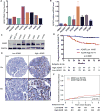
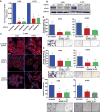
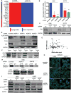
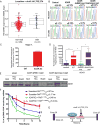
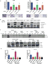
Large-scale, genome-wide studies report that RNA binding proteins are altered in cancers, but it is unclear how these proteins control tumor progression. We found that the RNA-editing protein ADAR (adenosine deaminase acting on double-stranded RNA) acted as a facilitator of lung adenocarcinoma (LUAD) progression through its ability to stabilize transcripts encoding focal adhesion kinase (FAK). In samples from 802 stage I LUAD patients, increased abundance of ADAR at both the mRNA and protein level correlated with tumor recurrence. Knocking down ADAR in LUAD cells suppressed their mesenchymal properties, migration, and invasion in culture. Analysis of gene expression patterns in LUAD cells identified ADAR-associated enrichment of a subset of genes involved in cell migration pathways; among these, FAK is the most notable gene whose expression was increased in the presence of ADAR. Molecular analyses revealed that ADAR posttranscriptionally increased FAK protein abundance by binding to the FAK transcript and editing a specific intronic site that resulted in the increased stabilization of FAK mRNA. Pharmacological inhibition of FAK blocked ADAR-induced invasiveness of LUAD cells, suggesting a potential therapeutic application for LUAD that has a high abundance of ADAR.
Copyright © 2017 The Authors, some rights reserved; exclusive licensee American Association for the Advancement of Science. No claim to original U.S. Government Works.
Conflict of interest statement
Figures

Similar articles
-
Global Transcriptome Analysis of RNA Abundance Regulation by ADAR in Lung Adenocarcinoma.EBioMedicine. 2018 Jan;27:167-175. doi: 10.1016/j.ebiom.2017.12.005. Epub 2017 Dec 6. EBioMedicine. 2018. PMID: 29273356 Free PMC article.
-
The RNA editing enzyme ADAR modulated by the rs1127317 genetic variant diminishes EGFR-TKIs efficiency in advanced lung adenocarcinoma.Life Sci. 2022 May 1;296:120408. doi: 10.1016/j.lfs.2022.120408. Epub 2022 Feb 22. Life Sci. 2022. PMID: 35202641
-
p53-inducible gene 3 promotes cell migration and invasion by activating the FAK/Src pathway in lung adenocarcinoma.Cancer Sci. 2018 Dec;109(12):3783-3793. doi: 10.1111/cas.13818. Epub 2018 Oct 26. Cancer Sci. 2018. PMID: 30281878 Free PMC article.
-
In cancer, A-to-I RNA editing can be the driver, the passenger, or the mechanic.Drug Resist Updat. 2017 May;32:16-22. doi: 10.1016/j.drup.2017.09.001. Epub 2017 Oct 4. Drug Resist Updat. 2017. PMID: 29145975 Review.
-
Mechanisms and implications of ADAR-mediated RNA editing in cancer.Cancer Lett. 2017 Dec 28;411:27-34. doi: 10.1016/j.canlet.2017.09.036. Epub 2017 Sep 30. Cancer Lett. 2017. PMID: 28974449 Review.
Cited by
-
Genetics of cell-type-specific post-transcriptional gene regulation during human neurogenesis.Am J Hum Genet. 2024 Sep 5;111(9):1877-1898. doi: 10.1016/j.ajhg.2024.07.015. Epub 2024 Aug 20. Am J Hum Genet. 2024. PMID: 39168119
-
ADAR1-mediated RNA-editing of 3'UTRs in breast cancer.Biol Res. 2018 Oct 5;51(1):36. doi: 10.1186/s40659-018-0185-4. Biol Res. 2018. PMID: 30290838 Free PMC article.
-
ADAR, the carcinogenesis mechanisms of ADAR and related clinical applications.Ann Transl Med. 2019 Nov;7(22):686. doi: 10.21037/atm.2019.11.06. Ann Transl Med. 2019. PMID: 31930087 Free PMC article. Review.
-
AGO2 Mediates MYC mRNA Stability in Hepatocellular Carcinoma.Mol Cancer Res. 2020 Apr;18(4):612-622. doi: 10.1158/1541-7786.MCR-19-0805. Epub 2020 Jan 15. Mol Cancer Res. 2020. PMID: 31941754 Free PMC article.
-
Role of RNA modifications in cancer.Nat Rev Cancer. 2020 Jun;20(6):303-322. doi: 10.1038/s41568-020-0253-2. Epub 2020 Apr 16. Nat Rev Cancer. 2020. PMID: 32300195 Review.
References
-
- Siegel RL, Miller KD, Jemal A. Cancer statistics, 2015. CA Cancer J Clin. 2015;65:5–29. - PubMed
-
- Pao W, Hutchinson KE. Chipping away at the lung cancer genome. Nat Med. 2012;18:349–351. - PubMed
-
- Fumagalli D, Gacquer D, Rothe F, Lefort A, Libert F, Brown D, Kheddoumi N, Shlien A, Konopka T, Salgado R, Larsimont D, Polyak K, Willard-Gallo K, Desmedt C, Piccart M, Abramowicz M, Campbell PJ, Sotiriou C, Detours V. Principles Governing A-to-I RNA Editing in the Breast Cancer Transcriptome. Cell Rep. 2015;13:277–289. - PMC - PubMed
-
- Han L, Diao L, Yu S, Xu X, Li J, Zhang R, Yang Y, Werner HM, Eterovic AK, Yuan Y, Li J, Nair N, Minelli R, Tsang YH, Cheung LW, Jeong KJ, Roszik J, Ju Z, Woodman SE, Lu Y, Scott KL, Li JB, Mills GB, Liang H. The Genomic Landscape and Clinical Relevance of A-to-I RNA Editing in Human Cancers. Cancer Cell. 2015;28:515–528. - PMC - PubMed
MeSH terms
Substances
Grants and funding
LinkOut - more resources
Full Text Sources
Other Literature Sources
Medical
Research Materials
Miscellaneous

