Histopathology of Middle East respiratory syndrome coronovirus (MERS-CoV) infection - clinicopathological and ultrastructural study
- PMID: 28858401
- PMCID: PMC7165512
- DOI: 10.1111/his.13379
Histopathology of Middle East respiratory syndrome coronovirus (MERS-CoV) infection - clinicopathological and ultrastructural study
Abstract
Aims: The pathogenesis, viral localization and histopathological features of Middle East respiratory syndrome - coronavirus (MERS-CoV) in humans are not described sufficiently. The aims of this study were to explore and define the spectrum of histological and ultrastructural pathological changes affecting various organs in a patient with MERS-CoV infection and represent a base of MERS-CoV histopathology.
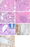
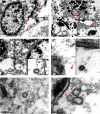
Methods and results: We analysed the post-mortem histopathological findings and investigated localisation of viral particles in the pulmonary and extrapulmonary tissue by transmission electron microscopic examination in a 33-year-old male patient of T cell lymphoma, who acquired MERS-CoV infection. Tissue needle biopsies were obtained from brain, heart, lung, liver, kidney and skeletal muscle. All samples were collected within 45 min from death to reduce tissue decomposition and artefact. Histopathological examination showed necrotising pneumonia, pulmonary diffuse alveolar damage, acute kidney injury, portal and lobular hepatitis and myositis with muscle atrophic changes. The brain and heart were histologically unremarkable. Ultrastructurally, viral particles were localised in the pneumocytes, pulmonary macrophages, renal proximal tubular epithelial cells and macrophages infiltrating the skeletal muscles.
Conclusion: The results highlight the pulmonary and extrapulmonary pathological changes of MERS-CoV infection and provide the first evidence of the viral presence in human renal tissue, which suggests tissue trophism for MERS-CoV in kidney.
Keywords: MERS-CoV; Middle East respiratory syndrome coronavirus; electron microscopy; extra pulmonary; histopathology; pulmonary; renal.
© 2017 John Wiley & Sons Ltd.
Conflict of interest statement
The authors declare no conflicts of interest.
Figures


Similar articles
-
Clinicopathologic, Immunohistochemical, and Ultrastructural Findings of a Fatal Case of Middle East Respiratory Syndrome Coronavirus Infection in the United Arab Emirates, April 2014.Am J Pathol. 2016 Mar;186(3):652-8. doi: 10.1016/j.ajpath.2015.10.024. Epub 2016 Feb 5. Am J Pathol. 2016. PMID: 26857507 Free PMC article.
-
Comparative pathology of rhesus macaque and common marmoset animal models with Middle East respiratory syndrome coronavirus.PLoS One. 2017 Feb 24;12(2):e0172093. doi: 10.1371/journal.pone.0172093. eCollection 2017. PLoS One. 2017. PMID: 28234937 Free PMC article.
-
Acute Respiratory Infection in Human Dipeptidyl Peptidase 4-Transgenic Mice Infected with Middle East Respiratory Syndrome Coronavirus.J Virol. 2019 Mar 5;93(6):e01818-18. doi: 10.1128/JVI.01818-18. Print 2019 Mar 15. J Virol. 2019. PMID: 30626685 Free PMC article.
-
Modulation of the immune response by Middle East respiratory syndrome coronavirus.J Cell Physiol. 2019 Mar;234(3):2143-2151. doi: 10.1002/jcp.27155. Epub 2018 Aug 26. J Cell Physiol. 2019. PMID: 30146782 Free PMC article. Review.
-
A Comparative Review of Animal Models of Middle East Respiratory Syndrome Coronavirus Infection.Vet Pathol. 2016 May;53(3):521-31. doi: 10.1177/0300985815620845. Epub 2016 Feb 11. Vet Pathol. 2016. PMID: 26869154 Review.
Cited by
-
Acute Liver Failure in a COVID-19 Patient Without any Preexisting Liver Disease.Cureus. 2020 Aug 26;12(8):e10045. doi: 10.7759/cureus.10045. Cureus. 2020. PMID: 32983735 Free PMC article.
-
Pathology and pathogenicity of severe acute respiratory syndrome coronavirus 2 (SARS-CoV-2).Exp Biol Med (Maywood). 2020 Sep;245(15):1299-1307. doi: 10.1177/1535370220942126. Epub 2020 Jul 7. Exp Biol Med (Maywood). 2020. PMID: 32635753 Free PMC article. Review.
-
Antifibrotics in COVID-19 Lung Disease: Let Us Stay Focused.Front Med (Lausanne). 2020 Sep 9;7:539. doi: 10.3389/fmed.2020.00539. eCollection 2020. Front Med (Lausanne). 2020. PMID: 33072773 Free PMC article.
-
A comparison of radiographic features between non-survivors and survivors from ICU.Eur J Radiol Open. 2021;8:100338. doi: 10.1016/j.ejro.2021.100338. Epub 2021 Apr 13. Eur J Radiol Open. 2021. PMID: 33869677 Free PMC article.
-
Differences in syncytia formation by SARS-CoV-2 variants modify host chromatin accessibility and cellular senescence via TP53.Cell Rep. 2023 Dec 26;42(12):113478. doi: 10.1016/j.celrep.2023.113478. Epub 2023 Nov 21. Cell Rep. 2023. PMID: 37991919 Free PMC article.
References
-
- Zaki AM, van Boheemen S, Bestebroer TM, et al Isolation of a novel coronavirus from a man with pneumonia in Saudi Arabia. N. Engl. J. Med. 2012; 367; 1814–1820. - PubMed
-
- World Health Organization . Middle East respiratory syndrome coronavirus (MERS‐CoV). Available at: https://doi.org/http://www.who.int/emergencies/mers-cov/en/ (accessed 14 July 2017).
-
- Arabi YM, Arifi AA, Balkhy HH, et al Clinical course and outcomes of critically ill patients with Middle East respiratory syndrome coronavirus infection. Ann. Intern. Med. 2014; 160; 389–397. - PubMed
Publication types
MeSH terms
LinkOut - more resources
Full Text Sources
Other Literature Sources

