. 2017 Jul 17;8(8):3671-3686.
doi: 10.1364/BOE.8.003671. eCollection 2017 Aug 1.

Whole blood clot optical clearing for nondestructive 3D imaging and quantitative analysis

Affiliations

Whole blood clot optical clearing for nondestructive 3D imaging and quantitative analysis

Peter Höök et al. Biomed Opt Express. .

Abstract

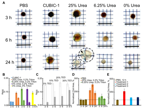
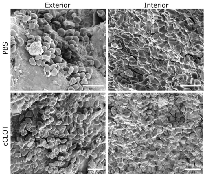
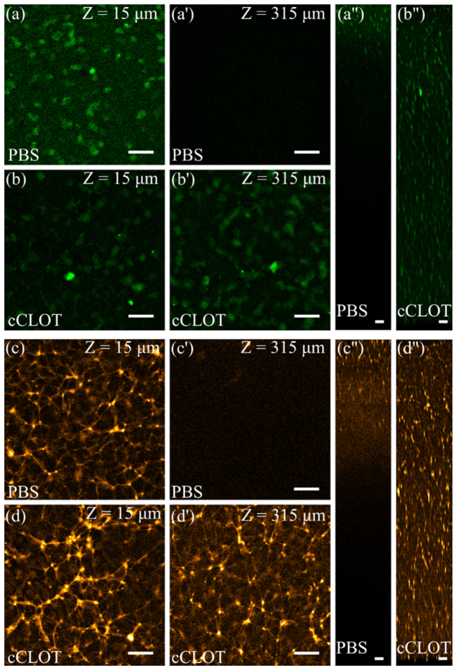
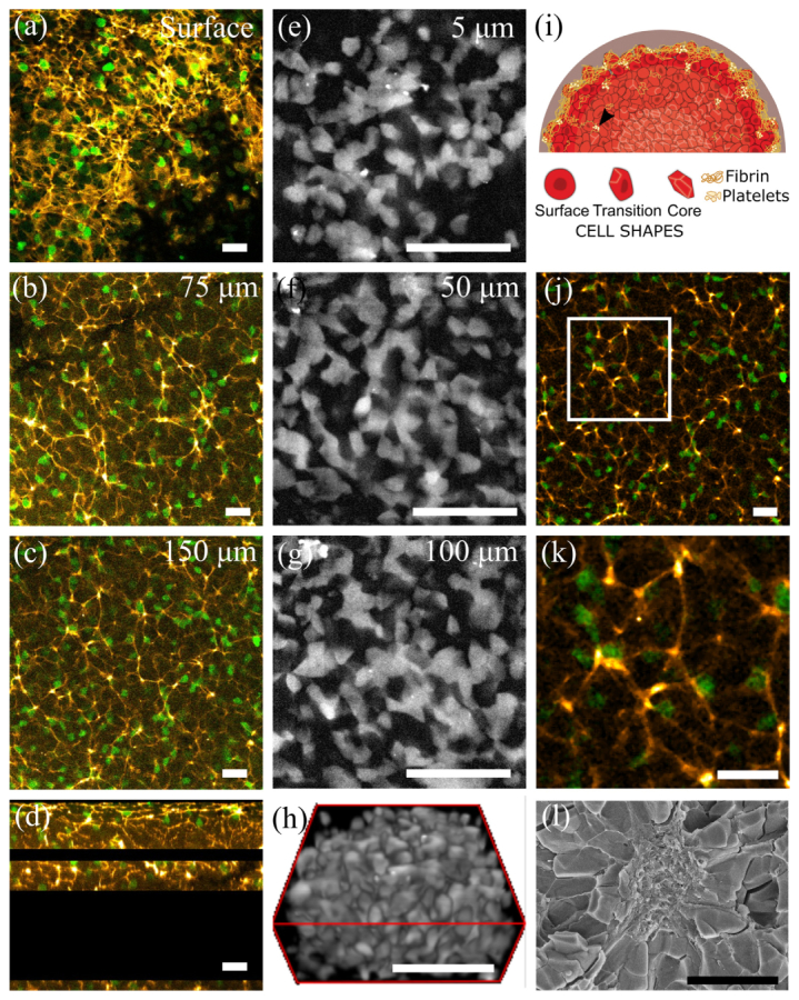
A technological revolution in both light and electron microscopy imaging now allows unprecedented views of clotting, especially in animal models of hemostasis and thrombosis. However, our understanding of three-dimensional high-resolution clot structure remains incomplete since most of our recent knowledge has come from studies of relatively small clots or thrombi, due to the optical impenetrability of clots beyond a few cell layers in depth. Here, we developed an optimized optical clearing method termed cCLOT that renders large whole blood clots transparent and allows confocal imaging as deep as one millimeter inside the clot. We have tested this method by investigating the 3D structure of clots made from reconstituted pre-labeled blood components yielding new information about the effects of clot contraction on erythrocytes. Although it has been shown recently that erythrocytes are compressed to form polyhedrocytes during clot contraction, observations of this phenomenon have been impeded by the inability to easily image inside clots. As an efficient and non-destructive method, cCLOT represents a powerful research tool in studying blood clot structure and mechanisms controlling clot morphology. Additionally, cCLOT optical clearing has the potential to facilitate imaging of ex vivo clots and thrombi derived from healthy or pathological conditions.

Keywords: (170.1790) Confocal microscopy; (170.3660) Light propagation in tissues; (170.3880) Medical and biological imaging; (170.6935) Tissue characterization.


Figures

Fig. 1
Fig. 1
Developing an optimized optical clearing method for whole blood clots. (a): Images of trans-illuminated PFA-fixed blood clots treated with PBS; CUBIC-1; or solutions containing 25% TED, 0.2% Triton X-100 and various concentrations of urea for 3, 6, and 24 h. 25% urea produced substantial clot expansion and damage. Inset in third column at 24 h: Close-up of fractured clot (arrowhead). Scale bar, 10 mm. (b): The rate of clot heme release was monitored by measuring the absorbance of clearing solutions at 600 nm following treatment at 3 hours and 6 hours with CUBIC-1 or solutions containing 25% TED and various concentrations of urea and Triton X-100. A 6.25% urea and 0.2% Triton X-100 solution significantly decolorized clots as compared to CUBIC-1 over 3 hours (p < 0.001) (c): Efficiency of heme release following treatment with 6.25% urea, 0.2% Triton X-100 and various concentrations of TED. Results are shown as average ± standard deviation (SD) of triplicate samples. TED of 25%-35% achieves significantly better heme clearance over a longer treatment period than 5% TED (6 h, p < 0.01). (d): Relative blood clot area measured at 3, 6 and 24 h. Dotted line represents 100%. A four-fold reduction in urea concentration (6.25%) produced transparent clots that were comparable in size to non-cleared control (Fig. 1(a), 1(d)) and significantly reduced the 6 h peak expansion (p < 0.05). (e): Excessive expansion of PFA-fixed clots following 24 hours of clearing with 6.25% urea (red, Treatment 1) could be reversed by an additional hour of treatment with PBS (light blue, Treatment 2) with a significantly reduced expansion (p < 0.02), followed by at least 1 hour of equilibration in CUBIC-2 (dark blue, Treatment 3). Clot area (%) for post-clearing solutions normalized to initial clot area. Dotted line represents 100%. Results reported as average ± standard deviation (SD) of triplicate samples.
Fig. 2
Fig. 2
Ultrastructural analysis of cCLOT-cleared blood clots. SEM images of the exterior and interior of blood clots treated with PBS or cCLOT for 24 hours. The structural arrangement with biconcave erythrocytes at the exterior and predominantly compressed polygonal-shaped erythrocytes at the interior of the clot is preserved after optical clearance. Scale bar, 10 μm.
Fig. 3
Fig. 3
PBS control vs. cCLOT-treated sample. (a, a’, a” and b, b’, b”): Clot erythrocytes visualized by two-photon imaging; 2% (v/v) of erythrocyte fraction relative to total reconstituted blood was stained with DRCT (Deep Red Cell Tracker). (a-a”): non-cleared control clot vs. (b-b”) cCLOT-cleared clot. (a”, b”): XZ orthogonal views. cCLOT enables visualization of erythrocyte shape and volume deep within the clot. (c, c’, c” and d, d’, d”): Clots stained for fibrin showing difference between non-cleared and cCLOT cleared clots. Note the extensive fibrin network at the outer edge and within the clot core (c”, d”): XZ orthogonal views of non-cleared vs. cCLOT-cleared clots demonstrate the ability of the cCLOT method to image fibrin networks throughout the clot and at depths that exceeds control clots by ~5-fold. Entire region depicted in a”, b”, c” and d” represents a depth of 739 μm. Fibrin aggregates can be seen as small, closely packed irregular structures from which fibrin fibers project. Imaged with 25x water objective (1.0 NA, 4x digital magnification) with 1 µm step z-slices. Scale bars are 25 μm.
Fig. 4
Fig. 4
Confocal microscopy of optically cleared clots. Left panels: Composite confocal images from a cCLOT-cleared clot in which fibrin is labeled with Alexa 564 fibrinogen (orange hot) and containing a 2% (v/v) of DRCT-labeled erythrocyte fraction relative to total reconstituted blood (green), (a): Clot surface, (b): 75 µm depth, (c): 150 µm depth, and (d): Orthogonal YZ view showing the Z-stacks obtained at 3 different depths of the clot. Gaps between Z-stacks were not imaged as a compromise to deal with the limited confocal scanning speed. Scale bars are 20 µm. Middle panels: Clots made with 20% (v/v) DRCT-labeled erythrocyte fraction relative to total reconstituted blood (grey) were imaged using a spinning disc confocal microscope with 60X objective lens: (e): 5 µm depth, (f): 50 µm depth, (g): 100 µm depth, and (h): 3D reconstruction of a 50 µm thick Z stack (0.1 µm steps). Scale bar, 50 µm. Panels e-h were imaged from a cCLOT solution containing 25% urea. Right panels: (i): Illustration of a blood clot with a platelet aggregate (black arrow) located deep in the clot. (j): cCLOT allows detection of red blood cells and fibrin aggregates deep within the clot (150 µm depth shown) erythrocytes and fibrin labeled as described in left panels (k): Inset from j, white square, showing magnified fibrin aggregate. (l) SEM of the exposed interior of a clot visualizing a platelet aggregate in the center of compressed erythrocytes. Note that associated fibrin fibers are not clearly visible. Scale bar, 10 µm.
Fig. 5
Fig. 5
Erythrocyte volumes in a contracted, cCLOT-cleared, clot at different depths (a): 3D view of clot showing sections used for erythrocyte volume calculations. Scale bar, 100 µm (b): Average volume of erythrocytes as a function of clot depth. The erythrocyte volume was measured from segmented images of z-stacks obtained at 1 µm steps in clots containing 2% v/v of DRCT-labeled erythrocyte fraction relative to total reconstituted blood (erythrocytes shown in green). Images for (a) and (b) were obtained using two photon microscopy with 25X water lens 4x digital magnification. Erythrocyte volumes in contracted versus non-contracted cCLOT cleared clots (c): Average volume of erythrocytes was measured for control non-treated reconstituted whole blood (WB) clots and clots made with blebbistatin treated reconstituted whole blood. The erythrocyte volume for (c) was measured from segmented images of z-stacks of clots containing 2% v/v of DRCT-labeled erythrocyte fraction relative to total reconstituted blood. Data for figure (c) extracted from images obtained using an Olympus confocal microscope with 60X oil lens. Z-stacks obtained at 0.1 µm steps.
Fig. 6
Fig. 6
Structural analysis of perturbed clots. Effects of the actomyosin inhibitor blebbistatin (Blebb) on blood clot contraction and fibrin network: (a): Normal clots contracted in the absence of blebbistatin (left, white arrow), versus non-contracted clots (right panels) made with blood pre-treated with blebbistatin (0.3 mM or 0.05 mM). Clots were made in cylindrical well chambers (5 mm diameter x 1 mm depth) on silicon gasket slides, which represent the geometry of cylindrical cross-sections. (b, c): Standard deviation projections of confocal z-stacks from contracted clots, (b), and clots where contraction was inhibited by 0.3 mM blebbistatin (c). Dramatic differences in fibrin aggregate distribution and architecture are apparent (red arrows). Projections represent a 25 μm slab sampled from ~15 μm under the clot surface and imaged on Olympus confocal configuration using 60x oil objective (d, e): Quantification of the number and size of fibrin aggregates in standard deviation projections of 25 μm slabs from 4 clots incubated with and without 0.3 mM blebbistatin. Contracted, control, clots show a significant difference in the number and size of fibrin aggregates indicative of a homogenously distributed fibrin network. Image (d) shows the average number of fibrin aggregates ± SD per region for 4 samples. Aggregates were differentiated from fibrin fibers. Image (e) shows the average area of fibrin aggregates ± SD per region for 4 samples. Quantified region at 60x represents an area of 179 x 179 μm. (g, h): Maximum intensity z-projection of a 25 μm slab (50 μm to 75 μm depth) of a contracted clot (g) and uncontracted clot formed in the presence of blebbistatin (h). Scale bars are 10 μm. Fibrin (orange hot) was imaged by incorporating Alexa 564-labeled human fibrinogen in reconstituted blood and contained 2% v/v of DRCT-labeled erythrocyte fraction relative to total reconstituted blood (erythrocytes not shown).
Fig. 7
Fig. 7
cCLOT optically clears human blood clots. (a, b): Confocal microscopy z-slices of a 3D fibrin network imaged at (a) 15 µm depth and (b) 315 µm depth. (c) XZ orthogonal view of the fibrin network inside the blood clot imaged with confocal microscopy. The entire clot depth imaged is 801 µm. Fibrin (orange hot) visualized by adding Alexa 564-labeled human fibrinogen into reconstituted blood. Scale bars in (a), (b) and (c) are 50 µm. (d, e): A human blood clot before (d) and after cCLOT clearing (e). Scale bars for (d) and (e) are 2 mm. (f-h) erythrocytes imaged at different depths within the clot. Clots containing 2% (v/v) of Alexa-488 WGA lectin labeled erythrocyte fraction relative to total reconstituted volume of blood. Note erythrocytes shape changes from biconcave cells near the clot surface (f) to polyhedrocytes located in the clot interior (h). Scale bars for (f), (g), and (h) are 25 µm. All confocal images were obtained with a 60X magnification objective.

Similar articles

Cited by

References

    1. Furie B., Furie B. C., “Mechanisms of Thrombus Formation,” N. Engl. J. Med. 359(9), 938–949 (2008).10.1056/NEJMra0801082 - DOI - PubMed
    1. Litvinov R. I., Farrell D. H., Weisel J. W., Bennett J. S., “The Platelet Integrin αIIbβ3 Differentially Interacts with Fibrin Versus Fibrinogen,” J. Biol. Chem. 291(15), 7858–7867 (2016).10.1074/jbc.M115.706861 - DOI - PMC - PubMed
    1. Clarke M., Spudich J. A., “Nonmuscle contractile proteins: the role of actin and myosin in cell motility and shape determination,” Annu. Rev. Biochem. 46(1), 797–822 (1977).10.1146/annurev.bi.46.070177.004053 - DOI - PubMed
    1. Lam W. A., Chaudhuri O., Crow A., Webster K. D., Li T. D., Kita A., Huang J., Fletcher D. A., “Mechanics and contraction dynamics of single platelets and implications for clot stiffening,” Nat. Mater. 10(1), 61–66 (2011).10.1038/nmat2903 - DOI - PMC - PubMed
    1. Muthard R. W., Diamond S. L., “Blood clots are rapidly assembled hemodynamic sensors: flow arrest triggers intraluminal thrombus contraction,” Arterioscler. Thromb. Vasc. Biol. 32(12), 2938–2945 (2012).10.1161/ATVBAHA.112.300312 - DOI - PMC - PubMed