Plk1 is essential for proper chromosome segregation during meiosis I/meiosis II transition in pig oocytes
- PMID: 28851440
- PMCID: PMC5575893
- DOI: 10.1186/s12958-017-0289-7
Plk1 is essential for proper chromosome segregation during meiosis I/meiosis II transition in pig oocytes
Abstract
Background: Polo-like kinase 1 (Plk1), as a characteristic regulator in meiosis, organizes multiple biological events of cell division. Although Plk1 has been implicated in various functions in somatic cell mitotic processes, considerably less is known regarding its function during the transition from metaphase I (MI) to metaphase II (MII) stage in oocyte meiotic progression.
Methods: In this study, the possible role of Plk1 during the MI-to-MII stage transition in pig oocytes was addressed. Initially, the spatiotemporal expression and subcellular localization pattern of Plk1 were revealed in pig oocytes from MI to MII stage using indirect immunofluorescence and confocal microscopy imaging techniques combined with western blot analyses. Moreover, a highly selective Plk1 inhibitor, GSK461364, was used to determine the potential role of Plk1 during this MI-to-MII transition progression.
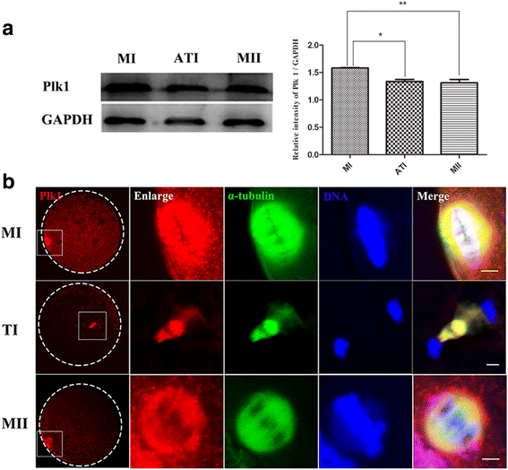
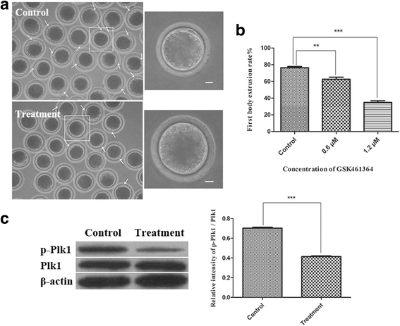
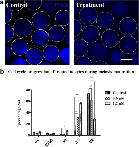
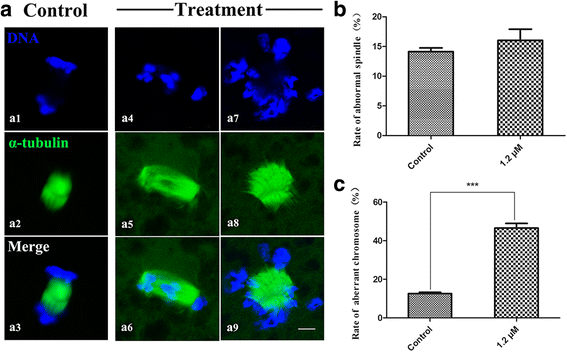
Results: Upon expression, Plk1 exhibited a specific dynamic intracellular localization, and co-localization of Plk1 with α-tubulin was revealed in the meiotic spindle of pig oocyte during the transition from MI to MII stage. GSK461364 treatment significantly blocked the first polar body (pbI) emission in a dose-dependent manner and resulted in a failure of meiotic maturation, with a larger percentage of the GSK461364-treated oocytes arresting in the anaphase-telophase I (ATI) stage. Further subcellular structure examination results showed that inhibition of Plk1 with GSK461364 had no visible effect on spindle assembly but caused a significantly higher proportion of the treated oocytes to have obvious defects in homologous chromosome segregation at ATI stage.
Conclusions: Thus, these results indicate that Plk1 plays an essential role during the meiosis I/meiosis II transition in porcine oocytes, and the regulation is associated with Plk1's effects on homologous chromosome segregation in the ATI stage.
Keywords: Chromosome segregation; Meiosis I/meiosis II transition; Oocyte; Pig; Polo-like 1.
Conflict of interest statement
Ethics approval and consent to participate
The animals used in this study and their care were according to the guidelines of Animal Research Institute Committee which is prescribed by Nanjing Agricultural University, China. The workers who executed the slaughtering complied with the pig slaughtering regulations (State Council of the People’s Republic of China, No. 666).
Consent for publication
Not applicable.
Competing interests
The authors declare no competing interests with respect to the authorship and/or publication of this article.
Publisher’s Note
Springer Nature remains neutral with regard to jurisdictional claims in published maps and institutional affiliations.
Figures
Similar articles
-
Multiple Roles of PLK1 in Mitosis and Meiosis.Cells. 2023 Jan 2;12(1):187. doi: 10.3390/cells12010187. Cells. 2023. PMID: 36611980 Free PMC article. Review.
-
Polo-like kinase 1 inhibition results in misaligned chromosomes and aberrant spindles in porcine oocytes during the first meiotic division.Reprod Domest Anim. 2018 Feb;53(1):256-265. doi: 10.1111/rda.13102. Epub 2017 Nov 16. Reprod Domest Anim. 2018. PMID: 29143380
-
Unique subcellular distribution of phosphorylated Plk1 (Ser137 and Thr210) in mouse oocytes during meiotic division and pPlk1(Ser137) involvement in spindle formation and REC8 cleavage.Cell Cycle. 2015;14(22):3566-79. doi: 10.1080/15384101.2015.1100770. Cell Cycle. 2015. PMID: 26654596 Free PMC article.
-
Multiple requirements of PLK1 during mouse oocyte maturation.PLoS One. 2015 Feb 6;10(2):e0116783. doi: 10.1371/journal.pone.0116783. eCollection 2015. PLoS One. 2015. PMID: 25658810 Free PMC article.
-
Polo-like kinase 1: target and regulator of anaphase-promoting complex/cyclosome-dependent proteolysis.Cancer Res. 2006 Jul 15;66(14):6895-8. doi: 10.1158/0008-5472.CAN-06-0358. Cancer Res. 2006. PMID: 16849530 Review.
Cited by
-
Multiple Roles of PLK1 in Mitosis and Meiosis.Cells. 2023 Jan 2;12(1):187. doi: 10.3390/cells12010187. Cells. 2023. PMID: 36611980 Free PMC article. Review.
-
TRIM8: a double-edged sword in glioblastoma with the power to heal or hurt.Cell Mol Biol Lett. 2023 Jan 23;28(1):6. doi: 10.1186/s11658-023-00418-z. Cell Mol Biol Lett. 2023. PMID: 36690946 Free PMC article. Review.
References
-
- Coticchio G, Dal Canto M, Mignini Renzini M, Guglielmo MC, Brambillasca F, Turchi D, Novara PV, Fadini R. Oocyte maturation: gamete-somatic cells interactions, meiotic resumption, cytoskeletal dynamics and cytoplasmic reorganization. Hum Reprod Update. 2015;21:427–454. doi: 10.1093/humupd/dmv011. - DOI - PubMed
MeSH terms
Substances
LinkOut - more resources
Full Text Sources
Other Literature Sources
Miscellaneous

