

Electron microscopy using the genetically encoded APEX2 tag in cultured mammalian cells
- PMID: 28796234
- PMCID: PMC5851282
- DOI: 10.1038/nprot.2017.065
Electron microscopy using the genetically encoded APEX2 tag in cultured mammalian cells
Abstract
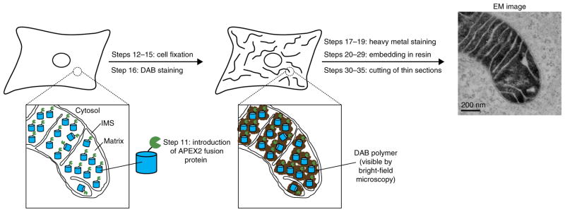
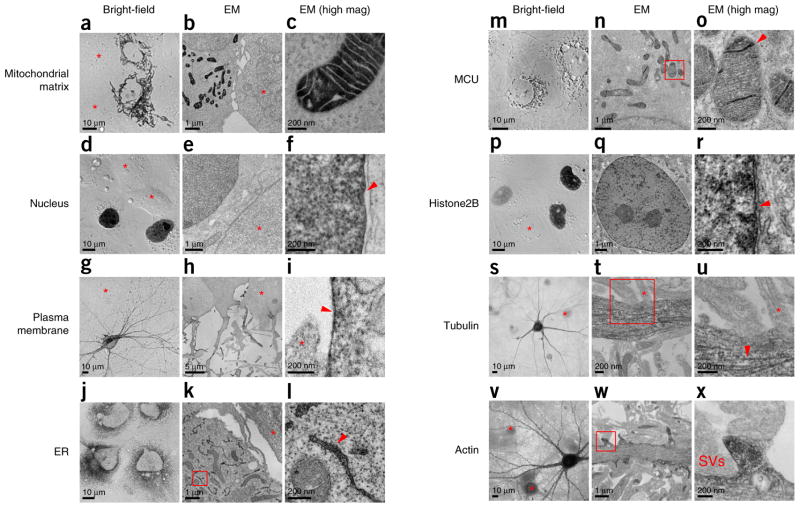
Electron microscopy (EM) is the premiere technique for high-resolution imaging of cellular ultrastructure. Unambiguous identification of specific proteins or cellular compartments in electron micrographs, however, remains challenging because of difficulties in delivering electron-dense contrast agents to specific subcellular targets within intact cells. We recently reported enhanced ascorbate peroxidase 2 (APEX2) as a broadly applicable genetic tag that generates EM contrast on a specific protein or subcellular compartment of interest. This protocol provides guidelines for designing and validating APEX2 fusion constructs, along with detailed instructions for cell culture, transfection, fixation, heavy-metal staining, embedding in resin, and EM imaging. Although this protocol focuses on EM in cultured mammalian cells, APEX2 is applicable to many cell types and contexts, including intact tissues and organisms, and is useful for numerous applications beyond EM, including live-cell proteomic mapping. This protocol, which describes procedures for sample preparation from cell monolayers and cell pellets, can be completed in 10 d, including time for APEX2 fusion construct validation, cell growth, and solidification of embedding resins. Notably, the only additional steps required relative to a standard EM sample preparation are cell transfection and a 2- to 45-min staining period with 3,3-diaminobenzidine (DAB) and hydrogen peroxide (H2O2).
Conflict of interest statement
Figures
Similar articles
-
Engineered ascorbate peroxidase as a genetically encoded reporter for electron microscopy.Nat Biotechnol. 2012 Nov;30(11):1143-8. doi: 10.1038/nbt.2375. Epub 2012 Oct 21. Nat Biotechnol. 2012. PMID: 23086203 Free PMC article.
-
A Validated Set of Ascorbate Peroxidase-Based Organelle Markers for Electron Microscopy of Saccharomyces cerevisiae.mSphere. 2022 Aug 31;7(4):e0010722. doi: 10.1128/msphere.00107-22. Epub 2022 Jun 21. mSphere. 2022. PMID: 35727034 Free PMC article.
-
Localization of Mitochondrial Nucleoids by Transmission Electron Microscopy Using the Transgenic Expression of the Mitochondrial Helicase Twinkle and APEX2.Methods Mol Biol. 2023;2615:173-188. doi: 10.1007/978-1-0716-2922-2_13. Methods Mol Biol. 2023. PMID: 36807792
-
Visualizing viral protein structures in cells using genetic probes for correlated light and electron microscopy.Methods. 2015 Nov 15;90:39-48. doi: 10.1016/j.ymeth.2015.06.002. Epub 2015 Jun 9. Methods. 2015. PMID: 26066760 Free PMC article. Review.
-
Combined video fluorescence and 3D electron microscopy.Methods Cell Biol. 2008;88:83-95. doi: 10.1016/S0091-679X(08)00405-6. Methods Cell Biol. 2008. PMID: 18617029 Review.
Cited by
-
Layer 5 of cortex innervates the thalamic reticular nucleus in mice.Proc Natl Acad Sci U S A. 2022 Sep 20;119(38):e2205209119. doi: 10.1073/pnas.2205209119. Epub 2022 Sep 12. Proc Natl Acad Sci U S A. 2022. PMID: 36095204 Free PMC article.
-
Microscopic Visualization of Cell-Cell Adhesion Complexes at Micro and Nanoscale.Front Cell Dev Biol. 2022 Apr 20;10:819534. doi: 10.3389/fcell.2022.819534. eCollection 2022. Front Cell Dev Biol. 2022. PMID: 35517500 Free PMC article. Review.
-
Dysregulation of Amyloid Precursor Protein Impairs Adipose Tissue Mitochondrial Function and Promotes Obesity.Nat Metab. 2019 Dec;1(12):1243-1257. doi: 10.1038/s42255-019-0149-1. Epub 2019 Dec 13. Nat Metab. 2019. PMID: 31984308 Free PMC article.
-
Volume electron microscopy.Nat Rev Methods Primers. 2022 Jul 7;2:51. doi: 10.1038/s43586-022-00131-9. Nat Rev Methods Primers. 2022. PMID: 37409324 Free PMC article.
-
Syntaxin 17 regulates the localization and function of PGAM5 in mitochondrial division and mitophagy.EMBO J. 2018 Nov 2;37(21):e98899. doi: 10.15252/embj.201798899. Epub 2018 Sep 20. EMBO J. 2018. PMID: 30237312 Free PMC article.
References
-
- Giepmans BN, Adams SR, Ellisman MH, Tsien RY. The fluorescent toolbox for assessing protein location and function. Science. 2006;312:217–224. - PubMed
-
- Fernandez-Suarez M, Ting AY. Fluorescent probes for super-resolution imaging in living cells. Nat Rev Mol Cell Biol. 2008;9:929–943. - PubMed
-
- Xu K, Shim S-H, Zhuang X. In: Far-Field Optical Nanoscopy. Tinnefeld P, Eggeling C, Hell SW, editors. Springer; 2015. pp. 27–64.
-
- Eggeling C, Hell SW. In: Far-Field Optical Nanoscopy. Tinnefeld P, Eggeling C, Hell SW, editors. Springer; 2015. pp. 3–25.
-
- De Mey J, Moeremans M, Geuens G, Nuydens R, De Brabander M. High resolution light and electron microscopic localization of tubulin with the IGS (immuno gold staining) method. Cell Biol Int Rep. 1981;5:889–899. - PubMed
MeSH terms
Substances
Grants and funding
LinkOut - more resources
Full Text Sources
Other Literature Sources
Research Materials
