. 2017 Sep 7;474(18):3207-3226.
doi: 10.1042/BCJ20161104.

Central catalytic domain of BRAP (RNF52) recognizes the types of ubiquitin chains and utilizes oligo-ubiquitin for ubiquitylation

Affiliations

Central catalytic domain of BRAP (RNF52) recognizes the types of ubiquitin chains and utilizes oligo-ubiquitin for ubiquitylation

Shisako Shoji et al. Biochem J. .

Abstract

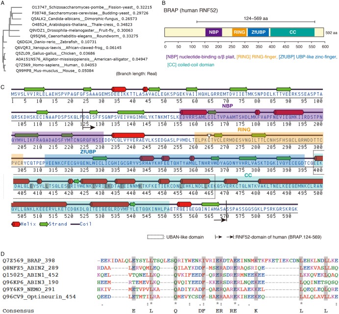
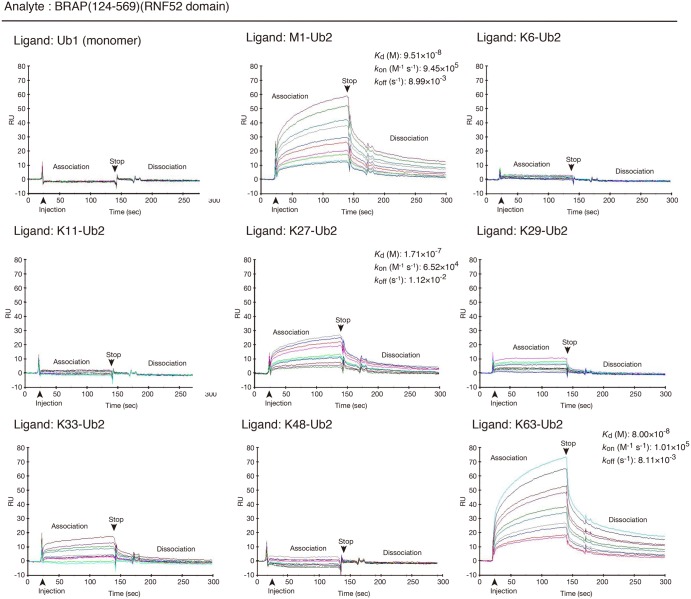
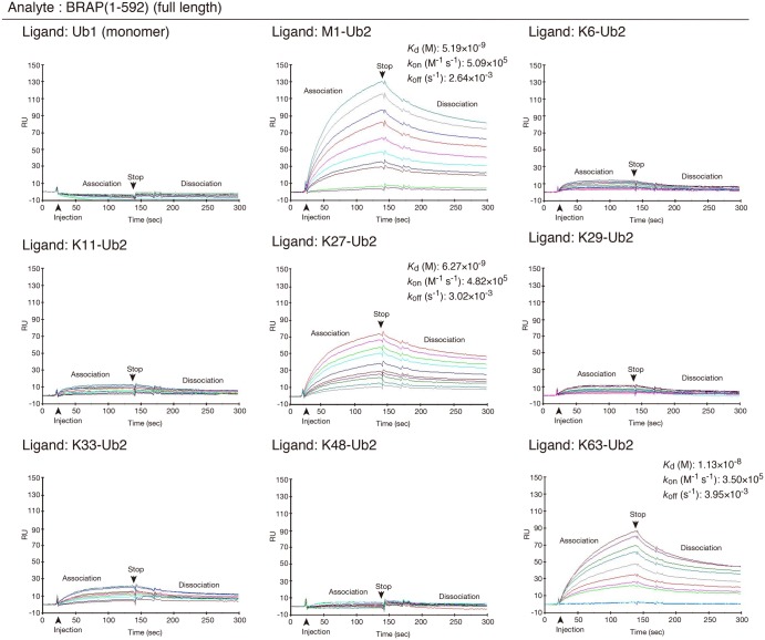
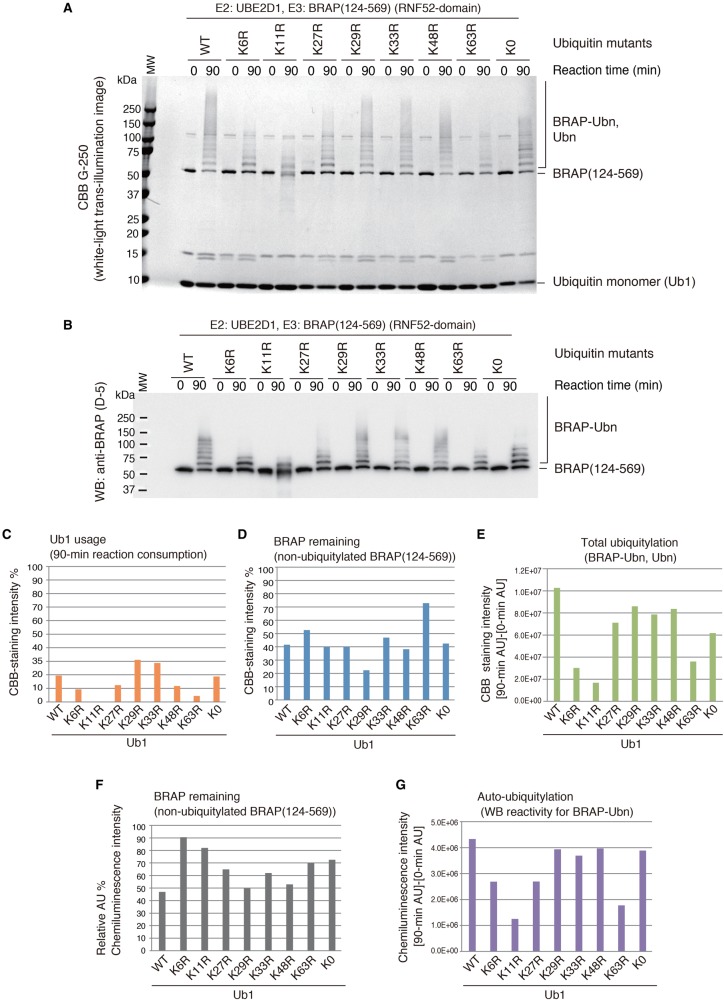
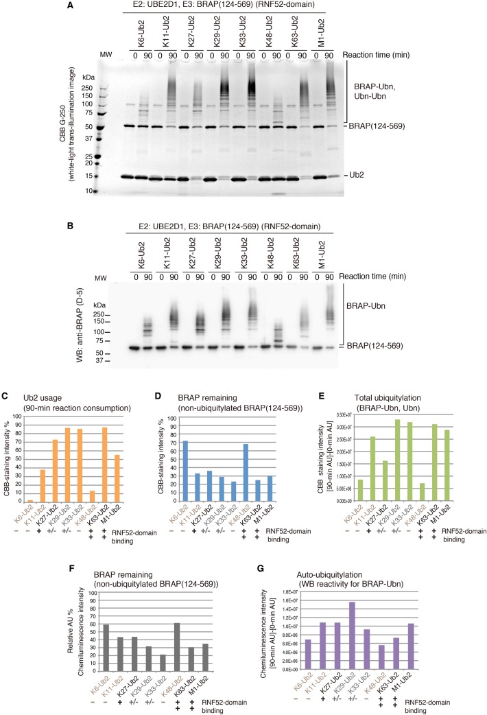
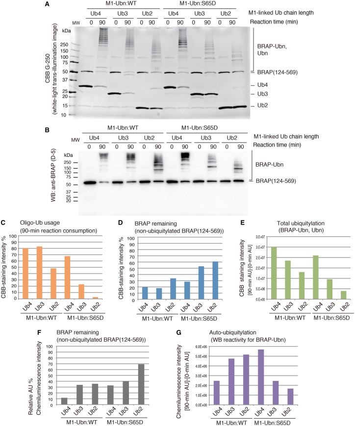
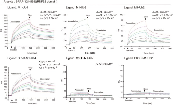
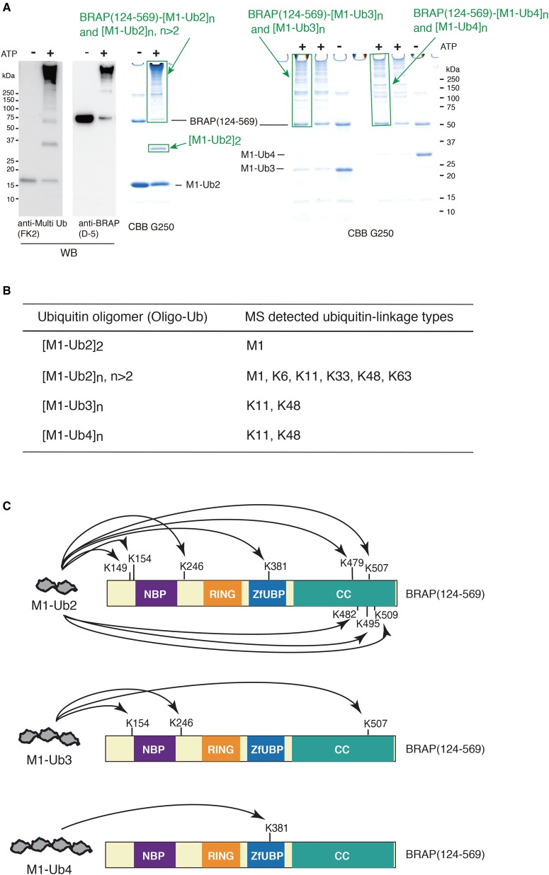
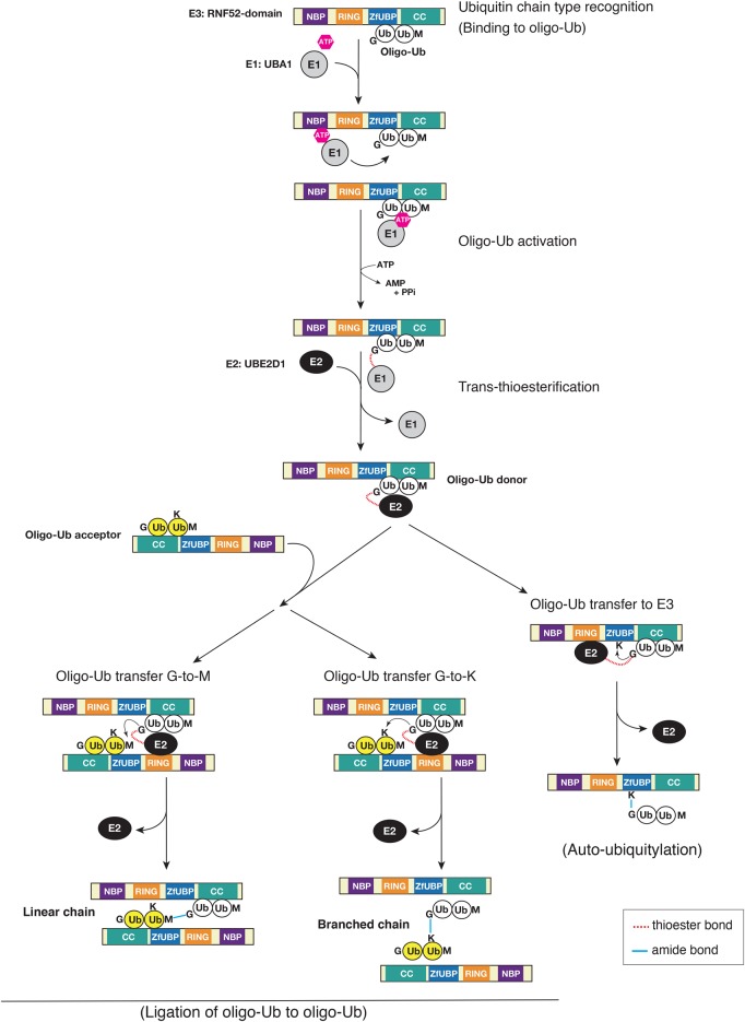
Really interesting new gene (RING)-finger protein 52 (RNF52), an E3 ubiquitin ligase, is found in eukaryotes from yeast to humans. Human RNF52 is known as breast cancer type 1 susceptibility protein (BRCA1)-associated protein 2 (BRAP or BRAP2). The central catalytic domain of BRAP comprises four subdomains: nucleotide-binding α/β plait (NBP), really interesting new gene (RING) zinc finger, ubiquitin-specific protease (UBP)-like zinc finger (ZfUBP), and coiled-coil (CC). This domain architecture is conserved in RNF52 orthologs; however, the domain's function in the ubiquitin system has not been delineated. In the present study, we discovered that the RNF52 domain, comprising NBP-RING-ZfUBP-CC, binds to ubiquitin chains (oligo-ubiquitin) but not to the ubiquitin monomers, and can utilize various ubiquitin chains for ubiquitylation and auto-ubiquitylation. The RNF52 domain preferentially bound to M1- and K63-linked di-ubiquitin chains, weakly to K27-linked chains, but not to K6-, K11-, or K48-linked chains. The binding preferences of the RNF52 domain for ubiquitin-linkage types corresponded to ubiquitin usage in the ubiquitylation reaction, except for K11-, K29-, and K33-linked chains. Additionally, the RNF52 domain directly ligated the intact M1-linked, tri-, and tetra-ubiquitin chains and recognized the structural alterations caused by the phosphomimetic mutation of these ubiquitin chains. Full-length BRAP had nearly the same specificity for the ubiquitin-chain types as the RNF52 domain alone. Mass spectrometry analysis of oligomeric ubiquitylation products, mediated by the RNF52 domain, revealed that the ubiquitin-linkage types and auto-ubiquitylation sites depend on the length of ubiquitin chains. Here, we propose a model for the oligomeric ubiquitylation process, controlled by the RNF52 domain, which is not a sequential assembly process involving monomers.

Keywords: BRAP; RNF52; ubiquitin ligases; ubiquitin oligomer; ubiquitin system; ubiquitin-chain assembly.


Conflict of interest statement

The Authors declare that there are no competing interests associated with the manuscript.

Figures

Figure 1.
Figure 1.. Characteristics of amino acid sequences of BRAP/RNF52.
(A) Phylogenetic tree of RNF52 amino acid sequences in eukaryotes, including mammals (Homo sapiens and Mus musculus), a bird (Gallus gallus), a reptile (Alligator mississippiensis), an amphibian (Xenopus laevis), a fish (Danio rerio), a plant (Arabidopsis thaliana), an insect (Drosophila melanogaster), a worm (Caenorhabditis elegans), and fungi (Candida albicans, Saccharomyces cerevisiae, and Schizosaccharomyces pombe); the tree was generated using the Clustal Omega program 1.2.1 (http://www.ebi.ac.uk/Tools/msa/clustalo/). UniProtKB accession codes are given before the species name. The amino acid sequences and multiple sequence alignment are shown in Supplementary Data S1. (B) Schematic diagram showing the conserved central catalytic domain (RNF52 domain) of BRAP; this domain contains four subdomains that are highly conserved in vertebrates: NBP, nucleotide-binding α/β plait; RING, really interesting new gene zinc finger; ZfUBP, ubiquitin-specific protease (UBP)-like zinc finger; CC, coiled-coil domain. (C) Predicted secondary structure and amino acid sequence of BRAP including the region at amino acid positions 124–569 that corresponds to the RNF52 domain. Inter Pro (http://www.ebi.ac.uk/interpro/) was used for the motif and domain searches. The program DSC (Discrimination of Protein Secondary Structure Class) [49] was used to predict secondary structures. The white downward-pointing triangle indicates the RING-finger motif mutation (C264). (D) Sequence alignment of a part of the BRAP-CC domain and the UBAN domains of human ABIN proteins (ABIN-1, ABIN-2, ABIN-3, NEMO, and optineurin). The highly conserved amino acids in the UBAN motif [50] are shaded in gray.
Figure 2.
Figure 2.. Protein–protein interaction profiles showing the binding of the RNF52 domain of BRAP to ubiquitin monomers and to all di-ubiquitin oligomer (Ub2) linkage types.
Surface plasmon resonance sensorgrams from the Biacore assay (i.e. protein–protein interaction profiles) are shown. Resonance signal, expressed as the response unit (RU), indicates the degree of binding between BRAP and the various ubiquitin molecules. The immobilized ligands (i.e. ubiquitin and the various di-ubiquitin oligomers) are shown in the top left of each panel. The analyte (applied as the mobile molecule to an immobilized ligand on the sensor chip) was BRAP (124–569) (RNF52 domain). The measured equilibrium dissociation constant (Kd), association rate constant (kon), and dissociation rate constant (koff), for each binding, are shown in the top right of each panel where possible.
Figure 3.
Figure 3.. Protein–protein interaction profiles showing the binding ability of full-length BRAP against ubiquitin monomers and all ubiquitin-linkage types of di-ubiquitin chains (Ub2).
Surface plasmon resonance sensorgrams from the Biacore assay (i.e. protein–protein interaction profiles) are shown. Resonance signal, expressed as the response unit (RU), indicates the degree of binding between BRAP and the various ubiquitin molecules. The immobilized ligands (i.e. ubiquitin and the various di-ubiquitin oligomers) are shown in the top left of each panel. The analyte (applied as the mobile molecule to an immobilized ligand on the sensor chip) was BRAP (1–592) (full-length). The measured equilibrium dissociation constant (Kd), association rate constant (kon), and dissociation rate constant (koff), for each binding, are shown in the top right of each panel where possible.
Figure 4.
Figure 4.. The E3 activity of the RNF52 domain of BRAP on ubiquitin monomer and its different lysine mutants.
Ubiquitylation assays of the BRAP (124–569) fragment (RNF52 domain of BRAP) in the presence of UBA1, UBE2D1, and WT ubiquitin or the ubiquitin mutants: K6R, K11R, K27R, K29R, K33R, K48R, K63R, and K0 (no lysine residue). (A) SDS–PAGE gels of the ubiquitylation reaction products stained with CBB G-250. (B) WB of the same samples as in (A) probed with an anti-BRAP antibody (clone D-5). (CG) The band signals from the CBB-stained gels and WB were quantified using the ImageQuant TL software. (C) Ub1 (ubiquitin monomer) usage was calculated by determining what percentage of the band had disappeared after a 90-min reaction, assuming that the band signal of the unshifted bands for the Ub1 in each lane at time 0 (zero) of the reaction was 100%. (D and F) The percentage of BRAP (124–569) remaining at the end of the reaction (i.e. the non-ubiquitylated BRAP present after a 90-min reaction) was calculated by assessing band signal intensity that corresponded to non-ubiquitylated BRAP (124–569) in each lane after a 90-min reaction, assuming that the band signal for the unshifted Ub1 bands in each lane, at reaction time 0, was 100%. (E) To assess total ubiquitylation, the intensity of the high molecular mass signals, detected in the CBB-stained gel after a 90-min reaction, was obtained, and the signals from the same area of the gel at reaction time 0 were subtracted as background. (G) To assess the production of auto-ubiquitylated BRAP, the signals detected in the high molecular mass area of the WB, after a 90-min reaction, were obtained, and the signals from the same area of the blot at reaction time 0 were subtracted as background. AU, arbitrary unit of the band signal intensity. These graphs show the average values obtained from two independent experiments.
Figure 5.
Figure 5.. Oligomeric ubiquitylation preference of the RNF52 domain of BRAP for different linkage types of di-ubiquitin chains.
Ubiquitylation assays of the BRAP (124–569) fragment (RNF52 domain of BRAP) in the presence of UBA1, UBE2D1, and K6-, K11-, K27-, K29-, K33-, K48-, and K63-linked di-ubiquitin chains (Ub2). (A) SDS–PAGE gels of the ubiquitylation reaction products stained with CBB G-250. (B) WB of the same samples as in (A) probed with an anti-BRAP antibody (clone D-5). (CG) The band signals from the CBB-stained gels and WB were quantified using the ImageQuant TL software. (C) Ub2 (di-ubiquitin chain) usage was calculated by determining what percentage of the band had disappeared after a 90-min reaction, assuming that the band signal of the unshifted bands for the Ub2 in each lane at reaction time 0 (zero) was 100%. (D and F) The percentage of BRAP (124–569), remaining at the end of the reaction (i.e. the non-ubiquitylated BRAP present after a 90-min reaction), was calculated by assessing band signal intensity that corresponded to non-ubiquitylated BRAP (124–569) in each lane after a 90-min reaction, assuming that the band signal for the unshifted Ub2 bands in each lane, at reaction time 0, was 100%. (E) To assess total ubiquitylation, the intensity of the high molecular mass signals, detected in the CBB-stained gel after a 90-min reaction, were obtained, and the signals from the same area of the gel at reaction time 0 were subtracted as background. (G) To assess the production of auto-ubiquitylated BRAP, the signals, detected in the high molecular mass area of the WB after a 90-min reaction, were obtained, and the signals from the same area of the blot at reaction time 0 were subtracted as background. AU, arbitrary unit of the band signal intensity. These graphs show the average values obtained from two independent experiments.
Figure 6.
Figure 6.. E3 activity of the RNF52 domain of BRAP against M1-linked di-, tri-, and tetra-ubiquitin chains (ubiquitin oligomers) and the phosphomimetic form that consists of S65D ubiquitin mutant.
Ubiquitylation assays of the BRAP (124–569) fragment (RNF52 domain of BRAP) in the presence of UBA1, UBE2D1, and M1-linked di-, tri-, and tetra-ubiquitin oligomers (Ub2, Ub3, and Ub4) or their phosphomimetic forms, which consist of the phosphorylation-mimic ubiquitin mutant S65D. (A) SDS–PAGE gels of the ubiquitylation reaction products stained with CBB G-250. (B) WB of the same samples as in (A) probed with an anti-BRAP antibody (clone D-5). (CG) The band signals on CBB-stained gels and WB were quantified using the ImageQuant TL software. (C) Oligo-Ub usage was calculated by determining what percentage of the band had disappeared after a 90-min reaction, assuming that the band signal of the unshifted bands for the ubiquitin oligomer in each lane at reaction time 0 (zero) was 100%. (D and F) The percentage of BRAP (124–569) remaining at the end of the reaction (i.e. the non-ubiquitylated BRAP present after a 90-min reaction) was calculated by assessing the band signal intensity that corresponded to non-ubiquitylated BRAP (124–569) in each lane after a 90-min reaction, assuming that the band signal for the unshifted ubiquitin oligomer bands in each lane at reaction time 0 was 100%. (E) To assess total ubiquitylation, the intensity of the high molecular mass signals, detected in the CBB-stained gel after a 90-min reaction, was obtained, and the signals from the same area of the gel at reaction time 0 were subtracted as background. (G) To assess the production of auto-ubiquitylated BRAP, the signals detected in the high molecular mass area of the WB, after a 90-min reaction, were obtained, and the signals from the same area of the blot at reaction time 0 were subtracted as background. AU, arbitrary unit of the band signal intensity. These graphs show the average values obtained from two independent experiments.
Figure 7.
Figure 7.. Protein–protein interaction profiles showing binding of the RNF52 domain of BRAP to M1-linked di-, tri-, and tetra-ubiquitin chains (ubiquitin oligomers).
Surface plasmon resonance sensorgrams from the Biacore assay (i.e. protein–protein interaction profiles) are shown. Resonance signal, expressed as response unit (RU), shows the degree of the binding of BRAP to the ubiquitin oligomers. The immobilized ligands [i.e. M1-linked di-, tri-, and tetra-ubiquitin oligomers (Ub2, Ub3, and Ub4), and their phosphomimetic forms that consisted of the S65D mutation] are shown in the top left of each panel. The analyte (applied as a mobile molecule to an immobilized ligand on the sensor chip) was BRAP (124–569) (RNF52 domain). The measured equilibrium dissociation constant (Kd), association rate constant (kon), and dissociation rate constant (koff), for each binding, are shown at the top right of each panel where possible.
Figure 8.
Figure 8.. Summary of mass spectrometry analysis of the products from BRAP (124–569) (RNF52 domain)-mediated in vitro ubiquitylation using M1-linked ubiquitin oligomers.
(A) Images of SDS–PAGE/CBB-stained gels, showing the gel slices used for mass spectrometry analysis, are highlighted using green boxes and labeled with a green arrow. These oligomeric ubiquitylation samples were taken after a 90-min reaction at 37°C, with or without ATP. The two left-hand side panels are WBs of the M1-Ub2 oligomeric ubiquitylation probed with either an anti-multi Ub (FK2) antibody or an anti-BRAP (D-5) antibody. (B) Variation in ubiquitin-chain-linkage types produced by the RNF52 domain [BRAP (124–569)]-mediated oligomeric ubiquitylation. (C) Schematic showing the effect of ubiquitin-chain length on auto-ubiquitylation. Mass spectra and details of the mass spectrometry analysis are provided in Supplementary Data S8 and S9.
Figure 9.
Figure 9.. Diagrammatic representation of oligomeric ubiquitylation by the central catalytic domain (RNF52 domain) of BRAP using M1-linked ubiquitin oligomers (oligo-Ub).
A proposed model for the mechanism of oligomeric ubiquitylation catalyzed by the RNF52 domain of BRAP with UBE2D1 (E2) and UBA1 (E1). Details are described in the Discussion section.

Similar articles

Cited by

References

    1. Hershko A. and Ciechanover A. (1998) The ubiquitin system. Ann. Rev. Biochem. 67, 425–479 doi:10.1146/annurev.biochem.67.1.425 - DOI - PubMed
    1. Pickart C.M. and Eddins M.J. (2004) Ubiquitin: structures, functions, mechanisms. Biochim. Biophys. Acta, Mol. Cell Res. 1695, 55–72 doi:10.1016/j.bbamcr.2004.09.019 - DOI - PubMed
    1. Akutsu M., Dikic I. and Bremm A. (2016) Ubiquitin chain diversity at a glance. J. Cell Sci. 129, 875–880 doi:10.1242/jcs.183954 - DOI - PubMed
    1. Komander D. and Rape M. (2012) The ubiquitin code. Ann. Rev. Biochem. 81, 203–229 doi:10.1146/annurev-biochem-060310-170328 - DOI - PubMed
    1. Weissman A.M., Shabek N. and Ciechanover A. (2011) The predator becomes the prey: regulating the ubiquitin system by ubiquitylation and degradation. Nat. Rev. Mol. Cell Biol. 12, 605–620 doi:10.1038/nrm3173 - DOI - PMC - PubMed

Publication types