. 2017 Aug 1;9(8):202.
doi: 10.3390/v9080202.

A Motif in the F Homomorph of Rabbit Haemorrhagic Disease Virus Polymerase Is Important for the Subcellular Localisation of the Protein and Its Ability to Induce Redistribution of Golgi Membranes

Affiliations

A Motif in the F Homomorph of Rabbit Haemorrhagic Disease Virus Polymerase Is Important for the Subcellular Localisation of the Protein and Its Ability to Induce Redistribution of Golgi Membranes

Nadya Urakova et al. Viruses. .

Abstract

Rabbit haemorrhagic disease virus (RHDV) is a calicivirus that infects and frequently kills rabbits. Previously, we showed that the RHDV RNA-dependent RNA polymerase (RdRp) is associated with distinct, but yet uncharacterised subcellular structures and is capable of inducing a redistribution of Golgi membranes. In this study, we identified a partially hidden hydrophobic motif that determines the subcellular localisation of recombinant RHDV RdRp in transfected cells. This novel motif, 189LLWGCDVGVAVCAAAVFHNICY210, is located within the F homomorph, between the conserved F3 and A motifs of the core RdRp domain. Amino acid substitutions that decrease the hydrophobicity of this motif reduced the ability of the protein to accumulate in multiple subcellular foci and to induce a rearrangement of the Golgi network. Furthermore, preliminary molecular dynamics simulations suggest that the RHDV RdRp could align with the negatively charged surfaces of biological membranes and undergo a conformational change involving the F homomorph. These changes would expose the newly identified hydrophobic motif so it could immerse itself into the outer leaflet of intracellular membranes.

Keywords: Golgi membranes; RHDV; RNA-dependent RNA polymerase; Rabbit haemorrhagic disease virus; caliciviruses.


Conflict of interest statement

The authors declare no conflict of interest.

Figures

Figure 1
Figure 1
Conserved motifs in RNA-dependent RNA polymerases (RdRps) of single-stranded, positive sense RNA viruses. Conserved sequence motifs A–G are coloured purple, green, pink, yellow, brown, dark blue, cyan, respectively, and their relative positions are shown in a schematic representation (a) and a ribbon diagram of the Rabbit haemorrhagic disease virus (RHDV) coding sequence (b). Conserved structural elements (homomorphs) that encompass the conserved sequence motifs A–G are shown in the context of the entire protein (c) and without other protein elements (d).
Figure 2
Figure 2
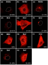
Expression of recombinant calicivirus RdRps in cultured cells. Rabbit kidney (RK-13) cells were transiently transfected with constructs encoding myc-tagged RdRp of Rabbit haemorrhagic disease virus (RHDV) (ac); Rabbit calicivirus (RCV) (df); Murine norovirus (MNV) (g); Human norovirus (NoV) (h); Feline calicivirus (FCV) (i) and Sapovirus (SaV) (j,k). Cells were fixed 24 h after transfection and recombinant RdRps were immunostained using myc-specific antibodies. Dotted lines indicate the outline of cell nuclei. Percentages indicate frequencies with which different localisation profiles were found.
Figure 3
Figure 3
Identification of a putative membrane interacting motif in the RHDV RdRp by sequence and structure analysis. Kyte–Doolittle hydrophobicity plots for RHDV RdRp (a) and MNV RdRp (b) sequences show the presence of a relatively hydrophobic motif unique to the RHDV RdRp (positions of the hydrophobic motif in RHDV and corresponding region in MNV are indicated by red arrows). Amino acid sequence and secondary structure of the putative membrane interacting motif of the RHDV RdRp (c) and the corresponding region in MNV (d). Location of proposed membrane interacting motif within the tertiary structure of the RHDV RdRp (e) and location of the corresponding region in MNV RdRp (f). The putative functional motif is shown in red, and the location of the F and G homomorphs is shown in dark blue and cyan, respectively. Protein Bank Database IDs of crystal structures of RdRps: 1KHW (RHDV) and 3UQS (MNV).
Figure 4
Figure 4
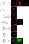
The newly identified hydrophobic motif is important for the subcellular localisation of RHDV RdRp and sufficient to change the subcellular localisation of GFP. RK-13 cells were transiently transfected with expression constructs coding for myc-tagged versions of the full-length RHDV RdRp (ac); the N-terminal part of the protein without the hydrophobic motif (d); the C-terminal part without the motif (e); the N-terminal part with the motif (f,g); the C-terminal part with the motif (h); or the motif (and a few flanking amino acids) without the remainder of the protein (i). Furthermore, cells were transfected with constructs in which the motif was fused to GFP (j); or control constructs for the expression of unaltered GFP (k). Cells were fixed 24 h after transfections and recombinant proteins were immunostained using anti-myc or anti-GFP antibodies. Schematic representations of the various recombinant proteins are shown to the left of the fluorescence images. A grey box indicates the position of the hydrophobic motif. M indicates an additional N-terminal methionine. Dotted lines indicate the outline of cell nuclei. Percentages indicate frequencies with which different localisation profiles were found.
Figure 5
Figure 5
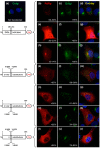
Amino acid substitutions decreasing the hydrophobicity of the newly identified motif change the subcellular localisation and the ability of the RHDV RdRp to induce Golgi membrane rearrangements. RK-13 cells were transfected with constructs encoding myc-tagged wild-type RHDV RdRp (bj) or constructs encoding versions of the protein in which valine (V) residues within the new hydrophobic motif had been changed to serine (S) residues: i.e., V195S and V197S (km); V199S and V204S (ns); and V195S, V197S, V199S, and V204S (tv). Cells were fixed 24 h after transfection, recombinant proteins and the Golgi membrane marker giantin were immunostained using myc-specific antibodies (shown in red) and anti-giantin antibodies (shown in green), respectively. Cell nuclei were stained using 4′,6-diamidino-2-phenylindole (DAPI, blue). Schematic representations of the various recombinant proteins are shown to the left of the fluorescence images. A grey box indicates the position of the hydrophobic motif. M indicates an additional N-terminal methionine. Compared with the giantin staining in untransfected control cells (a), the majority of cells expressing wild-type RdRp or proteins with the two N-terminal (relative to the hydrophobic domain) substitutions V195S and V197S showed a giantin staining indicative of rearranged Golgi membranes (c,f,i,l). RdRp variants with the C-terminal substitutions V199S and V204S were no longer able to change Golgi membrane in a about a third of cells (o,r) and variants with all four substitutions did not change Golgi membranes in the majority of cells (u).
Figure 6
Figure 6
Backbone root-mean-square deviation (RMSD) per residue over time for the 100-ns RHDV RdRp molecular dynamics trajectory. Red and green indicate stability and mobility, respectively. The hydrophobic motif is shown in yellow for structural perspective, but was stable throughout the simulation.
Figure 7
Figure 7
Structural features proposed to be involved in interactions with the Golgi membranes, with Loop 1 and Loop 2 shown in green, Loop 3 shown in orange and the hydrophobic motif shown in yellow. Val195, Val197, Val199 and Val204 on the hydrophobic motif are shown in blue stick representation. The three collinear lysines (Lys 149, Lys 153 and Lys 162) on Helix 1 are shown as cyan atom-in-stick representations.

Similar articles

Cited by

References

    1. Abrantes J., van der Loo W., Le Pendu J., Esteves P.J. Rabbit haemorrhagic disease (RHD) and Rabbit haemorrhagic disease virus (RHDV): A review. Vet. Res. 2012;43:12. doi: 10.1186/1297-9716-43-12. - DOI - PMC - PubMed
    1. Thiel H.-J., König M. Caliciviruses: An overview. Vet. Microbiol. 1999;69:55–62. doi: 10.1016/S0378-1135(99)00088-7. - DOI - PubMed
    1. Clarke I.N., Lambden P.R. The molecular biology of caliciviruses. J. Gen. Virol. 1997;78:291–301. doi: 10.1099/0022-1317-78-2-291. - DOI - PubMed
    1. Meyers G., Wirblich C., Thiel H.-J. Genomic and subgenomic RNAs of rabbit hemorrhagic disease virus are both protein-linked and packaged into particles. Virology. 1991;184:677–686. doi: 10.1016/0042-6822(91)90437-G. - DOI - PMC - PubMed
    1. Meyers G., Wirblich C., Thiel H.-J. Rabbit hemorrhagic disease virus—Molecular cloning and nucleotide sequencing of a calicivirus genome. Virology. 1991;184:664–676. doi: 10.1016/0042-6822(91)90436-F. - DOI - PubMed

MeSH terms

Substances