. 2017;16(17):1611-1621.
doi: 10.1080/15384101.2017.1356509. Epub 2017 Jul 27.

Kaposi's sarcoma-associated herpesvirus infection promotes differentiation and polarization of monocytes into tumor-associated macrophages

Affiliations

Kaposi's sarcoma-associated herpesvirus infection promotes differentiation and polarization of monocytes into tumor-associated macrophages

Natarajan Bhaskaran et al. Cell Cycle. 2017.

Abstract

Tumor associated macrophages (TAMs) promote angiogenesis, tumor invasion and metastasis, and suppression of anti-tumor immunity. These myeloid cells originate from monocytes, which differentiate into TAMs upon exposure to the local tumor microenvironment. We previously reported that Kaposi's sarcoma-associated herpes virus (KSHV) infection of endothelial cells induces the cytokine angiopoietin-2 (Ang-2) to promote migration of monocytes into tumors. Here we report that KSHV infection of endothelial cells induces additional cytokines including interleukin-6 (IL-6), interleukin-10 (IL-10), and interleukin-13 (IL-13) that drive monocytes to differentiate and polarize into TAMs. The KSHV-induced TAMs not only express TAM-specific markers such as CD-163 and legumain (LGMN) but also display a gene expression profile with characteristic features of viral infection. More importantly, KSHV-induced TAMs enhance tumor growth in nude mice. These results are consistent with the strong presence of TAMs in Kaposi's sarcoma (KS) tumors. Therefore, KSHV infection of endothelial cells generates a local microenvironment that not only promotes the recruitment of monocytes but also induces their differentiation and polarization into TAMs. These findings reveal a new mechanism of KSHV contribution to KS tumor development.

Keywords: KSHV; Kaposi's sarcoma; monocytes; tumor-associated macrophages.


Figures
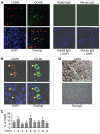
Figure 1.
Figure 1.
Detection of TAMs in KS tumor sections by immuno-fluorescence antibody staining (IFA). A, representative image of KS tumor sections co-stained with a rabbit anti-human LGMN polyclonal antibody and a mouse anti-human CD-68 monoclonal antibody, which were revealed with an Alexa Fluor®-633 conjugated goat anti-rabbit secondary antibody and a FITC-conjugated goat anti-mouse secondary antibody respectively. Rabbit IgG and mouse IgG were used as negative controls, and DAPI was used for nuclear staining of all cells. LGMN and CD-68 positive cells were imaged under a confocal microscope with a 10x objective. B, LGMN and CD-68 positive cells described in (A)that were imaged under a confocal microscope with a 40x oil objective. C, percentage (%) of TAMs in KS tumors. The numbers of LGMN+/CD-68+ cells and total cells per field under 40x oil objective from 4 fields per tumor were counted and used for the calculation of % of TAMs. D, representative image of KS tumor sections stained with a rat monoclonal antibody to KSHV latent protein LANA, which was revealed with a biotinylated secondary antibody and streptavidin-horseradish peroxidase, and DAB (3,3′-diaminobenzidine) detection system from Biolegend (San Diego, California, USA). Rat IgG was used as a negative control. Images were captured under a microscope (Carl Zeiss, Inc., Thornwood, NY).
Figure 2.
Figure 2.
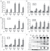
KSHV acute infection of HUVECs induces expression of cytokines that are known to promote differentiation and polarization of monocytes into M2 macrophages/TAMs. A, B, C, D, and E, relative levels of the mRNAs encoding IL-6, IL-10, IL-13, Ang-2, and VEGF in HUVECs infected with mock (M) or KSHV (K) at different hours post-infection (hpi), measured by qRT-PCR. Differences in mRNA levels between M and K with a P value < 0.05 are marked with a star. F, Western blot detection of IL-6, IL-10, IL-13, Ang-2, and VEGF proteins in the supernatants of mock (M) or KSHV (K)-infected HUVECs. The protein level of β-tubulin in cells corresponding to each supernatant was measured to demonstrate that equal numbers of cells were used for the comparison between mock (M) and KSHV (K) at time point (hpi).
Figure 3.
Figure 3.
Incubation of monocytes with supernatant from KSHV-infected HUVECs results in cells expressing TAMs-specific markers. A, relative levels of mRNAs encoding LGMN, CD-163, CD-206, and CD-11b in un-treated monocytes (Mono) and macrophages (ϕ) resulting from incubation with supernatant from mock or KSHV-infected HUVECs. Differences in mRNA levels between ϕ-mock (Mock) and ϕ-KSHV (KSHV) with a P value < 0.05 are marked with a star. B, Western blot detection of LGMN, CD-163, and α-actin in cells described in A. C, representative histograms of cells described in (A) that were stained with fluorescently labeled anti-CD-163 antibody or control isotype IgG and subsequently analyzed by flow cytometry as described in Materials and Methods. D, mean values from 2 independent experiments of the fluorescence intensities in cells described in (A)that were stained with fluorescently labeled anti-LGMN or CD-163 and analyzed by flow cytometry as shown in C.
Figure 4.
Figure 4.
KSHV-induced IL-6, IL-10, and IL-13 are responsible for inducing differentiation and polarization of monocytes into TAMs. A, representative flow cytometry histograms of ϕ-mock (Mock) and ϕ-KSHV (KSHV) generated by incubation of monocytes with supernatant from mock or KSHV-infected HUVECs in the presence of control IgG or neutralizing antibodies specific for human IL-6, IL-10, and IL-13, alone or in combination. The cells were stained with fluorescently labeled anti-CD-163 antibody or control isotype IgG and subsequently analyzed by flow cytometry. Similar experiment was also done on these cells with fluorescently labeled anti-LGMN antibody or control IgG. B, mean values from 2 independent experiments of the fluorescence intensities in cells that were stained with fluorescently labeled anti-LGMN or CD-163 and analyzed as described in A.
Figure 5.
Figure 5.
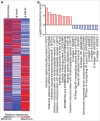
Transcription profile of KSHV-induced TAMs. A, transcription profiles of un-treated monocytes and macrophages resulting from incubation of monocytes with supernatant from mock (ϕ-mock) or KSHV-infected (ϕ-KSHV) HUVECs, generated by RNA-seq analysis, using monocytes from 3 healthy donors. B, lists of 10 most upregulated and 10 most downregulated transcripts in ϕ-KSHV, when compared with ϕ-mock.
Figure 6.
Figure 6.
Validation of RNA-seq data by qRT-PCR. A, mRNA levels of IFIT1, IFI27, and RSAD2, which are 3 of the 10 most upregulated transcripts in ϕ-KSHV, in the same RNA samples from un-treated monocytes, ϕ-mock, and ϕ-KSHV that were used for RNA-seq analysis. B, mRNA levels of GAGE1, GAGE12J, and GAGE8, 3 of the 10 most downregulated transcripts ϕ-KSHV, in the RNA samples described in A. C, mRNA levels of TAMs-specific markers LGMN, CD-163, and CD-206 in the RNA samples described in A. D, mRNA levels of MMP-9 and IL-10 in the RNA samples described in A. Differences between ϕ-mock and ϕ-KSHV with a P value < 0.05 are marked with a star.
Figure 7.
Figure 7.
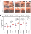
KSHV-induced TAMs enhance tumor growth in nude mice. A, representative images of tumors resulting from subcutaneous inoculation of equal numbers of (1 × 107/injection site) of TIVE-KSHV cells mixed with medium (control) or KSHV-induced TAMs (ϕ-KSHV) with a TIVE-KSHV to ϕ-KSHV ratio of 50 to 1 into nude mice at 6 weeks post-inoculation. B, average tumor volumes of the 2 groups of tumors described in (A) at different weeks post-inoculation. Statistically significant differences (P value < 0.05) are marked with 3 stars. NS, not significant; n, number of tumors per group.
Figure 8.
Figure 8.
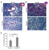
KSHV-induced TAMs enhance tumor angiogenesis. (A) and (B), immunochemical staining of blood vessels with an antibody specific for mouse endothelial cell marker CD-31 and ϕ-KSHV with a mouse anti-human CD-68 antibody in tumors resulting from subcutaneous inoculation of TIVE-KSHV cells mixed with medium (control) or KSHV-induced TAMs (ϕ-KSHV), which were collected 43 d upon inoculation. DAPI was used for nuclear staining of all cells. Enlarged representative blood vessels and ϕ-KSHV are indicated with an arrow. C, average numbers of vessels per field (10x magnification) from 3 fields per tumor and 6 tumors per group (n = 18). Significant differences (P value < 0.05) are marked with a star.

Similar articles

Cited by

References

    1. Chang Y, Cesarman E, Pessin MS, Lee F, Culpepper J, Knowles DM, Moore PS. Identification of herpesvirus-like DNA sequences in AIDS-associated Kaposi's sarcoma. Science. 1994;266:1865-69. doi:10.1126/science.7997879. PMID:7997879. - DOI - PubMed
    1. Boshoff C. Kaposi's sarcoma biology. IUBMB life. 2002;53:259-261. doi:10.1080/15216540212645. PMID:12121006. - DOI - PubMed
    1. Jones T, Ye F, Bedolla R, Huang Y, Meng J, Qian L, Pan H, Zhou F, Moody R, Wagner B, et al.. Direct and efficient cellular transformation of primary rat mesenchymal precursor cells by KSHV. J Clin Invest. 2012;122:1076-81. doi:10.1172/JCI58530. PMID:22293176. - DOI - PMC - PubMed
    1. Simonart T, Degraef C, Mosselmans R, Hermans P, Lunardi-Iskandar Y, Noel JC, Van Vooren JP, Parent D, Heenen M, Galand P. Early- and late-stage Kaposi's sarcoma-derived cells but not activated endothelial cells can invade de-epidermized dermis. J Invest Dermatol. 2001;116:679-85; http//dx.doi.org/10.1046/j.0022-202x.2001.doc.x. PMID:11348455. - DOI - PubMed
    1. Ensoli B, Sturzl M. Kaposi's sarcoma: a result of the interplay among inflammatory cytokines, angiogenic factors and viral agents. Cytokine Growth Factor Rev. 1998;9:63-83. doi:10.1016/S1359-6101(97)00037-3. PMID:9720757. - DOI - PubMed

MeSH terms