. 2017 Jul 25;20(4):935-948.
doi: 10.1016/j.celrep.2017.06.042.

Phospho-Rasputin Stabilization by Sec16 Is Required for Stress Granule Formation upon Amino Acid Starvation

Affiliations

Phospho-Rasputin Stabilization by Sec16 Is Required for Stress Granule Formation upon Amino Acid Starvation

Angelica Aguilera-Gomez et al. Cell Rep. .

Erratum in

Abstract

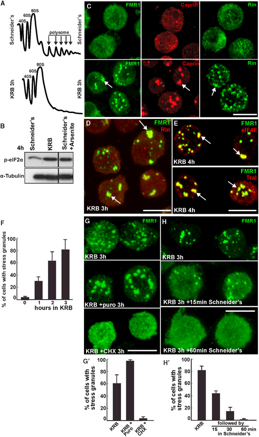
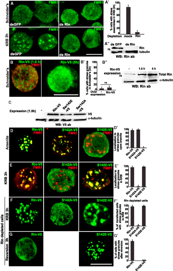
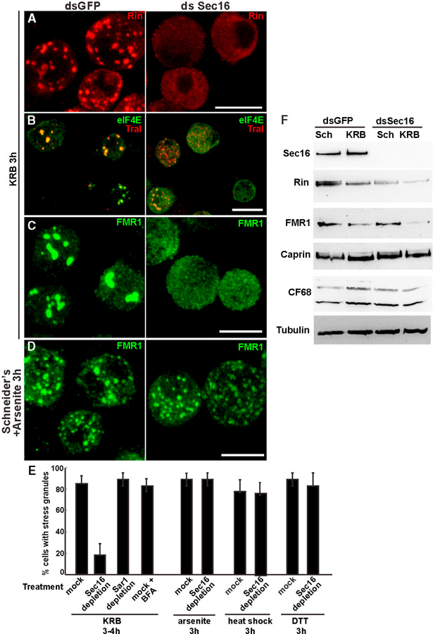
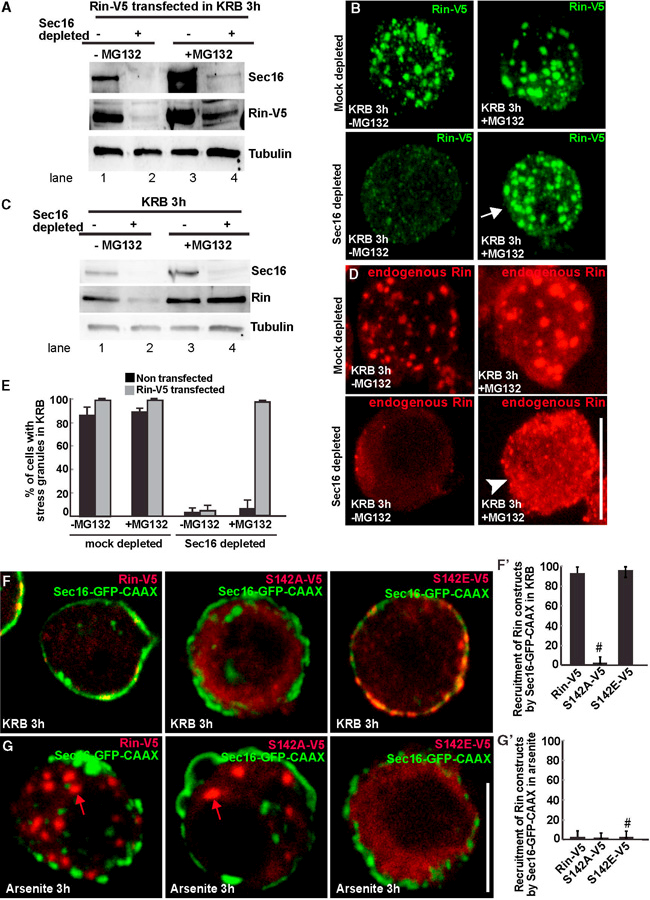
Most cellular stresses induce protein translation inhibition and stress granule formation. Here, using Drosophila S2 cells, we investigate the role of G3BP/Rasputin in this process. In contrast to arsenite treatment, where dephosphorylated Ser142 Rasputin is recruited to stress granules, we find that, upon amino acid starvation, only the phosphorylated Ser142 form is recruited. Furthermore, we identify Sec16, a component of the endoplasmic reticulum exit site, as a Rasputin interactor and stabilizer. Sec16 depletion results in Rasputin degradation and inhibition of stress granule formation. However, in the absence of Sec16, pharmacological stabilization of Rasputin is not enough to rescue the assembly of stress granules. This is because Sec16 specifically interacts with phosphorylated Ser142 Rasputin, the form required for stress granule formation upon amino acid starvation. Taken together, these results demonstrate that stress granule formation is fine-tuned by specific signaling cues that are unique to each stress. These results also expand the role of Sec16 as a stress response protein.

Keywords: Drosophila S2 cells; Rasputin; Sec16; amino acid starvation; arsenite; elF2 alpha; phosphorylation; protein stabilization; protein translation; protein transport in the secretory pathway; stress granules; stress response.


Figures

Figure 1.
Figure 1.. Amino Acid Starvation Leads to the Formation of Canonical Stress Granules
(A) Polysome profiles of Drosophila S2 cells grown in Schneider’s and incubated 4 hr in KRB. (B) Western blot visualization of elF2a phosphorylation in growing, KRB- and arsenite-treated cells. (C) Immunofluorescence (IF) visualization of endogenous FMR1, Caprin, and Rin in cells incubated in Schneider’s and KRB for 3 hr. Note that stress granules form (arrows). (D) IF co-localization of FMR1 and Rin. Note that they perfectly co-localize in stress granules (arrows). (E) IF co-localization of FMR1 and eIF4E and FMR1 and Tral in stress granules (arrows). (F) Kinetics of stress granule formation (marked by FMR1) in S2 cells incubated in KRB over the indicated time. (G) IF visualization of FMR1 upon KRB incubation, KRB supplemented with puromycin (puro) and cycloheximide (CHX), quantified in (G’). Note that stress granules do not form upon CHX incubation. (H) IF visualization of FMR1 upon KRB incubation followed by 15 and 60 min reversion in Schneider’s, quantified in (H’). Note that stress granules dissolve rapidly upon stress relief. Scale bars, 10 µm. Error bar, SEM.
Figure 2.
Figure 2.. Rin S142E, not S142A, Is Incorporated in Stress Granules upon Amino Acid Starvation
(A–A”) IF visualization of FMR1 in mock- (dsGFP) and Rin- (dsRin) depleted S2 cells (A and A” ) in growing conditions (Schneider’s) and upon amino acid starvation (KRB), quantified in (A’ ). Note that in Rin-depleted cells stress granules do not form. (B) Note that Rin-V5 overexpression (1.5 and 5 hr) does not induce stress granule formation even though the level of total Rin measured by western blot (using the anti-Rin antibody) increases (B”), quantified in (B’). (C) Western blot (using anti-V5 antibody) assessing the expression level of the V5-tagged Rin constructs used in (D)–(G). (D, D’) IF visualization of FRM1 (red) and overexpressed Rin-V5, S142A-V5, and S142E-V5 upon arsenite treatment for 3 hr (D). Note that S142A-V5 is steadily recruited into stress granule, while S142E is not. Quantified in D’. (E, E”) IF visualization of FRM1 (red) an-d overexpressed Rin-V5, S142A-V5, and S142E-V5 upon incubation in KRB for 3 hr (E). Note that S142E-V5 is steadily recruited into stress granule, while S142A is not. Quantified in E’. (F–F’) IF visualization of Rin-V5, S142A-V5, and S142E-V5 in cells depleted of endogenous Rin incubated in KRB for 3 hr (F). Note that Rin-V5 and S142E-V5 expression rescues stress granule formation, whereas S142A expression does not. Quantified in F’. (G–G’) IF visualization of Rin-V5 and S142E-V5 in Rin depleted cells after incubation in KRB for 3 hr followed by 3 hr incubation in Schneider’s (reversion) (G and quantified in G’ ). Note that Rin-V5 positive stress granules are completely reverted, whereas S142E positive stress granules are not. Scale bars, 10 µm. Error bar, SEM.
Figure 3.
Figure 3.. Sec16 Interacts with Rin
(A) IF visualization of FMR1 (green) and Sec16 (red) in cells in Schneider’s (A) and in KRB (A’). Note that Sec bodies (marked by Sec16) and stress granules form in close proximity to each other (A’’). (B) Immuno-EM localization of Sec16 (10 nm gold) and Tral (15 nm gold) in ultrathin sections of S2 cells incubated in KRB for 4 hr (B and B’). Note that Sec16 is mostly localized to Sec bodies (arrow) but also populates (red circles) stress granules (asterisks, marked by Tral). Note the mitochondria surrounding stress granules. (C) Pie chart representation of the 12 gene ontology groups representing the 149 interactors of endogenous Sec16 (see the Experimental Procedures and Table S2). (D) Western blot visualization of Rin following immunoprecipitation of endogenous Sec16 from growing (Schneider’s) and amino acid-starved (KRB 3 hr) S2 cells. (E–G and J) Confocal sections co-visualizing full-length Sec16-GFP-CAAX (E), DCter Sec16-GFP-CAAX (F), and Cter-GFP-CAAX (G) and endogenous Rin (red) upon amino acid starvation (KRB), quantified in (J). (H, I, and K) Confocal sections co-visualizing Sec16-GFP-CAAX with Rin-V5 (red, H) and with DRRM Rin-V5 (red, I) upon amino acid starvation (KRB), quantified in (K). Note that Rin-V5 is efficiently recruited to the plasma membrane by Sec16-CAAX, whereas DRRM-V5 is not and also does not form stress granules. Scale bars, 10 µm. Error bar, SD.
Figure 4.
Figure 4.. Sec16 Is Required for Stress Granule Formation Specifically upon Amino Acid Starvation
(A–C) IF visualization of endogenous Rin (A), Tral and eIF4E (B), and FMR1 (C) in mock- (dsGFP) and Sec16- (dsSec16) depleted cells upon 3 hr of incubation in KRB. Note that, in the absence of Sec16, stress granules do not form and Rin fluorescence is weak when compared to mock depleted cells. (D) IF visualization of endogenous FMR1 in mock- and Sec16-depleted cells upon arsenite treatment (3 hr). Note that stress granules form as efficiently in mock- and Sec16-depleted cells. (E) Quantification of Sec16-depleted cells treated with KRB, arsenite, heat shock, and DTT, expressed in percentage of cell with stress granules. (F) Western blot of Sec16, Rin, FMR1, Caprin, CF68, and tubulin in mock- and Sec16-depleted cells. Scale bars, 10 µm. Error bars, SEM.
Figure 5.
Figure 5.. Sec16 Prevents Rin Degradation by the Proteasome
(A) Western blot visualization of Rin-V5 (using an anti-V5 antibody) transfected in mock- and Sec16-depleted cells incubated in Schneider’s and KRB (3 hr), supplemented or not by MG132. Note that MG132 incubation partially rescues Rin-V5 protein level. (B) IF visualization of Rin-V5 transfected in mock- and Sec16-depleted cells in KRB, supplemented or not with MG132. Note that MG132 incubation completely rescues stress granule formation in Sec16-depleted cells. (C) Western blot visualization of endogenous Rin in mock- and Sec16-depleted cells in KRB, supplemented or not with MG132. Note that MG132 incubation completely rescues endogenous Rin protein level. (D) IF visualization of endogenous Rin in mock- and Sec16-depleted cells in KRB, supplemented or not with MG132. Note that MG132 incubation rescues endogenous Rin protein level (C), but it does not rescue stress granule formation. (E) Quantification of stress granule formation in Rin-V5 transfected and non-transfected cells in mock- and Sec16-depleted cells incubated in KRB, supplemented or not with MG132, expressed in percentage of cell profiles with stress granules. (F) Confocal section co-visualizing full-length Sec16-GFP-CAAX in cells expressing Rin-V5, S142A-V5, and S142E-V5 Rin mutants upon amino acid starvation (KRB) (F), quantified in F’ (expressed as percentage of Sec16-GFP-CAAX transfected cells). Note that both Rin-V5 and S142E-V5 steadily localize at the plasma membrane in cells expressing Sec16-GFP-CAAX, whereas S142A remains dispersed in the cytoplasm (marked by # in F’). (G) Confocal section co-visualizing full-length Sec16-GFP-CAAX in cells expressing Rin-V5, S142A-V5, and S142E-V5 Rin mutants upon arsenite treatment (G), quantified in G’ (expressed as percentage of Sec16-GFP-CAAX transfected cells). Note that none of the Rin variants are recruited by Sec16-GFP-CAAX. Furthermore, in agreement with Figures 2E and 2F, Rin-V5 and S142A form stress granules (red arrows). In contrast, S142E remains diffuse in the cytoplasm (marked by # in G’). Scale bars, 10 µm. Error bars, SD

Similar articles

Cited by

References

    1. Aguilera-Gomez A, van Oorschot MM, Veenendaal T, and Rabouille C (2016). In vivo vizualisation of mono-ADP-ribosylation by dPARP16 upon amino-acid starvation. eLife 5, e21475. - PMC - PubMed
    1. Anderson P, and Kedersha N (2002). Stressful initiations. J. Cell Sci 115, 3227–3234. - PubMed
    1. Anderson P, and Kedersha N (2006). RNA granules. J. Cell Biol 172, 803–808. - PMC - PubMed
    1. Anderson P, and Kedersha N (2008). Stress granules: the Tao of RNA triage. Trends Biochem. Sci 33, 141–150. - PubMed
    1. Aulas A, Stabile S, and Vande Velde C (2012). Endogenous TDP-43, but not FUS, contributes to stress granule assembly via G3BP. Mol. Neurodegener 7, 54. - PMC - PubMed

MeSH terms

LinkOut - more resources