

RIG-I expression in perifascicular myofibers is a reliable biomarker of dermatomyositis
- PMID: 28738907
- PMCID: PMC5525343
- DOI: 10.1186/s13075-017-1383-0
RIG-I expression in perifascicular myofibers is a reliable biomarker of dermatomyositis
Abstract
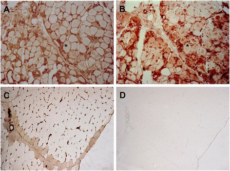
Background: Dermatomyositis (DM) is inflammatory myopathy or myositis characterized by muscle weakness and skin manifestations. In the differential diagnosis of DM the evaluation of the muscle biopsy is of importance among other parameters. Perifascicular atrophy in the muscle biopsy is considered a hallmark of DM. However, perifascicular atrophy is not observed in all patients with DM and, conversely, perifascicular atrophy can be observed in other myositis such as antisynthetase syndrome (ASS), complicating DM diagnosis. Retinoic acid inducible-gene I (RIG-I), a receptor of innate immunity that promotes type I interferon, was observed in perifascicular areas in DM. We compared the value of RIG-I expression with perifascicular atrophy as a biomarker of DM.
Methods: We studied by immunohistochemical analysis the expression of RIG-I and the presence of perifascicular atrophy in 115 coded muscle biopsies: 44 patients with DM, 18 with myositis with overlap, 8 with ASS, 27 with non-DM inflammatory myopathy (16 with polymyositis, 6 with inclusion body myositis, 5 with immune-mediated necrotizing myopathy), 8 with muscular dystrophy (4 with dysferlinopathy, 4 with fascioscapulohumeral muscle dystrophy) and 10 healthy controls.
Results: We found RIG-I-positive fibers in 50% of DM samples vs 11% in non-DM samples (p < 0.001). Interestingly, RIG-I staining identified 32% of DM patients without perifascicular atrophy (p = 0.007). RIG-I sensitivity was higher than perifascicular atrophy (p < 0.001). No differences in specificity between perifascicular atrophy and RIG-I staining were found (92% vs 88%). RIG-I staining was more reproducible than perifascicular atrophy (κ coefficient 0.52 vs 0.37).
Conclusions: The perifascicular pattern of RIG-I expression supports the diagnosis of DM. Of importance for clinical and therapeutic studies, the inclusion of RIG-I in the routine pathological staining of samples in inflammatory myopathy will allow us to gather more homogeneous subgroups of patients in terms of immunopathogenesis.
Keywords: Biomarker; Dermatomyositis; Inflammatory myopathies; Muscle biopsy; Perifascicular atrophy.
Conflict of interest statement
Consent for publication
Not applicable.
Competing interests
The authors declare that they have no competing interests.
Publisher’s Note
Springer Nature remains neutral with regard to jurisdictional claims in published maps and institutional affiliations.
Figures
Similar articles
-
Diagnostic potential of sarcoplasmic myxovirus resistance protein A expression in subsets of dermatomyositis.Neuropathol Appl Neurobiol. 2019 Aug;45(5):513-522. doi: 10.1111/nan.12519. Epub 2018 Nov 22. Neuropathol Appl Neurobiol. 2019. PMID: 30267437
-
Redefining dermatomyositis: a description of new diagnostic criteria that differentiate pure dermatomyositis from overlap myositis with dermatomyositis features.Medicine (Baltimore). 2014 Nov;93(24):318-332. doi: 10.1097/MD.0000000000000222. Medicine (Baltimore). 2014. PMID: 25500701 Free PMC article.
-
[Expression of retinoic acid-I nducible gene I in the muscle tissues of idiopathic inflammatory myopathies].Zhonghua Yi Xue Za Zhi. 2015 Jun 16;95(23):1823-8. Zhonghua Yi Xue Za Zhi. 2015. PMID: 26712399 Chinese.
-
Contribution of Complement, Microangiopathy and Inflammation in Idiopathic Inflammatory Myopathies.J Neuromuscul Dis. 2024;11(1):5-16. doi: 10.3233/JND-230168. J Neuromuscul Dis. 2024. PMID: 38143369 Free PMC article. Review.
-
[Inflammatory myopathies: diagnosis and classifications].Presse Med. 2009 Jul-Aug;38(7-8):1141-63. doi: 10.1016/j.lpm.2009.01.013. Epub 2009 Mar 17. Presse Med. 2009. PMID: 19282137 Review. French.
Cited by
-
Immunoproteasome subunit β5i promotes perifascicular muscle atrophy in dermatomyositis by upregulating RIG-I.RMD Open. 2023 Feb;9(1):e002818. doi: 10.1136/rmdopen-2022-002818. RMD Open. 2023. PMID: 36854567 Free PMC article.
-
UBXN9 governs GLUT4-mediated spatial confinement of RIG-I-like receptors and signaling.Res Sq [Preprint]. 2024 Jun 4:rs.3.rs-3373803. doi: 10.21203/rs.3.rs-3373803/v1. Res Sq. 2024. Update in: Nat Immunol. 2024 Dec;25(12):2234-2246. doi: 10.1038/s41590-024-02004-7. PMID: 38883790 Free PMC article. Updated. Preprint.
-
Sarcoplasmic Myxovirus Resistance Protein A: A Study of Expression in Idiopathic Inflammatory Myopathy.J Inflamm Res. 2023 Nov 20;16:5417-5426. doi: 10.2147/JIR.S433239. eCollection 2023. J Inflamm Res. 2023. PMID: 38026261 Free PMC article.
-
Pathophysiological Mechanisms and Treatment of Dermatomyositis and Immune Mediated Necrotizing Myopathies: A Focused Review.Int J Mol Sci. 2022 Apr 13;23(8):4301. doi: 10.3390/ijms23084301. Int J Mol Sci. 2022. PMID: 35457124 Free PMC article. Review.
-
Meta-Analysis of Polymyositis and Dermatomyositis Microarray Data Reveals Novel Genetic Biomarkers.Genes (Basel). 2019 Oct 30;10(11):864. doi: 10.3390/genes10110864. Genes (Basel). 2019. PMID: 31671645 Free PMC article.
References
-
- Troyanov Y, Targoff IN, Payette MP, Raynauld JP, Chartier S, Goulet JR, Bourre-Tessier J, Rich E, Grodzicky T, Fritzler MJ, et al. Redefining dermatomyositis: a description of new diagnostic criteria that differentiate pure dermatomyositis from overlap myositis with dermatomyositis features. Medicine (Baltimore) 2014;93(24):318–32. doi: 10.1097/MD.0000000000000222. - DOI - PMC - PubMed
-
- Aouizerate J, De Antonio M, Bassez G, Gherardi RK, Berenbaum F, Guillevin L, Berezne A, Valeyre D, Maisonobe T, Dubourg O, et al. Myofiber HLA-DR expression is a distinctive biomarker for antisynthetase-associated myopathy. Acta Neuropathol Commun. 2014;2:154. doi: 10.1186/s40478-014-0154-2. - DOI - PMC - PubMed
MeSH terms
Substances
LinkOut - more resources
Full Text Sources
Other Literature Sources
Miscellaneous
