

[Angiotensin-(1-7) protects cardiac myocytes against high glucose-induced injury by inhibiting ClC-3 chloride channels]
- PMID: 28736364
- PMCID: PMC6765525
- DOI: 10.3969/j.issn.1673-4254.2017.07.07
[Angiotensin-(1-7) protects cardiac myocytes against high glucose-induced injury by inhibiting ClC-3 chloride channels]
Abstract
Objective: To explore whether angiotensin-(1-7) [Ang-(1-7)] protects cardiac myocytes against high glucose (HG)-induced injury by inhibiting ClC-3 chloride channels.
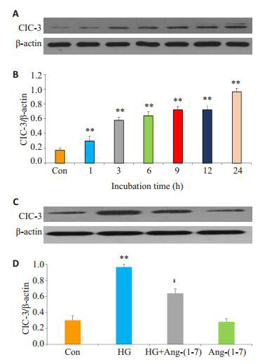
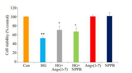
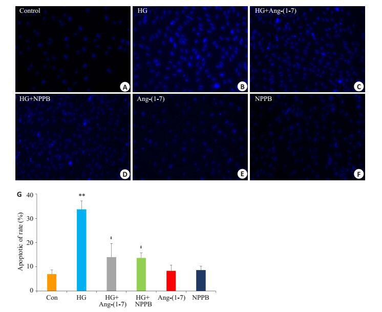
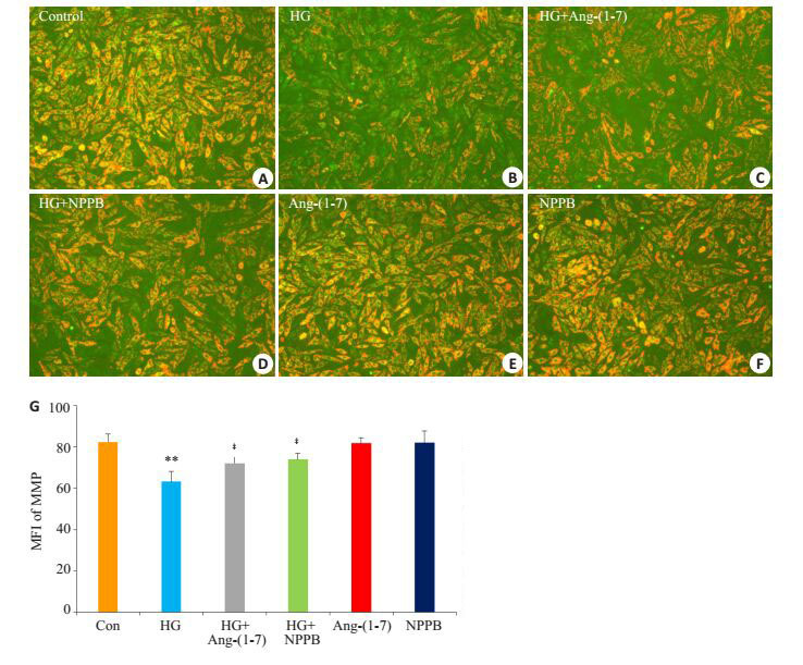
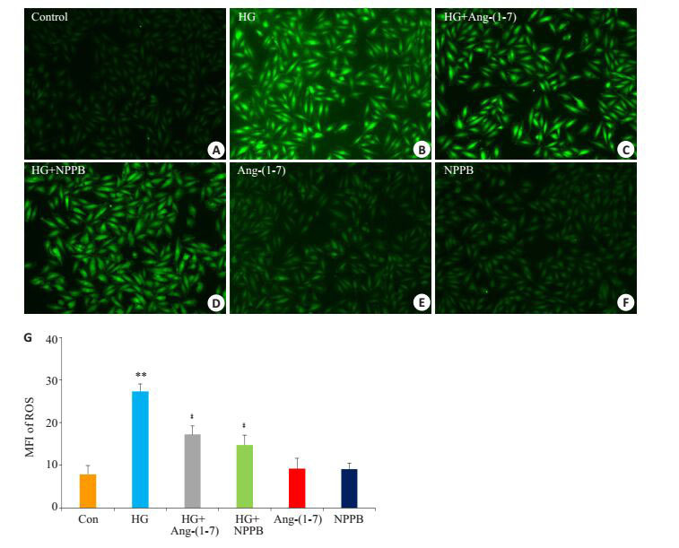
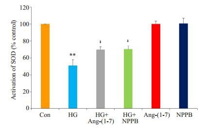
Method: H9c2 cardiac cells were exposed to 35 mmol/L glucose for 24 h to establish a cell injury model. The cells were treated with Ang-(1-7) or the inhibitor of chloride channel (NPPB) in the presence of HG for 24 h to observe the changes in HG-induced cell injury. Cell counter kit 8 (CCK-8) assay was used to test the cell viability, and the morphological changes of the apoptotic cells were detected using Hoechst 33258 staining and fluorescent microscopy. The intracellular level of reactive oxygen species (ROS) was examined by DCFH-DA staining, SOD activity in the culture medium was measured using commercial kits, and the mitochondrial membrane potential (MMP) of the cells was tested with rodamine 123 staining. The expression level of cardiac ClC-3 chloride channels was detected with Western blotting.
Results: Exposure of H9c2 cardiac cells to 35 mmol/L glucose for 24 h markedly enhanced the expressions of cardiac ClC-3 channel protein (P<0.01). Co-treatment of the cells with 1 µmol/L Ang-(1-7) and HG for 24 h significantly attenuated HG- induced upregulation of ClC-3 channel protein expression (P<0.01). Co-treatment of the cells exposed to HG with 1 µmol/L Ang-(1-7) or 100 µmol/L NPPB for 24 h obviously ameliorated HG-induced injuries as shown by increased cell viability, enhanced SOD activity, decreased number of apoptotic cells, and reduced intracellular ROS generation and loss of MMP (P<0.01).
Conclusion: ClC-3 channels are involved in HG-induced injury in cardiac cells. Ang-(1-7) protects cardiac cells against HG-induced injury by inhibiting ClC-3 channels.
目的: 探讨血管紧张素-(1-7)[Ang-(1-7)]能否通过调控ClC-3通道保护心肌细胞对抗高糖引起的损伤。
方法: 应用35 mmol/L葡萄糖处理H9c2心肌细胞24 h建立损伤模型。Ang-(1-7)或氯通道抑制剂与心肌细胞共处理24 h观察对高糖诱发的心肌细胞损伤的影响。应用细胞计数试剂盒8检测细胞存活率;Hoechst33258染色荧光显微镜照相术检测凋亡细胞的形态学改变;双氯荧光素染色荧光显微镜照相术测定细胞内活性氧水平;超氧化物歧化酶试剂盒测定活性;罗丹明123染色荧光显微镜照相术检测线粒体膜电位;Western blot法测定心肌细胞ClC-3通道蛋白的表达水平。
结果: 35 mmol/L葡萄糖处理H9c2心肌细胞24 h明显地增加ClC-3通道蛋白的表达水平(P < 0.01);1 μmol/LAng(-1-7)与葡萄糖共处理心肌细胞24 h显著地抑制葡萄糖对ClC-3通道蛋白表达的上调作用(P < 0.01);1 μmol/LAng(-1-7)或100 μmol/L氯通道抑制剂氯通道抑制剂与葡萄糖共处理心肌细胞24 h减轻葡萄糖引起的损伤作用,表现为增加细胞存活率和超氧化物歧化酶活性,减小细胞凋亡数量,胞内活性氧水平及线粒体膜电位丢失(P < 0.01)。
结论: ClC-3通道参与葡萄糖引起的心肌细胞损伤;Ang-(1-7)通过抑制ClC-3通道保护心肌细胞对抗葡萄糖引起的损伤。
Figures
Similar articles
-
Angiotensin-(1-7) protects cardiomyocytes against high glucose-induced injuries through inhibiting reactive oxygen species-activated leptin-p38 mitogen-activated protein kinase/extracellular signal-regulated protein kinase 1/2 pathways, but not the leptin-c-Jun N-terminal kinase pathway in vitro.J Diabetes Investig. 2017 Jul;8(4):434-445. doi: 10.1111/jdi.12603. Epub 2017 Feb 28. J Diabetes Investig. 2017. PMID: 27896943 Free PMC article.
-
Protective effect of angiotensin-(1-7) against hyperglycaemia-induced injury in H9c2 cardiomyoblast cells via the PI3K̸Akt signaling pathway.Int J Mol Med. 2018 Mar;41(3):1283-1292. doi: 10.3892/ijmm.2017.3322. Epub 2017 Dec 15. Int J Mol Med. 2018. PMID: 29286068 Free PMC article.
-
The Opening of ATP-Sensitive K+ Channels Protects H9c2 Cardiac Cells Against the High Glucose-Induced Injury and Inflammation by Inhibiting the ROS-TLR4-Necroptosis Pathway.Cell Physiol Biochem. 2017;41(3):1020-1034. doi: 10.1159/000461391. Epub 2017 Feb 22. Cell Physiol Biochem. 2017. PMID: 28291959
-
ATP-sensitive K⁺ channels contribute to the protective effects of exogenous hydrogen sulfide against high glucose-induced injury in H9c2 cardiac cells.Int J Mol Med. 2016 Mar;37(3):763-72. doi: 10.3892/ijmm.2016.2467. Epub 2016 Jan 25. Int J Mol Med. 2016. PMID: 26820501
-
Exogenous H2S protects H9c2 cardiac cells against high glucose-induced injury and inflammation by inhibiting the activation of the NF-κB and IL-1β pathways.Int J Mol Med. 2015 Jan;35(1):177-86. doi: 10.3892/ijmm.2014.2007. Epub 2014 Nov 19. Int J Mol Med. 2015. PMID: 25412187
References
-
- Xu W, Chen J, Lin J, et al. Exogenous H2S protects H9c2 cardiac cells against high glucose-induced injury and inflammation by inhibiting the activation of the NF-κB and IL-1β pathways. https://www.ncbi.nlm.nih.gov/pubmed/25412187. Int J Mol Med. 2015;35(1):177–86. [Xu W, Chen J, Lin J, et al. Exogenous H2S protects H9c2 cardiac cells against high glucose-induced injury and inflammation by inhibiting the activation of the NF-κB and IL-1β pathways [J]. Int J Mol Med, 2015, 35(1): 177-86.] - PubMed
-
- Lei Y, Xu Q, Zeng B, et al. Angiotensin-(1-7) protects cardiomyocytes against high glucose-induced injuries through inhibiting reactive oxygen species-activated leptin-p38 mitogenactivated protein kinase/extracellular signal-regulated protein kinase 1/2 pathways, but not the leptin-c-Jun N-terminal kinase pathway in vitro. https://www.ncbi.nlm.nih.gov/pubmed/27896943. J Diabetes Investig. 2016;29(11):12603. [Lei Y, Xu Q, Zeng B, et al. Angiotensin-(1-7) protects cardiomyocytes against high glucose-induced injuries through inhibiting reactive oxygen species-activated leptin-p38 mitogenactivated protein kinase/extracellular signal-regulated protein kinase 1/2 pathways, but not the leptin-c-Jun N-terminal kinase pathway in vitro [J]. J Diabetes Investig, 2016, 29(11): 12603.] - PMC - PubMed
-
- Xu WM, Wu W, Chen JF, et al. Exogenous Hydrogen sulfide protects H9c2 cardiac cells against high glucose-induced injury by inhibiting the activities of the p38 MAPK and ERK1/2 pathways. http://www.ncbi.nlm.nih.gov/pubmed/23912965. Int J Mol Med. 2013;32(4):917–25. [Xu WM, Wu W, Chen JF, et al. Exogenous Hydrogen sulfide protects H9c2 cardiac cells against high glucose-induced injury by inhibiting the activities of the p38 MAPK and ERK1/2 pathways [J]. Int J Mol Med, 2013, 32(4): 917-25.] - PubMed
-
- Fuentes-Antrás J, Ioan AM, Tuáón J, et al. Activation of toll-like receptors and inflammasome complexes in the diabetic cardiomyopathy-associated inflammation. http://www.ncbi.nlm.nih.gov/pubmed/24744784. Int J Endocrinol. 2014;(6):847827. [Fuentes-Antrás J, Ioan AM, Tuáón J, et al. Activation of toll-like receptors and inflammasome complexes in the diabetic cardiomyopathy-associated inflammation [J]. Int J Endocrinol, 2014 (6): 847827.] - PMC - PubMed
-
- Verkerk AO, Veldkamp MW, Bouman LN, et al. Calcium-activated Cl(-) current contributes to delayed afterdepolarizations in single Purkinje and ventricular myocytes. Circulation. 2000;101(22):2639–44. doi: 10.1161/01.CIR.101.22.2639. [Verkerk AO, Veldkamp MW, Bouman LN, et al. Calcium-activated Cl(-) current contributes to delayed afterdepolarizations in single Purkinje and ventricular myocytes[J]. Circulation, 2000, 101(22): 2639-44.] - DOI - PubMed
Publication types
Grants and funding
LinkOut - more resources
Full Text Sources
Miscellaneous