. 2018 May 1;314(5):F773-F787.
doi: 10.1152/ajprenal.00260.2017. Epub 2017 Jul 19.

ErbB4 deletion accelerates renal fibrosis following renal injury

Affiliations

ErbB4 deletion accelerates renal fibrosis following renal injury

Fenghua Zeng et al. Am J Physiol Renal Physiol. .

Abstract

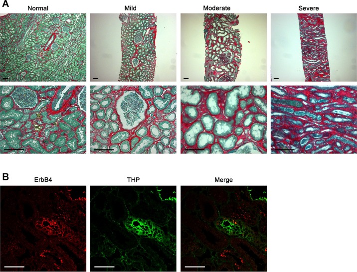
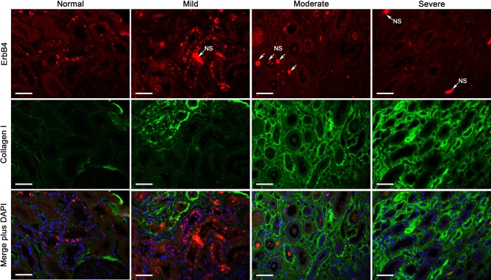
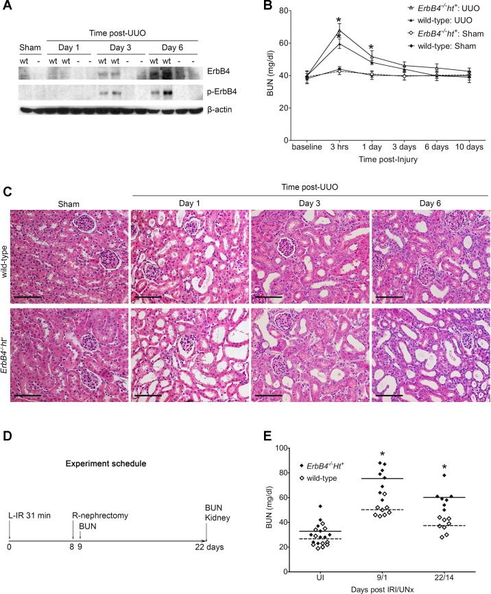
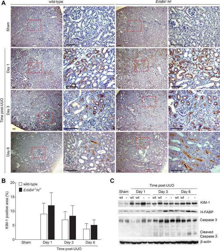
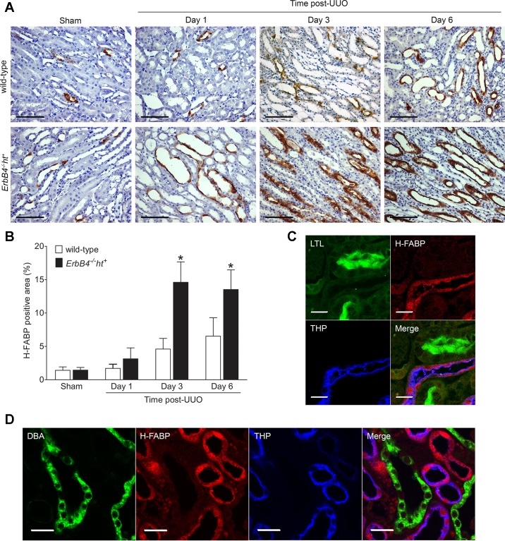
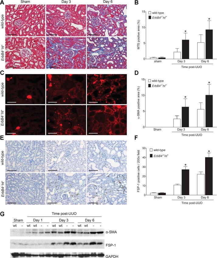
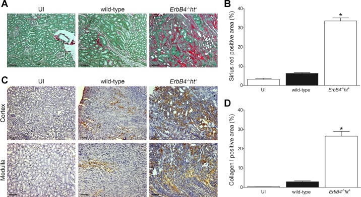
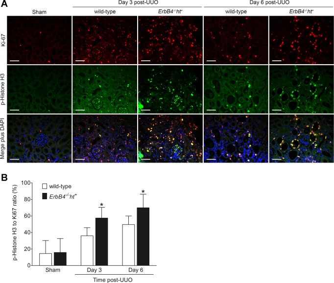
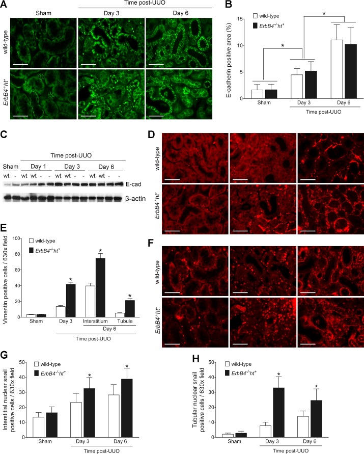
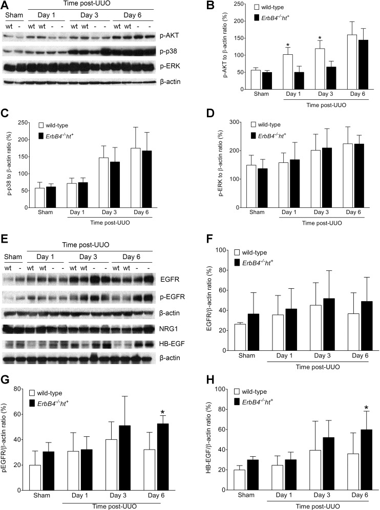
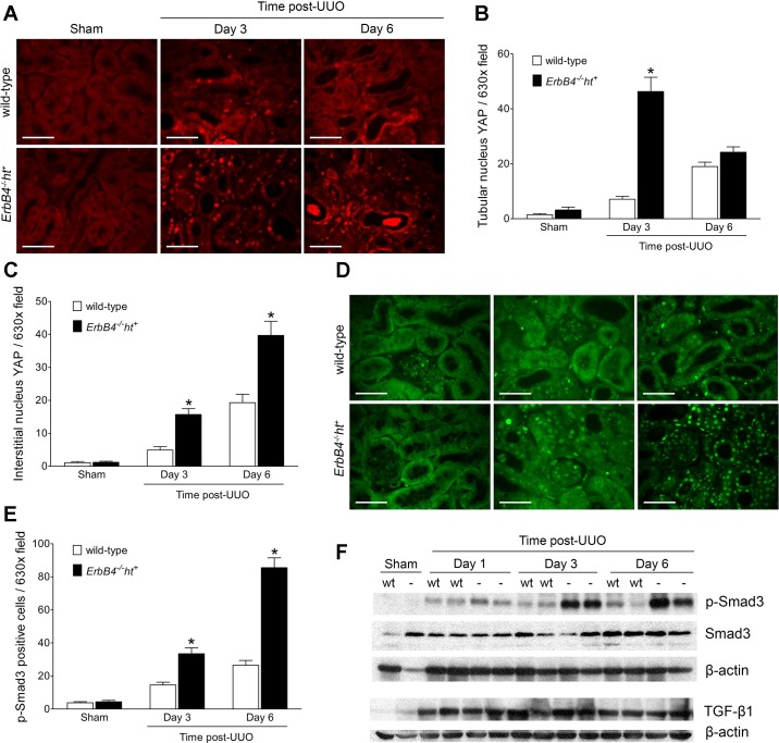
Tubulointerstitial fibrosis (TIF) is a prominent factor in the progression of chronic kidney disease regardless of etiology. Avian erythroblastic leukemia viral oncogene homolog 4 (ErbB4) expression levels were inversely correlated to renal fibrosis in human fibrotic kidneys. In both unilateral ureteral obstruction (UUO) and ischemia-reperfusion injury followed by uninephrectomy (IRI/UNx) mouse models, expression levels of ErbB4 were elevated in the early stage of renal injury. Using mice with global ErbB4 deletion except for transgenic rescue in cardiac tissue ( ErbB4-/-ht+), we determined that UUO induced similar injury in proximal tubules compared with wild-type mice but more severe injury in distal nephrons. TIF was apparent earlier and was more pronounced following UUO in ErbB4-/-ht+ mice. With ErbB4 deletion, UUO injury inhibited protein kinase B phosphorylation and increased the percentage of cells in G2/M arrest. There was also increased nuclear immunostaining of yes-associated protein and increased expression of phospho-Mothers against decapentaplegic homolog 3, snail1, and vimentin. These results indicate that ErbB4 deletion accelerates the development and progression of renal fibrosis in obstructive nephropathy. Similar results were found in a mouse IRI/UNx model. In conclusion, increased expression of ErbB4 in the early stages of renal injury may reflect a compensatory effect to lessen tubulointerstitial injury.

Keywords: avian erythroblastic leukemia viral oncogene homolog 4; cell cycle; epithelial dedifferentiation; tubulointerstitial fibrosis.


Figures

Fig. 1.
Fig. 1.
Validation of the degree of renal fibrosis and avian erythroblastic leukemia viral oncogene homolog 4 (ErbB4) localization in human kidney biopsies. A: sirius red/fast green staining to validate the degree of renal fibrosis in human samples. Scale bar, 100 µm. B: low levels of ErbB4 (red) were expressed in normal human kidneys; some was detected in thick ascending limb, as indicated by the colocalization with Tamm-Horsfall glycoprotein (THP). Scale bar, 50 µm.
Fig. 2.
Fig. 2.
ErbB4 expression in normal and fibrotic human kidneys. Low levels of ErbB4 (red) were expressed in normal human kidneys, with increased expression in the mild renal fibrotic kidneys but decreased expression in the moderate and severe fibrotic kidneys, as indicated by collagen I immunostaining (green). NS, nonspecific staining. Representative images from n = 3 mice in each group are shown. Scale bar, 50 µm.
Fig. 3.
Fig. 3.
Characterization of ErbB4 deletion on changes of renal morphology and function following injury. A: expression and activation of ErbB4 levels after injury detected by immunoblotting. In wild-type sham-operated kidneys, minimum levels of ErbB4 were detected without activation form [phosphorylated (p)-ErbB4]. After unilateral ureteral obstruction (UUO), ErbB4 levels increased gradually from day 3 to day 6 post-UUO, concomitant with increased levels of p-ErbB4 in wild-type controls. ErbB4 expression was not detected in either sham or UUO kidneys from ErbB4−/−ht+ mice. wt, Wild type; −, ErbB4−/−ht+. B: blood urea nitrogen (BUN) level after UUO injury. Compared with sham, mice undergoing UUO had increased BUN levels after 3 h of injury in both groups. After UUO (1 day), only ErbB4−/−ht+ mice still had significantly elevated levels of BUN; n = 6–8 in each time point of each group. *P < 0.05 compared with sham. C: hematoxylin and eosin (H&E) staining. No morphological changes were detected in sham-operated kidneys in either group. Compared with wild-type controls, kidneys with ErbB4 deletion had more dilated tubules 1 day after UUO and increased interstitial area 3 and 6 days post-UUO. Scale bar, 100 µm. D: experimental schedule for ischemia-reperfusion injury followed by uninephrectomy (IRI/UNx). E: BUN levels post-IRI/UNx injury. Compared with the uninjured mice (UI), IRI/UNx injury increased BUN levels in both wild-type and ErbB4 deletion mice after 1 or 14 days of injury (P < 0.05). In IRI/UNx groups, compared with the wild type, ErbB4 deletion further increased BUN levels to a significant degree with slow recovery at 14 days after UNx. *P < 0.05; n = 7–8 in each group.
Fig. 4.
Fig. 4.
Effects of ErbB4 deletion on tubular injury after UUO. A: kidney injury molecule-1 (KIM-1) immunohistochemistry staining. In sham-operated kidneys, no KIM-1 immunostaining was detected in either group. Post-UUO injury (1 day), KIM-1 immunostaining was dramatically increased in the S3 segments and was also detected in S1 and S2 segments at 3 and 6 days post-UUO in both groups. Scale bar, 100 µm. B: quantification of KIM-1-positive immunostaining area of A using Image J software. Values represent means ± SE from 20 random fields of 4–6 kidneys in each group. C: expression levels of kidney injury marker proteins by immunoblotting. KIM-1 expression levels increased early in the UUO injury by 1 day post-UUO and remained elevated at days 3 and 6 post-UUO with no significant differences between wild-type and ErbB4−/−ht+. Elevated heart fatty acid-binding protein (H-FABP) levels were detected 3 days post-UUO injury and remained elevated at 6 days post-UUO in ErbB4−/−ht+ mouse kidneys compared with wild-type kidneys at the same time points. Baseline caspase-3 expression was detected in sham-operated kidneys. After UUO, kidneys from ErbB4−/−ht+ mice showed slightly higher levels of total caspase-3 at 1 and 3 days post-UUO, whereas elevated cleaved caspase-3 was only detected at 6 days post-UUO. Images represent 3 independent experiments of 6 samples from each group.
Fig. 5.
Fig. 5.
H-FABP localization and its expression levels after UUO injury. A: expression of H-FABP after UUO injury. Compared with the wild type, ErbB4−/−ht+ kidneys showed increased H-FABP immunoreactivity 1 day after UUO and had further increases at 3 and 6 days post-UUO. Scale bar, 100 µm. B: quantification of H-FABP-positive immunostaining area of A using Image J software. Values represent means ± SE from 20 random fields of 4–6 kidneys in each group. *P < 0.05 compared with the wild type at the same time point. C and D: immunofluorescence staining of H-FABP with marker proteins. H-FABP colocalized with THP, a marker for thick ascending limb and distal convoluted tubule. H-FABP did not colocalize with Lotus tetragonolobus lectin (LTL), a marker for proximal tube, or DBA, a marker for collecting duct. Scale bar, 20 µm.
Fig. 6.
Fig. 6.
ErbB4 deletion accelerated renal interstitial fibrosis after UUO injury. Paraffin-embedded kidney sections from wild-type (n = 6) or ErbB4−/−ht+ (n = 8) mice were stained with Masson’s trichrome (A) to identify collagen fibers (scale bar, 100 µm), α-smooth muscle actin (α-SMA, C) to identify activated interstitial myofibroblasts (scale bar, 50 µm), and fibroblast-specific protein 1 (FSP-1, E), a marker for fibroblasts (scale bar, 100 µm). Representative images are shown, and positive staining was quantified from 20 random fields using ImageJ software. The corresponding results are shown in B, D, and F. Values represent means ± SE. *P < 0.05 compared with the wild type at the same time point. G: α-SMA and FSP-1 expression by immunoblotting. Compared with kidneys from the wild type, kidneys with ErbB4 deletion showed elevated expression levels of α-SMA and FSP-1 starting at 3 days post-UUO injury and remained high at 6 days post-UUO.
Fig. 7.
Fig. 7.
ErbB4 deletion accelerated renal interstitial fibrosis after IRI/UNx injury. A: sirius red/fast green staining on renal kidney post-22 days of initial IRI/UNx injury. Compared with the uninjured or wild-type kidneys, ErbB4 deletion kidney showed extensive sirius red staining. Scale bar, 100 µm. C: collagen I immunostaining in IRI/UNx kidneys. Scale bar, 100 µm. Representative images are shown, and positive staining was quantified from 20 random fields using ImageJ software. The corresponding results are shown in B and D. Values represent means ± SE. *P < 0.05 compared with the wild type and uninjured.
Fig. 8.
Fig. 8.
ErbB4 deletion increased G2/M arrest after UUO injury. A: representative images from sham-operated kidneys in either group (n = 6) and 3 and 6 days post-UUO kidneys from wild-type (n = 6) and ErbB4−/−ht+ mice (n = 8) stained with Ki-67 (red), p-Histone H3 (green), and DAPI (blue). Scale bar, 50 µm. B: p-Histone H3 and Ki-67-positive cells were counted using ImageJ software from at least 20 random fields. p-Histone H3-to-Ki-67 ratio was used to evaluate cells in G2/M phase. Values represent means ± SE. *P < 0.05 compared with the wild type at the same time point.
Fig. 9.
Fig. 9.
ErbB4 deletion induced renal tubular epithelial dedifferentiation after UUO injury. Sham or UUO-operated kidney sections were stained with anti-E-cadherin (E-cad, A), an epithelial marker, and anti-vimentin (D) and Snail 1 (F), mesenchymal marker. Scale bar, 50 µm. Positive immunostaining areas or cell numbers were calculated using ImageJ software and represented as means ± SE (B, E, G, and H). *P < 0.05 compared with the wild type at the same time point. C: E-cadherin expression levels by immunoblotting. Images represent results from 3 independent experiments.
Fig. 10.
Fig. 10.
Signaling alterations after UUO injury. A: representative images from 3 independent experiments of MAP kinase protein expression levels by immunoblotting. B–D: densitometric analysis of immunoblotting in A expressed as ratios to β-actin. Values represent means ± SE. *P < 0.05 compared with the wild type. E: representative images of epidermal growth factor receptor (EGFR) expression, activation, and ligand level changes by Western blots. F–H: densitometric analysis of immunoblotting in E expressed as ratios to β-actin. Values represent means ± SE. *P < 0.05 compared with the wild type.
Fig. 11.
Fig. 11.
ErbB4 deletion increased yes-associated protein (YAP) and p-Mothers against decapentaplegic homolog 3 (Smad3) nuclear expression after UUO. A: immunofluorescence staining of YAP. Scale bar, 50 µm. B and C: quantification of YAP-positive cells from at least 20 random fields using ImageJ. Compared with the wild type, obstructed kidneys with ErbB4 deletion showed more YAP nuclear staining in both tubular epithelium and interstitium as early as 3 days post-UUO. At 6 days post-UUO, much more YAP nuclear-positive cells were located in the interstitium. Values represent means ± SE. *P < 0.05 compared with the wild type. D: immunofluorescence staining of p-Smad3. Scale bar, 50 µm. E: quantification of p-Smad3-positive cells. Compared with the wild type, obstructed kidneys with ErbB4 deletion showed more p-Smad3 nuclear staining starting 3 days post-UUO and increased continuously at 6 days post-UUO. Values represent means ± SE from at least 20 random fields. *P < 0.05 compared with the wild type. F: p-Smad3, Smad3, and TGF-β1 expression by immunoblotting. Images represent results from 3 independent experiments.

Similar articles

Cited by

References

    1. Basu S, Totty NF, Irwin MS, Sudol M, Downward J. Akt phosphorylates the Yes-associated protein, YAP, to induce interaction with 14-3-3 and attenuation of p73-mediated apoptosis. Mol Cell 11: 11–23, 2003. doi:10.1016/S1097-2765(02)00776-1. - DOI - PubMed
    1. Böger CA, Sedor JR. GWAS of diabetic nephropathy: is the GENIE out of the bottle? PLoS Genet 8: e1002989, 2012. doi:10.1371/journal.pgen.1002989. - DOI - PMC - PubMed
    1. Boukhalfa G, Desmoulière A, Rondeau E, Gabbiani G, Sraer JD. Relationship between alpha-smooth muscle actin expression and fibrotic changes in human kidney. Exp Nephrol 4: 241–247, 1996. - PubMed
    1. Chen J, Chen JK, Nagai K, Plieth D, Tan M, Lee TC, Threadgill DW, Neilson EG, Harris RC. EGFR signaling promotes TGFβ-dependent renal fibrosis. J Am Soc Nephrol 23: 215–224, 2012. doi:10.1681/ASN.2011070645. - DOI - PMC - PubMed
    1. Docherty NG, Calvo IF, Quinlan MR, Pérez-Barriocanal F, McGuire BB, Fitzpatrick JM, Watson RW. Increased E-cadherin expression in the ligated kidney following unilateral ureteric obstruction. Kidney Int 75: 205–213, 2009. doi:10.1038/ki.2008.482. - DOI - PubMed

Publication types

MeSH terms