. 2017 Sep 4;216(9):2827-2841.
doi: 10.1083/jcb.201606043. Epub 2017 Jul 17.

Outer nuclear membrane protein Kuduk modulates the LINC complex and nuclear envelope architecture

Affiliations

Outer nuclear membrane protein Kuduk modulates the LINC complex and nuclear envelope architecture

Zhao-Ying Ding et al. J Cell Biol. .

Abstract

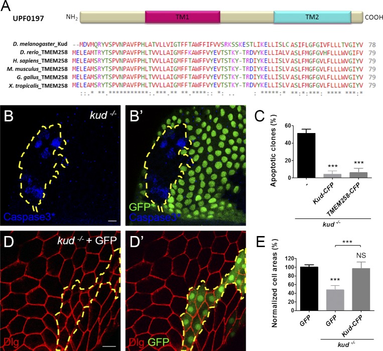
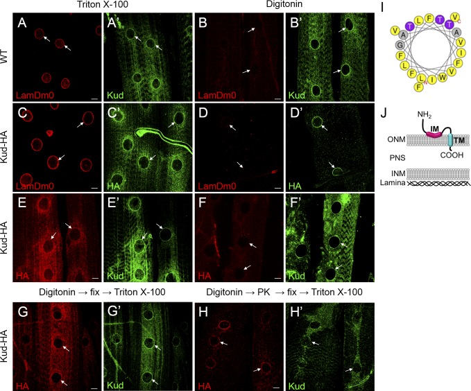
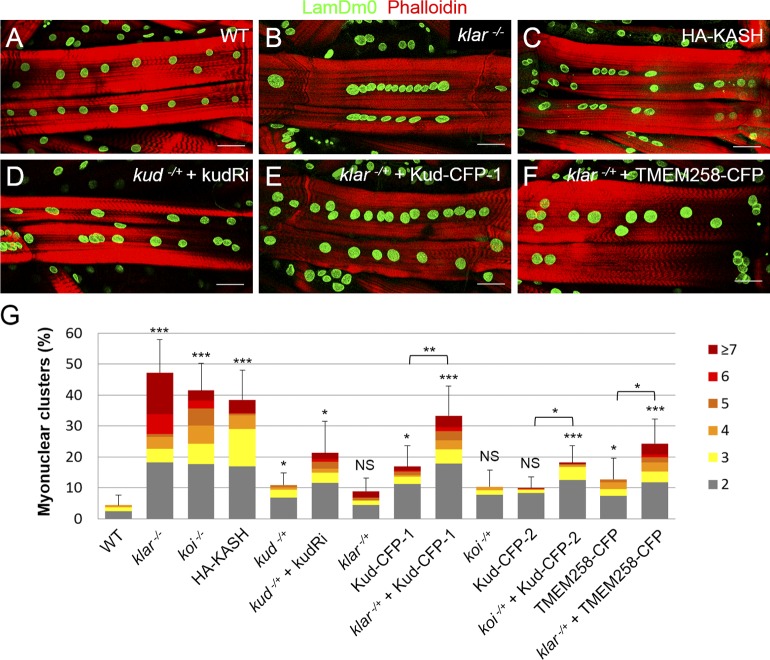
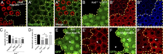
Linker of nucleoskeleton and cytoskeleton (LINC) complexes spanning the nuclear envelope (NE) contribute to nucleocytoskeletal force transduction. A few NE proteins have been found to regulate the LINC complex. In this study, we identify one, Kuduk (Kud), which can reside at the outer nuclear membrane and is required for the development of Drosophila melanogaster ovarian follicles and NE morphology of myonuclei. Kud associates with LINC complex components in an evolutionarily conserved manner. Loss of Kud increases the level but impairs functioning of the LINC complex. Overexpression of Kud suppresses NE targeting of cytoskeleton-free LINC complexes. Thus, Kud acts as a quality control mechanism for LINC-mediated nucleocytoskeletal connections. Genetic data indicate that Kud also functions independently of the LINC complex. Overexpression of the human orthologue TMEM258 in Drosophila proved functional conservation. These findings expand our understanding of the regulation of LINC complexes and NE architecture.


Figures

Figure 1.
Figure 1.
The evolutionarily conserved protein Kud is required for ovarian follicle development. (A) UPF0197 proteins have been conserved in evolution; TM1 and TM2 are putative TMs. The NCBI accession numbers of the UPD0197 proteins from top to bottom are Q9VVA8, Q6PBS6, P61165, P61166, Q76LT9, and Q6DDB3. Asterisks indicate conserved residues, colons indicate residues with strongly similar properties, and periods indicate residues with weakly similar properties. (B and B’) Apoptotic cells were marked with activated caspase 3 (Caspase3*), and the kud mutant cells lacking GFP expression are outlined by dashed lines. (C) Quantification of the clones containing apoptotic cells: n = 78, 153, and 78 from left to right. (D and D′) The boundaries of follicle cells were visualized by the plasma membrane protein Disc large (Dlg in red), and the kud mutant cells expressing GFP are outlined with dashed lines. Bars, 10 µm. (E) Quantification of normalized cell areas: n (number of clones) = 9, 13, and 15 from left to right. ***, P < 0.001. Error bars indicate means ± SD.
Figure 2.
Figure 2.
Conserved subcellular localizations at the NE and in the cytoplasm. (A) TMEM258 is membrane associated. Western blot (WB) of soluble (Sol) and membrane (Mem) fractions of TMEM258-Myc–expressing 293T cells probed with anti-GAPDH (cytoplasm), anti-Calnexin (ER and ONM), anti-Emerin (INM), and anti-Myc antibodies. (B–C′′) The overexpressed TMEM258-Myc in 293T cells (B) and the endogenous Kud in ovarian follicle cells (C) locate at the cytoplasm and colocalize with NE marker lamins (B′ and C′, arrows). Merges are shown in B′′ and C′′. Bars, 10 µm.
Figure 3.
Figure 3.
Kud spans the ONM. (A–F′) Larval muscles of indicated genotypes were permeabilized with Triton X-100 or digitonin as indicated. The muscles were stained with antibodies against LamDm0 (A, B, C, and D), Kud (A′, B′, E′, and F′), or HA (C′, D′, E, and F). Bars, 10 µm. (G–H) Proteinase K protection assays of intact nuclei were performed. Kud-HA–expressing larval muscles were permeabilized with digitonin and were treated in the absence (G) or presence of proteinase K (PK; H). The tissues were then immunostained with anti-HA and anti-Kud antibodies in the presence of Triton X-100. The N terminus anti-Kud signals were highly reduced, whereas the HA signal of the C terminus remained (white arrows). Arrows indicate nuclei. (I) Helical projection of aa 19–39 of Kud. Color code for residues: gray, alanine and glycine; purple, threonine; yellow, hydrophobic. The polar residues defined by HeliQuest (http://heliquest.ipmc.cnrs.fr) include threonine and glycine. (J) A schematic diagram of the possible topology of Kud.
Figure 4.
Figure 4.
Kud associates with LINC complex components in an evolutionarily conserved manner. (A) Kud colocalizes with Klar (A′ and A′′) at the NE (arrow) of larval muscles. Bar, 10 µm. (B–D) CoIP experiments were performed with the transfected S2 cell lysates (B) and with the transfected 293T cell lysates (C and D). (B) Kud was associated with the KASH domains of Msp300 and Klar, respectively. The interactions were specific because the coIP experiments did not detect the NPC protein Nup62 or the ER marker Cnx99A. (C) Nesprin 1 and Nesprin 2 associated with TMEM258, but deletion of the KASH domain abolished this interaction. (D) Association between TMEM258, SUN1, and the KASH domain. The association is specific because the coIP experiments did not detect the INM marker Emerin, the ER marker STIM1, or the chromatin-binding protein barrier to autointegration factor (BAF). IgG was used as negative control. Each coIP experiment was repeated three times. IP, immunoprecipitation; WB, Western blot.
Figure 5.
Figure 5.
Kud regulates myonuclear positioning through the LINC complex. (A–F) Positions of myonuclei were marked with LamDm0, and muscle fibers were marked with phalloidin. Bars, 50 µm. (G) Quantification of nuclear clustering: n = 19, 17, 18, 20, 20, 31, 9, 26, 29, 18, 26, 29, 38, and 29 from left to right. Corresponding numbers of myonuclei in any cluster are indicated by different colors (right). *, P < 0.05; **, P < 0.01; ***, P < 0.001. Error bars indicate means ± SD.
Figure 6.
Figure 6.
Depletion of Kud increases LINC complexes to impair the cells. (A and A′) kud mutant cells lacking the nuclear marker His-RFP (A) are outlined by dashed lines. The expression level of Msp300 was visualized by msp300–GFP-trap (A′). (A′′) Merged image. (B–B′′) The expression levels of Klar (B′) and Koi (B′′) were increased in kud mutant cells, which are outlined by dashed lines (GFP positive). (C) Quantification of the clones containing apoptotic cells: n = 53, 72, 86, 104, and 117 from left to right. (D) Quantification of normalized cell areas: n (number of clones) = 9, 13, 16, and 10 from left to right. *, P < 0.05; **, P < 0.01; ***, P < 0.001. Error bars indicate means ± SD. (E–F′) The kud mutant cells coexpressing Myc-tagged Klar-N and GFP are outlined by dashed lines. The Klar antibody recognizes a C-terminal region of Klar that Klar-N lacks. The overexpressed Klar-N (E’) and endogenous Klar (F′) localized at plasma membrane are indicated (arrows). The NE distributions of endogenous Klar are indistinguishable in control (white arrowheads) and in kud mutant cells (yellow arrowheads; F and F′). Bars, 10 µm.
Figure 7.
Figure 7.
Kud suppresses the anchorage of cytoskeleton-free KASH proteins at the NE. (A and A′) HA-KASH–expressing cells are outlined by dashed lines. The NE distribution of Klar was lower in these cells (yellow arrows) than that in control cells (white arrows). (B) Quantification of the clones containing apoptotic cells: n = 78, 43, and 84 from left to right. *, P < 0.05. Error bars indicate means ± SD. (C–D′) HA-KASH was coexpressed with mCD8::GFP (C) and Kud-CFP (D), respectively. The KASH domain was found at the NE in control cells (C′) but was dispersed from the NE in Kud-CFP–overexpressing cells (D′). Arrows indicate the NE. (E and E′) The NE distribution of Klar (E′) is indistinguishable in Kud-CFP–overexpressing cells (outlined by dashed lines; yellow arrows) and in adjacent control cells (white arrows). Bars, 10 µm.
Figure 8.
Figure 8.
Kud might down-regulate the level of LINC complex components. (A, B, and G) RT-PCR (A) and Western blotting (WB; B and G) were performed with larval muscle or whole larval lysates. Numbers shown are means ± SD. The normalized mRNA level of klar in third and second instar larvae of WT and in kud mutants was equivalent (A). The normalized protein levels of Klar in WT, kud, and transheterozygote mutants over the deficient condition are shown in B. IP, immunoprecipitation. (C–D′) Atg1 and GFP were coexpressed in follicles (outlined by dashed lines; C and D). In control cells (white arrows), Klar (C′) and Koi (D′) were present at the NE, and the expression was reduced by Atg1 overexpression (yellow arrows). (E and E′) The kud mutant cells (GFP positive) are outlined with dashed lines. The lysosomes and autolysosomes in cytoplasm were marked using LysoTracker (Ltr). (F) Quantification of LysoTracker-positive dots per cell. n = 29 and 41. Error bars indicate means ± SD. (G) The normalized protein levels of Atg8a are shown. Bars, 10 µm. *, P < 0.05; ***, P < 0.001.
Figure 9.
Figure 9.
Domain studies for the Kud–KASH association and the function of Kud. (A) Schematic diagram showing Kud variants. GA, glycine and alanine; NrgTM, the TM domain of Nrg; UzipTM, the TM domain of Uzip. (B and C) CoIP of Klar’s KASH domain and Kud variants expressed in S2 cells. The interactions were specific because the coIP did not detect the NPC protein Nup62 (B). The efficiency with which Kud variants were coimmunoprecipitated with the KASH domain was quantified by three independent experiments (C). Data shown are means ± SEM. IP, immunoprecipitation; WB, Western blot. (D–E′) Subcellular localization of Kud and KudTM-U. Ring-shaped structures, suggestive of the NE, appeared in larval muscle expressing Kud (D) but not in KudTM-U-expressing muscle (E). LamDm0 marked the NE (E′). Bars, 10 µm. (F) Percentages of cells with the increased Klar. (G) Percentages of clones containing apoptotic cells. The numbers in the bars are cells (F) and clones (G) scored. *, P < 0.05; ***, P < 0.001.
Figure 10.
Figure 10.
Kud regulates myonuclear morphology. (A–D, F–K′, and M–N′) Nuclei were marked with LamDm0. (A–D) The indicated myonuclei are magnified below each image. (E) Quantification of the nuclear roundness, represented by the CR (see the Quantification section in Materials and methods). n > 200 nuclei for each genotype. (F–K′ and M–N′) Confocal microscopy cross sections of the regions indicated by dashed arrows are shown below each image. The bubble-shaped nuclear foci were attached to the NE (arrows). (H–I′) In klar mutant myonuclei, the INM marker Koi (H′) but not the NPC proteins (I′) were found at LamDm0 foci. (J–K′) In Kud-depleted myonuclei, Koi (J′) and the ONM marker KASH domain (K′) were found at the LamDm0 foci. (L) Quantification of nuclear foci. (M–N′) In Kud-overexpressing myonuclei, Koi (M′) and the ONM marker Kud-CFP (N′) were found at the LamDm0 foci. Bars, 10 µm. (O) Quantification of nuclear foci size. Red lines in the scatter dot plots show the mean values. *, P < 0.05; ***, P < 0.001.

Similar articles

Cited by

References

    1. Adachi, N., Karanjawala Z.E., Matsuzaki Y., Koyama H., and Lieber M.R.. 2002. Two overlapping divergent transcription units in the human genome: The FEN1/C11orf10 locus. OMICS. 6:273–279. 10.1089/15362310260256927 - DOI - PubMed
    1. Attali, R., Warwar N., Israel A., Gurt I., McNally E., Puckelwartz M., Glick B., Nevo Y., Ben-Neriah Z., and Melki J.. 2009. Mutation of SYNE-1, encoding an essential component of the nuclear lamina, is responsible for autosomal recessive arthrogryposis. Hum. Mol. Genet. 18:3462–3469. 10.1093/hmg/ddp290 - DOI - PubMed
    1. Barth, J.M., Szabad J., Hafen E., and Köhler K.. 2011. Autophagy in Drosophila ovaries is induced by starvation and is required for oogenesis. Cell Death Differ. 18:915–924. 10.1038/cdd.2010.157 - DOI - PMC - PubMed
    1. Bochenek, G., Häsler R., El Mokhtari N.E., König I.R., Loos B.G., Jepsen S., Rosenstiel P., Schreiber S., and Schaefer A.S.. 2013. The large non-coding RNA ANRIL, which is associated with atherosclerosis, periodontitis and several forms of cancer, regulates ADIPOR1, VAMP3 and C11ORF10. Hum. Mol. Genet. 22:4516–4527. 10.1093/hmg/ddt299 - DOI - PubMed
    1. Boni, A., Politi A.Z., Strnad P., Xiang W., Hossain M.J., and Ellenberg J.. 2015. Live imaging and modeling of inner nuclear membrane targeting reveals its molecular requirements in mammalian cells. J. Cell Biol. 209:705–720. 10.1083/jcb.201409133 - DOI - PMC - PubMed

MeSH terms

Associated data