. 2017 Aug 2;25(8):1974-1987.
doi: 10.1016/j.ymthe.2017.01.008. Epub 2017 Jul 15.

Strategic Targeting of Multiple BMP Receptors Prevents Trauma-Induced Heterotopic Ossification

Affiliations

Strategic Targeting of Multiple BMP Receptors Prevents Trauma-Induced Heterotopic Ossification

Shailesh Agarwal et al. Mol Ther. .

Abstract

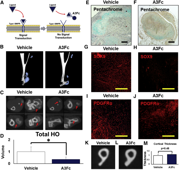
Trauma-induced heterotopic ossification (tHO) is a condition of pathologic wound healing, defined by the progressive formation of ectopic bone in soft tissue following severe burns or trauma. Because previous studies have shown that genetic variants of HO, such as fibrodysplasia ossificans progressiva (FOP), are caused by hyperactivating mutations of the type I bone morphogenetic protein receptor (T1-BMPR) ACVR1/ALK2, studies evaluating therapies for HO have been directed primarily toward drugs for this specific receptor. However, patients with tHO do not carry known T1-BMPR mutations. Here we show that, although BMP signaling is required for tHO, no single T1-BMPR (ACVR1/ALK2, BMPR1a/ALK3, or BMPR1b/ALK6) alone is necessary for this disease, suggesting that these receptors have functional redundancy in the setting of tHO. By utilizing two different classes of BMP signaling inhibitors, we developed a translational approach to treatment, integrating treatment choice with existing diagnostic options. Our treatment paradigm balances either immediate therapy with reduced risk for adverse effects (Alk3-Fc) or delayed therapy with improved patient selection but greater risk for adverse effects (LDN-212854).

Keywords: BMP receptors; BMP signaling; stem cells.


Figures

Figure 1
Figure 1
LDN19 Is a Potent Anti-inflammatory, Anti-proliferative, and Myelosuppressive Agent (A) Flow cytometry gating strategy for tissue-derived neutrophils (Ly6G+CD11b+F4/80) and macrophages (Ly6G-CD11b+F4/80+) in vehicle- and LDN19-treated mice. (B) LDN19 significantly reduces tenotomy-site neutrophils 5 days after injury (normalized count, 0.01 versus 1.0; p = 0.0002; n ≥ 3/group). (C) LDN19 significantly reduces tenotomy-site macrophages 5 days after injury (normalized count, 0.04 versus 1.0; p = 0.0003; n ≥ 3/group). (D) Representative neutrophil (Ly6G) immunostaining in a vehicle-treated hindlimb 5 days after injury. (E) Representative neutrophil (Ly6G) immunostaining in an LDN19-treated hindlimb 5 days after injury. (F) Representative macrophage (F4/80) immunostaining in a vehicle-treated hindlimb 5 days after injury. (G) Representative macrophage (F4/80) immunostaining in an LDN19-treated hindlimb 5 days after injury. (H) LDN19 significantly reduces overall cellularity at the tenotomy site 5 days after injury (normalized count, 0.04 versus 1.0; p = 0.001; n ≥ 3/group), including macrophages (dark blue), neutrophils (light blue), and non-leukocytes (white), on the basis of flow cytometry. (I) Representative mesenchymal cell (PDGFRα) immunostaining in a vehicle-treated hindlimb 5 days after injury. (J) Representative mesenchymal cell (PDGFRα) immunostaining in an LDN19-treated hindlimb 5 days after injury. (K) Representative immunostaining for proliferation (Ki67) in a vehicle-treated hindlimb 5 days after injury. (L) Representative immunostaining for proliferation (Ki67) in an LDN19-treated hindlimb 5 days after injury. (M) BrdU proliferation assay showing that LDN19 significantly reduces wild-type mesenchymal cell proliferation in vitro. (N) BrDU proliferation assay showing that LDN19 significantly reduces proliferation of Acvr1 knockout mesenchymal cells in vitro. *p < 0.05. p values are listed for all non-significant findings. All flow cytometry findings were normalized to vehicle-treated controls as indicated in Materials and Methods. Scale bars, 200 μm. Error bars represent one SD.
Figure 2
Figure 2
Postnatal Loss of ACVR1/ALK2 Does Not Eliminate tHO (A) Experimental design for tamoxifen-inducible Acvr1 knockout mice (Acvr1 tmKO: Ub.creERT/Acvr1fl/fl). (B) Representative 3D microCT reconstructions obtained 9 weeks after injury, showing tHO (blue) at the tenotomy site of Acvr1 littermate control and Acvr1 tmKO mice 9 weeks after injury. (C) Representative serial cross-sections obtained 9 weeks after injury showing tHO (red arrow) at the tenotomy site of Acvr1 littermate control and Acvr1 tmKO mice 9 weeks after injury. (D) Genetic loss of Acvr1 does not eliminate tHO 9 weeks after injury (normalized volume, 0.53 versus 1.0; p = 0.09; n ≥ 3/group). (E) Representative pentachrome image confirming the presence of cartilage marked by Alcian blue in a littermate control mouse 3 weeks after injury. (F) Representative pentachrome image confirming the presence of cartilage marked by Alcian blue in an Acvr1 tmKO mouse 3 weeks after injury. (G) Representative chondrocyte (SOX9) immunostaining confirming the presence of chondrocytes in a littermate control mouse 3 weeks after injury. (H) Representative chondrocyte (SOX9) immunostaining confirming the presence of chondrocytes in an Acvr1 tmKO mouse 3 weeks after injury. (I) Representative mesenchymal cell (PDGFRα) immunostaining confirming the presence of mesenchymal cells in a littermate control mouse 5 days after injury. (J) Representative mesenchymal cell (PDGFRα) immunostaining confirming the presence of mesenchymal cells in an Acvr1 tmKO mouse 5 days after injury. *p < 0.05. p values are listed for all non-significant findings. All volumes were normalized to tamoxifen-treated littermate controls as indicated in Materials and Methods. Scale bars, 200 μm. Error bars represent one SD.
Figure 3
Figure 3
Postnatal Loss of BMPR1a/ALK3 or BMPR1b/ALK6 Knockout Alone Is Unable to Eliminate tHO (A) Experimental design for tamoxifen-inducible Bmpr1a knockout mice (Bmpr1a tmKO: Ub.creERT/Bmpr1afl/fl) and for Bmpr1b knockout (Bmpr1b−/−) mice. (B) Representative 3D microCT reconstructions showing HO (blue) at the tenotomy site of littermate control and Bmpr1a tmKO mice 9 weeks after injury. (C) Representative serial cross-sections showing HO (red arrow) at the tenotomy site of littermate control and Bmpr1a tmKO mice 9 weeks after injury. (D) Genetic loss of Bmpr1a does not substantially or significantly reduce tHO 9 weeks after injury (normalized volume, 1.43 versus 1.0; p = 0.27; n ≥ 3/group). (E) Representative pentachrome image confirming the presence of cartilage marked by Alcian blue in a littermate control mouse 3 weeks after injury. (F) Representative pentachrome image confirming the presence of cartilage marked by Alcian blue in a Bmpr1a tmKO mouse 3 weeks after injury. (G) Representative 3D microCT reconstructions showing HO (blue) at the tenotomy site of littermate control and Bmpr1b−/− mice 9 weeks after injury. (H) Representative serial cross-sections showing HO (red arrow) at the tenotomy site of littermate control and Bmpr1b−/− mice 9 weeks after injury. (I) Genetic loss of Bmpr1b does not substantially or significantly reduce tHO 9 weeks after injury (normalized volume, 0.81 versus 1.0; p = 0.72; n ≥ 3/group). (J) Representative pentachrome image confirming the presence of cartilage marked by Alcian blue in a Bmpr1b littermate control mouse 3 weeks after injury. (K) Representative pentachrome image confirming the presence of cartilage marked by Alcian blue in a Bmpr1b−/− mouse 3 weeks after injury. *p < 0.05. p values are listed for all non-significant findings. All volumes were normalized to respective littermate controls as indicated in Materials and Methods. Scale bars, 200 μm. Error bars represent one SD.
Figure 4
Figure 4
Combined Postnatal Loss of ACVR1/ALK2 and BMPR1a/ALK3 Significantly Reduces tHO but Is Lethal (A) Experimental design for tamoxifen-inducible Acvr1 and Bmpr1a double knockout mice (Acvr1;Bmpr1a tmKO: Ub.creERT/Acvr1fll/fl/Bmpr1afl/fl). (B) Representative whole-body photos of Acvr1/Bmpr1a littermate control and mutant mice showing hair loss and cachexia in the Acvr1;Bmpr1a tmKO mouse. (C) A significant proportion (7 of 9) of Acvr1/Bmpr1a tmKO mice died within the first 6 weeks after tamoxifen induction. (D) Representative 3D microCT reconstructions showing HO (blue) at the tenotomy site of an Acvr1;Bmpr1a littermate control mouse and absent HO in the Acvr1;Bmpr1a tmKO mouse 9 weeks after injury. (E) Representative serial cross-sections showing HO (red arrow) at the tenotomy site of Acvr1;Bmpr1a littermate control and Acvr1;Bmpr1a tmKO mice 9 weeks after injury. (F) Genetic loss of Acvr1 and Bmpr1a significantly reduces tHO 9 weeks after injury (normalized volume, 0.06 versus 1.0; p = 0.025; n ≥ 3 for littermate control and n = 2 for double knockout). (G) Representative pentachrome image showing the histologic presence of bone and cartilage at the tenotomy site of an Acvr1;Bmpr1a littermate control mouse 9 weeks after injury. (H) Representative pentachrome image showing a markedly reduced presence of cartilage at the tenotomy site of an Acvr1;Bmpr1a tmKO mouse 9 weeks after injury. (I) Representative pentachrome image confirming the presence of cartilage (Alcian blue) at the tenotomy site of an Acvr1;Bmpr1a littermate control mouse 3 weeks after injury. (J) Representative pentachrome image confirming the relative absence of cartilage at the tenotomy site of an Acvr1;Bmpr1a tmKO mouse 3 weeks after injury. *p < 0.05. p values are listed for all non-significant findings. All volumes were normalized to tamoxifen-treated littermate controls as indicated in Materials and Methods. Scale bars, 200 μm. Error bars represent 1 SD.
Figure 5
Figure 5
Long-Term Treatment with A3Fc Significantly Reduces tHO (A) A3Fc functions as a BMP ligand trap. (B) Representative 3D microCT reconstructions showing HO (blue) at the tenotomy site of vehicle- and A3Fc-treated mice 9 weeks after injury. (C) Representative serial cross-sections showing HO (red arrow) at the tenotomy site of vehicle- and A3Fc-treated mice 9 weeks after injury. (D) Long-term treatment with A3Fc (daily 2 mg/kg i.p. for 6 weeks) significantly reduces tHO (normalized volume, 0.35 versus 1.0; p = 0.001; n ≥ 3/group). (E) Representative pentachrome image confirming the presence of cartilage marked by Alcian blue in a vehicle-treated mouse 3 weeks after injury. (F) Representative pentachrome image showing a relative decrease in cartilage marked by Alcian blue in an A3Fc-treated mouse 3 weeks after injury. (G) Representative chondrocyte (SOX9) immunostaining confirming the presence of chondrocytes in a vehicle-treated mouse 3 weeks after injury. (H) Representative chondrocyte (SOX9) immunostaining confirming the near absence of chondrocytes in an A3Fc-treated mouse 3 weeks after injury. (I) Representative mesenchymal cell (PDGFRα) immunostaining confirming the presence of mesenchymal cells in a vehicle-treated mouse 5 days after injury. (J) Representative mesenchymal cell (PDGFRα) immunostaining confirming the presence of mesenchymal cells in an A3Fc-treated mouse 5 days after injury. (K) Representative microCT cross-section showing the tibia-fibula confluence of the injured hindlimb in vehicle-treated mice 9 weeks after injury. (L) Representative microCT cross-section showing the tibia-fibula confluence of the injured hindlimb in A3Fc-treated mice 9 weeks after injury. (M) A3Fc does not reduce the cortical thickness of the injured hindlimb. *p < 0.05. p values are listed for all non-significant findings. All volumes were normalized to vehicle-treated controls as indicated in Materials and Methods. Scale bars, 200 μm. Error bars represent one SD.
Figure 6
Figure 6
A Translational Approach to Preventing tHO by Targeting BMP Signaling (A) Experimental design for short-term daily treatment initiated immediately after injury, during weeks 2–4 after injury, when spectroscopic evidence of cartilage and mineral deposition is observed, or during weeks 4–6 after injury, when radiographic evidence of the ossified lesion is observed (the red dashed arrow indicates no treatment; the gray dashed arrow indicates post-treatment; the solid black arrow indicates active treatment). (B) Representative 3D microCT reconstructions obtained 9 weeks after injury showing HO (blue) at the tenotomy site of mice treated with daily A3Fc during weeks 0–2, weeks 2–4, or weeks 4–6 after injury. (C) Short-term treatment with A3Fc (daily 2 mg/kg i.p. for 2 weeks) during weeks 0–2 significantly reduces tHO (normalized volume, 0.43 versus 1.0; p = 0.021; n ≥ 3/group) but not during weeks 2–4 (normalized volume, 0.63 versus 1.0; p = 0.25; n ≥ 3/group) or weeks 4–6 (normalized volume, 0.87 versus 1.0; p = 0.90; n ≥ 3/group). (D) Representative 3D microCT reconstructions obtained 9 weeks after injury showing HO (blue) at the tenotomy site of mice treated with daily LDN21 (6 mg/kg i.p.) for 6 weeks initiated immediately after injury. (E) Long-term treatment with LDN21 initiated immediately after injury significantly decreases tHO (normalized volume, 0.34 versus 1.0; p = 0.005; n ≥ 3/group). (F) Representative 3D microCT reconstructions obtained 9 weeks after injury showing HO (blue) at the tenotomy site of mice treated with daily LDN21 during weeks 0–2, weeks 2–4, or weeks 4–6 after injury. (G) Short-term treatment with LDN21 (daily 2 mg/kg i.p. for 2 weeks) during weeks 0–2 (normalized volume, 0.51 versus 1.0; p = 0.043; n ≥ 3/group) or weeks 2–4 (normalized volume, 0.38 versus 1.0; p = 0.032; n ≥ 3/group) but not during weeks 4–6 (normalized volume, 0.57 versus 1.0; p = 0.18; n ≥ 3/group) significantly reduces tHO. *p < 0.05. p values are listed for all non-significant findings. All volumes were normalized to the control (vehicle-treated) as indicated in Materials and Methods. Scale bars, 200 μm. Error bars represent one SD.

Similar articles

Cited by

References

    1. Forsberg J.A., Potter B.K. Heterotopic ossification in wartime wounds. J. Surg. Orthop. Adv. 2010;19:54–61. - PubMed
    1. Forsberg J.A., Pepek J.M., Wagner S., Wilson K., Flint J., Andersen R.C., Tadaki D., Gage F.A., Stojadinovic A., Elster E.A. Heterotopic ossification in high-energy wartime extremity injuries: prevalence and risk factors. J. Bone Joint Surg. Am. 2009;91:1084–1091. - PubMed
    1. Potter B.K., Forsberg J.A., Davis T.A., Evans K.N., Hawksworth J.S., Tadaki D., Brown T.S., Crane N.J., Burns T.C., O’Brien F.P., Elster E.A. Heterotopic ossification following combat-related trauma. J. Bone Joint Surg. Am. 2010;92(Suppl 2):74–89. - PubMed
    1. Alfieri K.A., Forsberg J.A., Potter B.K. Blast injuries and heterotopic ossification. Bone Joint Res. 2012;1:192–197. - PMC - PubMed
    1. Kung T.A., Jebson P.J., Cederna P.S. An individualized approach to severe elbow burn contractures. Plast. Reconstr. Surg. 2012;129:663e–673e. - PubMed

Publication types

MeSH terms