. 2017 Nov 9;36(45):6306-6314.
doi: 10.1038/onc.2017.232. Epub 2017 Jul 17.

Inhibition of WNT signaling attenuates self-renewal of SHH-subgroup medulloblastoma

Affiliations

Inhibition of WNT signaling attenuates self-renewal of SHH-subgroup medulloblastoma

J Rodriguez-Blanco et al. Oncogene. .

Abstract

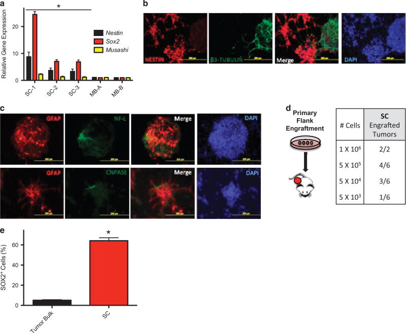
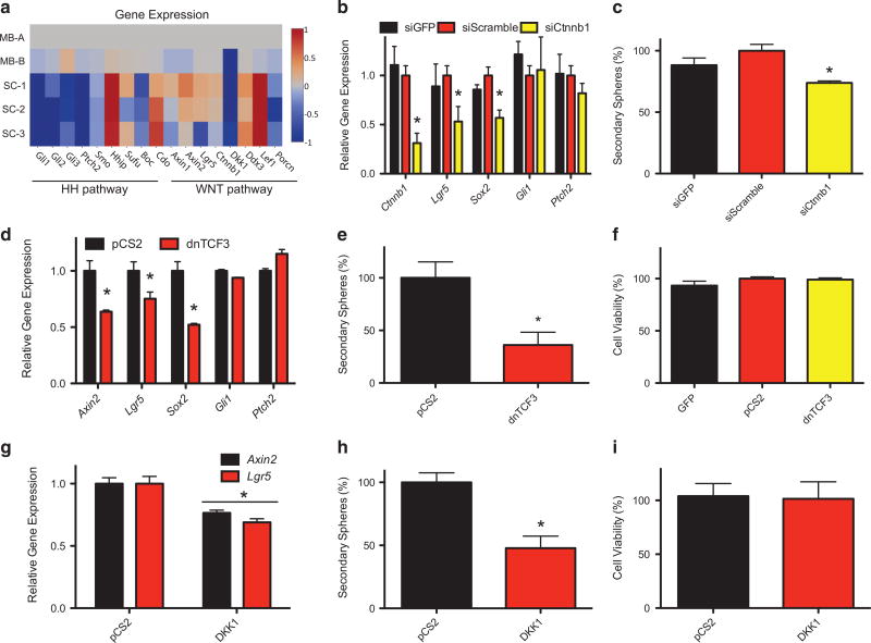
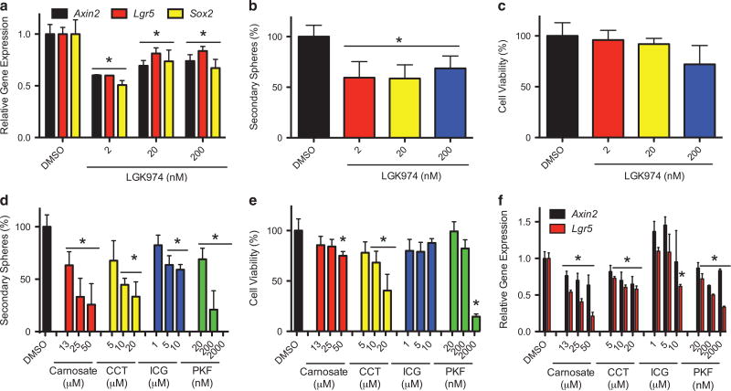
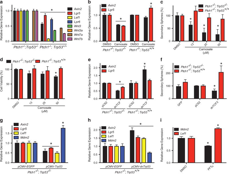
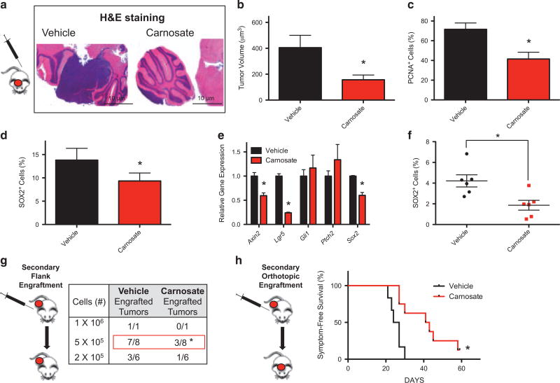
The SMOOTHENED inhibitor vismodegib is FDA approved for advanced basal cell carcinoma (BCC), and shows promise in clinical trials for SONIC HEDGEHOG (SHH)-subgroup medulloblastoma (MB) patients. Clinical experience with BCC patients shows that continuous exposure to vismodegib is necessary to prevent tumor recurrence, suggesting the existence of a vismodegib-resistant reservoir of tumor-propagating cells. We isolated such tumor-propagating cells from a mouse model of SHH-subgroup MB and grew them as sphere cultures. These cultures were enriched for the MB progenitor marker SOX2 and formed tumors in vivo. Moreover, while their ability to self-renew was resistant to SHH inhibitors, as has been previously suggested, this self-renewal was instead WNT-dependent. We show here that loss of Trp53 activates canonical WNT signaling in these SOX2-enriched cultures. Importantly, a small molecule WNT inhibitor was able to reduce the propagation and growth of SHH-subgroup MB in vivo, in an on-target manner, leading to increased survival. Our results imply that the tumor-propagating cells driving the growth of bulk SHH-dependent MB are themselves WNT dependent. Further, our data suggest combination therapy with WNT and SHH inhibitors as a therapeutic strategy in patients with SHH-subgroup MB, in order to decrease the tumor recurrence commonly observed in patients treated with vismodegib.


Conflict of interest statement

CONFLICT OF INTEREST

The authors declare no conflict of interest.

Figures

Figure 1
Figure 1
Medulloblastoma sphere cultures display stem cell properties. (a) The expression of the indicated progenitor cell biomarkers, from three independently derived Trp53-deficient MB sphere cultures (SC-1, 2 and 3) and two primary MBs (MB-A and MB-B), was determined and then normalized to that of MB-A. (b) SCs were immunostained for neuronal progenitor biomarkers (NESTIN and β3-TUBULIN). (c) The level of SC pluripotency was determined by examining the biomarkers GLIAL FRIBRILLARY ACIDIC PROTEIN (GFAP), NEUROFILAMENT-LARGE (NF-L) or CYCLIC-NUCLEOTIDE-PHOSPHODIESTERASE (CNPASE). Representative images of immunostained SC-2 are shown. (d) The indicated numbers of viable SC-2 cells were implanted subcutaneously into the flanks of immunocompromised mice. The frequency and number of tumor engraftments is shown. (e) The percentage of SOX2+ cells in MB flank tumors and SCs was determined by FACS analysis.
Figure 2
Figure 2
The self-renewal of medulloblastoma sphere cultures is dependent on WNT activity. (a) A heat map representing the relative gene expression of SHH (Gli1, Gli2, Gli3, Ptch2, Smo, Hhip, Sufu, Boc, Cdo) and WNT pathway-related genes (Axin1, Axin2, Lgr5, Ctnnb1, Dkk1, Ddx3, Lef1, Porcn), as determined in the indicated Trp53-deficient SCs and Trp53-deficient MB tissues (MB-A and MB-B). The color scale was calculated using Log2 transformed gene expression data normalized to their expression in MB-A. (b) SC-2 were transfected with the indicated siRNA smart pools, and the expression of the indicated genes determined 72 h later. (c) The ability of SC-2 to form secondary spheres 72 h after transfection with the indicated siRNA smart pool was determined. (d) SC-2 were transfected with a plasmid expressing a dnTCF3 construct or a control plasmid (pCS2), and the expression of the indicated genes determined 48 h later. (e) The ability of SC-2 to form secondary spheres was determined 48 h after transfection with a dnTCF3 expression plasmid or control plasmid (pCS2). (f) SC-2 was transfected with a plasmid expressing a dnTCF3 construct or control plasmid (pCS2) and cell viability determined 5 days later using an MTT reduction assay. (g) SC-2 was transfected with a plasmid expressing DKK1 or control plasmid (pCS2) and the expression of WNT target genes determined. (h) The ability of SC-2 to form secondary spheres was determined 48 h after transfection with a plasmid expressing DKK1 or control plasmid (pCS2). (i) SC-2 was transfected with a plasmid expressing DKK1 or control plasmid (pCS2) and cell viability determined 5 days later using an MTT reduction assay. Results were normalized to that from pCS2 transfected cultures unless otherwise indicated. MTT, 3-(4,5-dimethyl-2-thiazolyl) 2,5-diphenyl-2H-tetrazolium bromide.
Figure 3
Figure 3
Small-molecule WNT inhibitors attenuate the self-renewal of medulloblastoma sphere cultures. (a) SC-2 were treated with the indicated concentrations of the PORCUPINE inhibitor LGK974, and the expression of the indicated genes determined 72 h later. (b) The ability of SC-2 to form secondary spheres was determined following incubation (72 h) with the indicated concentrations of LGK974. (c) SC-2 were incubated with the indicated concentrations of LGK974, and cell viability determined 5 days later using an MTT reduction assay. (d) The ability of SC-2 to form secondary spheres was determined following incubation (24 h) with the indicated concentrations of TCF/β-CATENIN inhibitors (carnosate, CCT-031374 hydrobromide [CCT], ICG-001 [ICG] or PKF-115-584 [PKF]). (e) SC-2 were incubated for 72 h with the indicated concentrations of TCF/β-CATENIN inhibitors, and cell viability determined 5 days later using an MTT reduction assay. (f) SC-2 were treated with the indicated TCF/β-CATENIN inhibitors for 24 h. The expression of the indicated genes was determined. Results were normalized to dimethyl sulfoxide control. MTT, 3-(4,5-dimethyl-2-thiazolyl) 2,5-diphenyl-2H-tetrazolium bromide.
Figure 4
Figure 4
MPC enriched cultures expressing wild-type Trp53 are not WNT-dependent. (a) The expression of the indicated genes was determined in Ptch1−/−;Trp53−/− or Ptch1−/−;Trp53+/+ SCs, and normalized to the data from Ptch1−/−;Trp53−/− cultures. (b) SC cultures were treated with 25 µM carnosate for 24 h, and the expression of the indicated WNT target genes determined. (c) The ability of the SCs to form secondary spheres, following incubation (24 h) with the indicated concentrations of carnosate, was determined. (d) The indicated SCs were incubated for 3 days with the various concentrations of carnosate, and cell viability determined 5 days later using an MTT reduction assay. The results were normalized to those from the dimethyl sulfoxide control. (e) SCs were transfected with a plasmid expressing a dnTCF3 construct or a control plasmid (pCS2), and the expression of the indicated WNT target genes determined 48 h later. (f) The ability of SCs to form secondary spheres was determined 48 h after transfection with plasmids expressing dnTCF3, a control plasmid (pCS2) or GFP. Results were normalized to those from the pCS2 control. (g) Ptch1−/−;Trp53−/− SCs were transfected with plasmids expressing wild-type P53 (pCMV-Trp53), or an enhanced green fluorescent protein (EGFP) (pCMV-EGFP) control vector, and the expression of the indicated genes determined 72 h later. (h) Ptch1−/−; Trp53+/+ SCs were transfected with plasmids expressing dominant negative P53 (pCMV-dnTrp53) or EGFP (pCMV-EGFP), and the expression of the indicated genes determined 72 h later. The results were normalized to those from the EGFP expressing cultures. (i) Ptch1−/−;Trp53+/+ SCs were incubated with the P53/MDM2 inhibitor Pifithrin-α (PFT-α) for 24 h, and the expression of the indicated genes determined. The results were normalized to those from the dimethyl sulfoxide control. MTT, 3-(4,5-dimethyl-2-thiazolyl) 2,5-diphenyl-2H-tetrazolium bromide.
Figure 5
Figure 5
The propagation and growth of Trp53-deficient, SHH-subgroup medulloblastoma is WNT-dependent. Trp53 mutant, Ptch1-driven MB tissue, which had never been cultured ex vivo, was orthotopically implanted into the cerebellum of immunocompromised mice. These mice were then treated with carnosate or vehicle for 20 days, or until they developed MB symptoms. The mice were then killed and their brains collected. (a) Representative hematoxylin eosin staining of orthotopic MB tissue from a carnosate or vehicle-treated mouse is shown. (b) The volume of residual orthotopic tumors from these mice was calculated, and the mean volume and s.e.m. shown. (c) The residual orthotopic tumors from these mice were immunostained for the proliferation biomarker PCNA, and the numbers of PCNA+ cells quantified and normalized to total cell number per field (%). (d) The residual orthotopic tumors from these mice were immunostained for the MPC biomarker SOX2, and the numbers of SOX2+ cells quantified and normalized to total cell number per field (%). (e) MB tumor tissue was subcutaneously implanted into the flanks of immunocompromised mice, and treated daily with of carnosate, or vehicle control, for 8 days. The expression of the indicated genes in residual flank tumors was then determined. (f) The enrichment of SOX2+ cells in residual flank tissue, from carnosate or vehicle-treated mice, was determined by FACS analysis. (g) The indicated number of viable cells from residual MB tissue was transplanted into additional immunocompromised mice, and the frequency of tumor engraftment determined. (h) Equal numbers of viable cells from residual flank MB tissue were implanted into the cerebellum of additional immunocompromised mice, and MB symptom-free survival monitored for 60 days. Results were normalized to vehicle control.

Similar articles

Cited by

References

    1. Louis DN, Ohgaki H, Wiestler OD, Cavenee WK, Burger PC, Jouvet A, et al. The 2007 WHO classification of tumours of the central nervous system. Acta Neuropathol. 2007;114:97–109. - PMC - PubMed
    1. Crawford JR, MacDonald TJ, Packer RJ. Medulloblastoma in childhood: new biological advances. Lancet Neurol. 2007;6:1073–1085. - PubMed
    1. Gibson P, Tong Y, Robinson G, Thompson MC, Currle DS, Eden C, et al. Subtypes of medulloblastoma have distinct developmental origins. Nature. 2010;468:1095–1099. - PMC - PubMed
    1. Northcott Pa, Dubuc AM, Pfister S, Taylor MD. Molecular subgroups of medulloblastoma. Expert Rev Neurother. 2012;12:871–884. - PMC - PubMed
    1. Remke M, Ramaswamy V, Taylor MD. Medulloblastoma molecular dissection: the way toward targeted therapy. Curr Opin Oncol. 2013;25:674–681. - PubMed

Publication types

MeSH terms