

Longitudinal Changes in White Matter Fractional Anisotropy in Adult-Onset Niemann-Pick Disease Type C Patients Treated with Miglustat
- PMID: 28710748
- PMCID: PMC5953894
- DOI: 10.1007/8904_2017_42
Longitudinal Changes in White Matter Fractional Anisotropy in Adult-Onset Niemann-Pick Disease Type C Patients Treated with Miglustat
Abstract
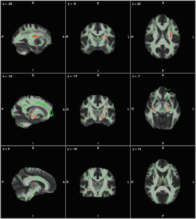
Niemann-Pick disease type C (NPC) is a rare neurometabolic disorder resulting in impaired intracellular lipid trafficking. The only disease-modifying treatment currently available is miglustat, an iminosugar that inhibits the accumulation of lipid metabolites in neurons and other cells. This longitudinal diffusion tensor imaging (DTI) study examined how the rate of white matter change differed between treated and non-treated adult-onset NPC patient groups. Nine adult-onset NPC patients (seven undergoing treatment with miglustat, two not treated) underwent DTI neuroimaging. Rates of change in white matter structure as indexed by Tract-Based Spatial Statistics (TBSS) of fractional anisotropy were compared between treated and untreated patients. Treated patients were found to have a significantly slower rate of white matter change in the corticospinal tracts, the thalamic radiation and the inferior longitudinal fasciculus. This is further evidence that miglustat treatment may have a protective effect on white matter structure in the adult-onset form of the disease.
Keywords: Diffusion tensor imaging; Fractional anisotropy; Miglustat; Niemann-Pick type C; White matter.
Figures
Similar articles
-
Miglustat in Niemann-Pick disease type C patients: a review.Orphanet J Rare Dis. 2018 Aug 15;13(1):140. doi: 10.1186/s13023-018-0844-0. Orphanet J Rare Dis. 2018. PMID: 30111334 Free PMC article. Review.
-
Longitudinal changes in cerebellar and subcortical volumes in adult-onset Niemann-Pick disease type C patients treated with miglustat.J Neurol. 2015 Sep;262(9):2106-14. doi: 10.1007/s00415-015-7819-z. Epub 2015 Jun 20. J Neurol. 2015. PMID: 26092521
-
Evolution of structural neuroimaging biomarkers in a series of adult patients with Niemann-Pick type C under treatment.Orphanet J Rare Dis. 2017 Feb 2;12(1):22. doi: 10.1186/s13023-017-0579-3. Orphanet J Rare Dis. 2017. PMID: 28148276 Free PMC article.
-
Early miglustat therapy in infantile Niemann-Pick disease type C.Pediatr Neurol. 2012 Jul;47(1):40-3. doi: 10.1016/j.pediatrneurol.2012.04.005. Pediatr Neurol. 2012. PMID: 22704015
-
The role of diffusion tensor imaging and fractional anisotropy in the evaluation of patients with idiopathic normal pressure hydrocephalus: a literature review.Neurosurg Focus. 2016 Sep;41(3):E12. doi: 10.3171/2016.6.FOCUS16192. Neurosurg Focus. 2016. PMID: 27581308 Review.
Cited by
-
Psychiatric and Cognitive Symptoms Associated with Niemann-Pick Type C Disease: Neurobiology and Management.CNS Drugs. 2019 Feb;33(2):125-142. doi: 10.1007/s40263-018-0599-0. CNS Drugs. 2019. PMID: 30632019 Review.
-
Hepatic and neuronal phenotype of NPC1-/- mice.Heliyon. 2019 Mar 14;5(3):e01293. doi: 10.1016/j.heliyon.2019.e01293. eCollection 2019 Mar. Heliyon. 2019. PMID: 30923761 Free PMC article.
-
Lysosomal diseases: Overview on current diagnosis and treatment.Genet Mol Biol. 2019;42(1 suppl 1):165-177. doi: 10.1590/1678-4685-GMB-2018-0159. Epub 2019 Apr 25. Genet Mol Biol. 2019. PMID: 31067291 Free PMC article.
-
Neurodegeneration in Niemann-Pick Type C Disease: An Updated Review on Pharmacological and Non-Pharmacological Approaches to Counteract Brain and Cognitive Impairment.Int J Mol Sci. 2021 Jun 20;22(12):6600. doi: 10.3390/ijms22126600. Int J Mol Sci. 2021. PMID: 34202978 Free PMC article. Review.
-
Miglustat in Niemann-Pick disease type C patients: a review.Orphanet J Rare Dis. 2018 Aug 15;13(1):140. doi: 10.1186/s13023-018-0844-0. Orphanet J Rare Dis. 2018. PMID: 30111334 Free PMC article. Review.
References
LinkOut - more resources
Full Text Sources
Other Literature Sources
