

P-Rex1 Expression in Invasive Breast Cancer in relation to Receptor Status and Distant Metastatic Site
- PMID: 28698809
- PMCID: PMC5494073
- DOI: 10.1155/2017/4537532
P-Rex1 Expression in Invasive Breast Cancer in relation to Receptor Status and Distant Metastatic Site
Abstract
Background: Phosphatidylinositol-3,4,5-trisphosphate-dependent Rac exchange factor 1 (P-Rex1) has been implicated in cancer growth, metastasis, and response to phosphatidylinositol 3-kinase (PI3K) inhibitor therapy. The aim of this study was to determine whether P-Rex1 expression differs between primary and metastatic human breast tumors and between breast cancer subtypes.
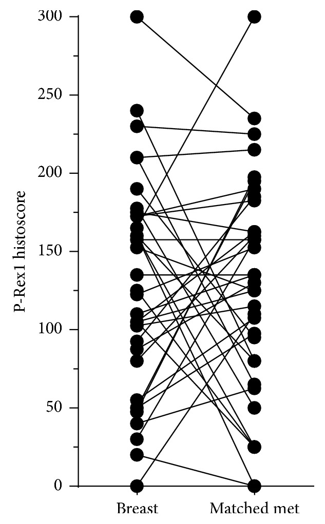
Design: P-Rex1 expression was measured in 133 specimens by immunohistochemistry: 40 and 42 primary breast tumors from patients who did versus did not develop metastasis, respectively, and 51 breast-derived tumors from metastatic sites (36 of which had matching primary tumors available for analysis).
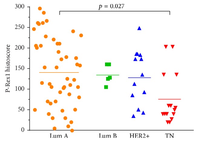
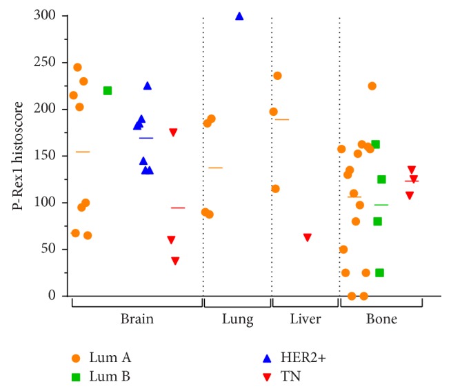
Results: Primary breast tumors showed significant differences in P-Rex1 expression based on receptor subtype. ER+ and HER2+ primary tumors showed higher P-Rex1 expression than primary triple-negative tumors. HER2+ metastases from all sites showed significantly higher P-Rex1 expression compared to other metastatic receptor subtypes. Solid organ (i.e., brain, lung, and liver) metastases showed higher P-Rex1 expression compared to bone metastases.
Conclusions: P-Rex1 expression is increased in ER+ and HER2+ breast cancers compared to triple-negative tumors. P-Rex1 may be differentially expressed in metastatic tumors based on site and receptor status. The role of P-Rex1 in the development of breast cancer metastases and as a predictive biomarker of therapeutic response warrants further investigation.
Figures
Similar articles
-
Phosphatidylinositol-3,4,5-trisphosphate dependent Rac exchange factor 1 is a diagnostic and prognostic biomarker for hepatocellular carcinoma.World J Clin Cases. 2020 Sep 6;8(17):3774-3785. doi: 10.12998/wjcc.v8.i17.3774. World J Clin Cases. 2020. PMID: 32953853 Free PMC article.
-
Breast carcinoma subtypes show different patterns of metastatic behavior.Virchows Arch. 2017 Mar;470(3):275-283. doi: 10.1007/s00428-017-2065-7. Epub 2017 Jan 19. Virchows Arch. 2017. PMID: 28101678
-
Organotropism and prognostic marker discordance in distant metastases of breast carcinoma: fact or fiction? A clinicopathologic analysis.Hum Pathol. 2012 Mar;43(3):398-404. doi: 10.1016/j.humpath.2011.05.009. Epub 2011 Aug 12. Hum Pathol. 2012. PMID: 21840040
-
Hormone Receptor Status and HER2 Expression in Primary Breast Cancer Compared With Synchronous Axillary Metastases or Recurrent Metastatic Disease.Clin Breast Cancer. 2015 Oct;15(5):307-12. doi: 10.1016/j.clbc.2015.03.010. Epub 2015 Mar 25. Clin Breast Cancer. 2015. PMID: 25922284 Review.
-
Potential prognostic tumor biomarkers in triple-negative breast carcinoma.Beijing Da Xue Xue Bao Yi Xue Ban. 2012 Oct 18;44(5):666-72. Beijing Da Xue Xue Bao Yi Xue Ban. 2012. PMID: 23073572 Review.
Cited by
-
Exosomes: A Promising Avenue for the Diagnosis of Breast Cancer.Technol Cancer Res Treat. 2019 Jan 1;18:1533033818821421. doi: 10.1177/1533033818821421. Technol Cancer Res Treat. 2019. PMID: 30760122 Free PMC article. Review.
-
Targeting Rac and Cdc42 GEFs in Metastatic Cancer.Front Cell Dev Biol. 2020 Apr 8;8:201. doi: 10.3389/fcell.2020.00201. eCollection 2020. Front Cell Dev Biol. 2020. PMID: 32322580 Free PMC article. Review.
-
Mammary molecular portraits reveal lineage-specific features and progenitor cell vulnerabilities.J Cell Biol. 2018 Aug 6;217(8):2951-2974. doi: 10.1083/jcb.201804042. Epub 2018 Jun 19. J Cell Biol. 2018. PMID: 29921600 Free PMC article.
-
Feedback regulation between phosphatidylinositol-3,4,5-trisphosphate dependent Rac exchange factor 1 and transforming growth factor β1 and prognostic value in gastric cancer.World J Gastroenterol. 2020 Jan 7;26(1):21-34. doi: 10.3748/wjg.v26.i1.21. World J Gastroenterol. 2020. PMID: 31933512 Free PMC article.
-
Phosphatidylinositol-3,4,5-trisphosphate dependent Rac exchange factor 1 is a diagnostic and prognostic biomarker for hepatocellular carcinoma.World J Clin Cases. 2020 Sep 6;8(17):3774-3785. doi: 10.12998/wjcc.v8.i17.3774. World J Clin Cases. 2020. PMID: 32953853 Free PMC article.
References
-
- Blows F. M., Driver K. E., Schmidt M. K., et al. Subtyping of breast cancer by immunohistochemistry to investigate a relationship between subtype and short and long term survival: a collaborative analysis of data for 10,159 cases from 12 studies. PLoS Medicine. 2010;7(5) doi: 10.1371/journal.pmed.1000279. - DOI - PMC - PubMed
-
- American_Cancer_Society. Cancer Facts and Figures 2016. Atlanta: American Cancer Society, 2016.
Grants and funding
LinkOut - more resources
Full Text Sources
Other Literature Sources
Research Materials
Miscellaneous
