

Characterization of the Extracellular Matrix of Normal and Diseased Tissues Using Proteomics
- PMID: 28675934
- PMCID: PMC8078728
- DOI: 10.1021/acs.jproteome.7b00191
Characterization of the Extracellular Matrix of Normal and Diseased Tissues Using Proteomics
Abstract
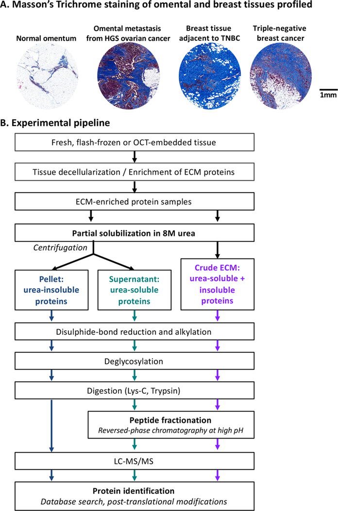
The extracellular matrix (ECM) is a complex meshwork of insoluble fibrillar proteins and signaling factors interacting together to provide architectural and instructional cues to the surrounding cells. Alterations in ECM organization or composition and excessive ECM deposition have been observed in diseases such as fibrosis, cardiovascular diseases, and cancer. We provide here optimized protocols to solubilize ECM proteins from normal or tumor tissues, digest the proteins into peptides, analyze ECM peptides by mass spectrometry, and interpret the mass spectrometric data. In addition, we present here two novel R-script-based web tools allowing rapid annotation and relative quantification of ECM proteins, peptides, and intensity/abundance in mass spectrometric data output files. We illustrate this protocol with ECMs obtained from two pairs of tissues, which differ in ECM content and cellularity: triple-negative breast cancer and adjacent mammary tissue, and omental metastasis from high-grade serous ovarian cancer and normal omentum. The complete proteomics data set generated in this study has been deposited to the public repository ProteomeXchange with the data set identifier: PXD005554.
Keywords: collagens; extracellular matrix; hydroxylation; mass-spectrometry-based proteomics; matrisome; microenvironment.
Figures
Similar articles
-
Exploring the extracellular matrix in health and disease using proteomics.Essays Biochem. 2019 Sep 13;63(3):417-432. doi: 10.1042/EBC20190001. Print 2019 Sep 13. Essays Biochem. 2019. PMID: 31462529 Review.
-
SWATH mass spectrometry as a tool for quantitative profiling of the matrisome.J Proteomics. 2018 Oct 30;189:11-22. doi: 10.1016/j.jprot.2018.02.026. Epub 2018 Mar 1. J Proteomics. 2018. PMID: 29501709 Free PMC article.
-
Comparison of extracellular matrix enrichment protocols for the improved characterization of the skin matrisome by mass spectrometry.J Proteomics. 2022 Jan 16;251:104397. doi: 10.1016/j.jprot.2021.104397. Epub 2021 Oct 20. J Proteomics. 2022. PMID: 34678517
-
Characterization of glomerular extracellular matrix by proteomic analysis of laser-captured microdissected glomeruli.Kidney Int. 2017 Feb;91(2):501-511. doi: 10.1016/j.kint.2016.09.044. Epub 2016 Dec 15. Kidney Int. 2017. PMID: 27988214 Free PMC article.
-
Ten Years of Extracellular Matrix Proteomics: Accomplishments, Challenges, and Future Perspectives.Mol Cell Proteomics. 2023 Apr;22(4):100528. doi: 10.1016/j.mcpro.2023.100528. Epub 2023 Mar 12. Mol Cell Proteomics. 2023. PMID: 36918099 Free PMC article. Review.
Cited by
-
Omentum-derived matrix enables the study of metastatic ovarian cancer and stromal cell functions in a physiologically relevant environment.Matrix Biol Plus. 2023 Nov 22;19-20:100136. doi: 10.1016/j.mbplus.2023.100136. eCollection 2023 Dec. Matrix Biol Plus. 2023. PMID: 38223308 Free PMC article.
-
Porcine Breast Extracellular Matrix Hydrogel for Spatial Tissue Culture.Int J Mol Sci. 2018 Sep 25;19(10):2912. doi: 10.3390/ijms19102912. Int J Mol Sci. 2018. PMID: 30257480 Free PMC article.
-
Regional and disease specific human lung extracellular matrix composition.Biomaterials. 2023 Feb;293:121960. doi: 10.1016/j.biomaterials.2022.121960. Epub 2022 Dec 24. Biomaterials. 2023. PMID: 36580718 Free PMC article.
-
Perlecan Knockdown Significantly Alters Extracellular Matrix Composition and Organization During Cartilage Development.Mol Cell Proteomics. 2020 Jul;19(7):1220-1235. doi: 10.1074/mcp.RA120.001998. Epub 2020 May 7. Mol Cell Proteomics. 2020. PMID: 32381549 Free PMC article.
-
Molecular analysis of the extracellular microenvironment: from form to function.FEBS Lett. 2024 Mar;598(6):602-620. doi: 10.1002/1873-3468.14852. Epub 2024 Mar 21. FEBS Lett. 2024. PMID: 38509768 Review.
References
-
- Hynes RO; Yamada KM Extracellular Matrix Biology; Cold Spring Harbor Perspectives in Biology; Cold Spring Harbor Laboratory Press: Cold Spring Harbor, NY, 2012.
Publication types
MeSH terms
Substances
Grants and funding
LinkOut - more resources
Full Text Sources
Other Literature Sources
Medical
Molecular Biology Databases
