. 2017 Oct:248:116-129.
doi: 10.1016/j.jviromet.2017.06.017. Epub 2017 Jul 1.

A Luciferase Immunoprecipitation System (LIPS) assay for profiling human norovirus antibodies

Affiliations

A Luciferase Immunoprecipitation System (LIPS) assay for profiling human norovirus antibodies

Christine M Tin et al. J Virol Methods. 2017 Oct.

Abstract

A luciferase immunoprecipitation systems (LIPS) assay was developed to define the antigenic specificity and titer of antibodies directed against human norovirus (HuNoV). Recombinant proteins, expressed by plasmid constructs encoding Renilla luciferase (Ruc) fused to the full-length HuNoV major capsid protein (VP1) (Ruc-antigen), were generated for ten HuNoV strains. In addition, subdomain constructs Ruc-Shell (S) and Ruc-Protruding (P) were engineered for a representative GII.4 norovirus (strain GII.4/2006b). The LIPS assay measured antibody levels in a well-defined panel of HuNoV-specific sera, and the results were compared to an ELISA standard. In hyperimmune sera, the LIPS produced titers similar to or higher than those measured by the ELISA of HuNoV-specific antibodies. The specificity of antibodies in various sera was profiled by LIPS with a panel of diverse Ruc-antigens containing full-length HuNoV VP1 proteins or VP1 subdomains, and the assay detected both specific and cross-reactive antibodies. Competition assays, in which antibodies were pre-incubated with one or more intact VLPs representing different genotypes, proved useful in further assessment of the antibody specificity detected by LIPS in complex polyclonal sera. The profiling of HuNoV-specific antibodies in the high-throughput LIPS format may prove useful in defining the strength or specificity of the adaptive immune response following natural infection or vaccination.

Keywords: ELISA; Human norovirus; LIPS assay; Serum antibody.


Figures

Figure 1
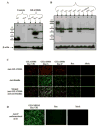
Figure 1. Expression of Ruc-antigen fusion proteins by COS1 cells confirmed by (A, B) western blot and (C, D) immunofluorescence
(A) Western blot of anti-Renilla antibody stained against untransfected, mock lysates and lysates of cells expressing Ruc, or Ruc-VP1, Ruc-S, or Ruc-P of a representative HuNoV strain (GII.4/2006b). (B) Western blot of anti-Renilla antibody stained against a panel of Ruc-VP1 antigen derived from multiple HuNoV strains. (C) Immunofluorescence stains of cells transfected with GII.4/2006b pRuc constructs. Untransfected cells were used as the negative control. Anti-Renilla antibody or hyperimmune guinea pig serum raised against the GII.4/2006b was used to detect fusion protein expression. (D) Immunofluorescence stain of cells transfected with GII.4/MD145 pRuc-VP1. pRuc was used as the negative control. A monoclonal antibody specific to a conformational epitope in the P domain was used to detect protein expression.
Figure 2
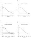
Figure 2. HuNoV-specific serum IgG antibody titers of (A, B) anti-GII.4/2006b or (C, D) anti-GII.4/MD2004 hyperimmune guinea pig serum measured by the (A, C) LIPS or (B, D) ELISA, using 107 RLU of GII.4/2006b Ruc-VP1 or 100 ng VLPs
Sera were diluted four-fold. Measurements were represented as the geometric mean RLUs (LIPS) or OD (ELISA) for two or more independent runs. Pooled, unimmunized guinea pig sera and the respective antigen were used as the negative control. The solid, horizontal black line represents the baseline, and was calculated as the average RLUs or OD of the negative control data. The dashed, horizontal black line represents the cutoff value, which was calculated as the average RLUs or OD of the negative control data plus three standard deviations. Error bars represent geometric standard deviation for multiple independent runs (n ≥ 2). Error bars are not shown for data points if shorter than the height of the symbol. The Synergy luminometer was used for these LIPS data.
Figure 3
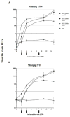
Figure 3. LIPS analysis of HuNoV-specific serum IgG antibody responses against GII.4/2006b Ruc-VP1, Ruc-S, and Ruc-P antigens of immunized minipigs identified as (A) 1584, or (B) 2718
HuNoV-specific antibody detection was presented as the geometric mean fold rise of RLUs of two or more independent LIPS assay runs for each minipig, and plotted on a logarithmic scale. Background was determined as the RLU of protein A/G beads, negative control pre-immunization sera, and the respective Ruc-antigen. The fold rise was calculated by dividing the RLU value of tested serum samples by the background. The black, horizontal dashed line represents the cutoff value for positive HuNoV-specific antibody detection. The cutoff value was calculated as the geometric mean fold rise of serum samples (n = 4) of a mock immunized pig plus 3 standard deviations. Error bars represent the geometric standard deviation of the mean fold rise for two or more independent runs. Error bars are not shown for data points if shorter than the height of the symbol. The black arrows represent the PIW at which booster doses were given. The Berthold luminometer was used for these LIPS data.
Figure 4
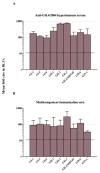
Figure 4. Preliminary assessment of LIPS assay specificity in (A) anti-GII.4/2004 hyperimmune, or (B) multicomponent immunization sera
HuNoV-specific serum IgG antibodies against Ruc-VP1 of 9 different HuNoV strains were measured in hyper- (guinea pig) and multicomponent (minipig) immunization sera. HuNoV-specific antibody detection was presented as the geometric mean fold rise of RLUs of two or more independent LIPS assay runs, and plotted on a logarithmic scale. Background was determined as the RLUs of protein A/G beads, negative control pre-immunization sera, and each Ruc-VP1 antigen. The fold rise was calculated by dividing the RLU value of tested serum samples by the background. The cutoff value was calculated as the geometric mean fold rise of mock immunized minipig serum samples from multiple independent runs (n = 2 to 16) plus three standard deviations. Error bars represent geometric standard deviation of the mean fold rise for two or more independent runs. The black, horizontal dashed line represents the cutoff value for positive HuNoV-specific antibody detection. The Synergy luminometer was used for these LIPS data.
Figure 5
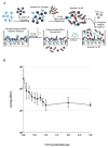
Figure 5. Competitive LIPS assay setup
(A) For the competitive LIPS assay, VLPs were added to the serum mixture (40 μL of buffer A, 10 μL of the 1:10 diluted serum) overnight at 4 degrees prior to the addition of Ruc-antigen. 107 RLUs of Ruc-antigen was then added and incubated for 1 hr at RT. The mixture was transferred to filter plates pre-coated with protein A/G beads. Protein beads precipitate the serum antibodies, and subsequent washes filter the unbound VLP and unbound Ruc-antigen through the wells. Coelenterazine substrate is added, and only antibody complexes with Ruc-antigen luminesce. (B) Different concentrations of GI.1 VLPs were titrated against serum samples from two immunized minipigs at terminal bleed. The detection of HuNoV-specific antibodies was measured using the GI.1 Ruc-VP1 LIPS assay. HuNoV-specific antibody detection was presented as the geometric mean of RLUs of serum samples (n = 2) of the two immunized minipigs for more than 4 independent LIPS assay runs, and plotted on a logarithmic scale. Error bars represent the geometric standard deviation of mean RLUs for multiple independent runs (n = 4 to 16). The Synergy luminometer was used for these LIPS data.
Figure 6
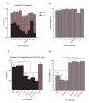
Figure 6. Competitive LIPS assay (A, C) and percent inhibition analyses (B, D) of HuNoV-specific serum IgG antibodies against (A) Ruc-VP1 of 9 different HuNoV strains and the homologous VLP, or (C) GI.1 Ruc-VP1 antigen and different combinations of GI VLPs
HuNoV-specific antibody detection was presented as the arithmetic mean of RLUs of more than four independent LIPS assay runs for each minipig at terminal bleed PIW 15, and plotted on a logarithmic scale. Background was determined as the RLU of protein A/G beads, pre-immunization minipig serum samples, and each Ruc-VP1 antigen and VLP. Average RLUs were calculated by subtracting the background RLUs from RLUs of the tested sample. The cutoff value was calculated as the average RLUs of mock immunized minipig serum samples incubated with each Ruc-VP1 antigen and VLP, plus three standard deviations. The black, horizontal dashed line represents the cutoff value for positive HuNoV-specific antibody detection, and error bars represent standard error of the arithmetic mean RLUs (n = 4 to 16 independent runs). An asterisk represents statistically significant differences (p < 0.05) in mean RLUs among groups by Tukey’s multiple comparisons test in one-way ANOVA. The Synergy luminometer was used for these LIPS data.

Similar articles

Cited by

References

    1. Atmar RL, Bernstein DI, Lyon GM, Treanor JJ, Al-Ibrahim MS, Graham DY, Vinje J, Jiang X, Gregoricus N, Frenck RW, Moe CL, Chen WH, Ferreira J, Barrett J, Opekun AR, Estes MK, Borkowski A, Baehner F, Goodwin R, Edmonds A, Mendelman PM. Serological correlates of protection against a GII.4 norovirus. Clin Vaccine Immunol. 2015;22:923–9. doi: 10.1128/cvi.00196-15. - DOI - PMC - PubMed
    1. Belliot G, Noel JS, Li JF, Seto Y, Humphrey CD, Ando T, Glass RI, Monroe SS. Characterization of capsid genes, expressed in the baculovirus system, of three new genetically distinct strains of Norwalk-like viruses. J Clin Microbiol. 2001;39:4288–95. doi: 10.1128/jcm.39.12.4288-4295.2001. - DOI - PMC - PubMed
    1. Bertolotti-Ciarlet A, Crawford SE, Hutson AM, Estes MK. The 3′ end of Norwalk virus mRNA contains determinants that regulate the expression and stability of the viral capsid protein VP1: a novel function for the VP2 protein. J Virol. 2003;77:11603–15. - PMC - PubMed
    1. Bok K, Abente EJ, Realpe-Quintero M, Mitra T, Sosnovtsev SV, Kapikian AZ, Green KY. Evolutionary dynamics of GII.4 noroviruses over a 34-year period. J Virol. 2009;83:11890–901. doi: 10.1128/jvi.00864-09. - DOI - PMC - PubMed
    1. Bok K, Parra GI, Mitra T, Abente E, Shaver CK, Boon D, Engle R, Yu C, Kapikian AZ, Sosnovtsev SV, Purcell RH, Green KY. Chimpanzees as an animal model for human norovirus infection and vaccine development. Proc Natl Acad Sci. 2011;108:325–30. doi: 10.1073/pnas.1014577107. - DOI - PMC - PubMed

Publication types

MeSH terms