. 2017 Jul 1;17(1):460.
doi: 10.1186/s12885-017-3430-2.

Inhibition of HDAC6 activity through interaction with RanBPM and its associated CTLH complex

Affiliations

Inhibition of HDAC6 activity through interaction with RanBPM and its associated CTLH complex

Louisa M Salemi et al. BMC Cancer. .

Abstract

Background: Histone deacetylase 6 (HDAC6) is a microtubule-associated deacetylase that promotes many cellular processes that lead to cell transformation and tumour development. We previously documented an interaction between Ran-Binding Protein M (RanBPM) and HDAC6 and found that RanBPM expression inhibits HDAC6 activity. RanBPM is part of a putative E3 ubiquitin ligase complex, termed the C-terminal to LisH (CTLH) complex. Here, we investigated the involvement of the CTLH complex on HDAC6 inhibition and assessed the outcome of this regulation on the cellular motility induced by HDAC6.

Methods: Cell lines (Hela, HEK293 and immortalized mouse embryonic fibroblasts) stably or transiently downregulated for several components of the CTLH complex were employed for the assays used in this study. Interactions of HDAC6, RanBPM and muskelin were assessed by co-immunoprecipitations. Quantifications of western blot analyses were employed to evaluate acetylated α-tubulin levels. Confocal microscopy analyses were used to determine microtubule association of HDAC6 and CTLH complex members. Cell migration was evaluated using wound healing assays.

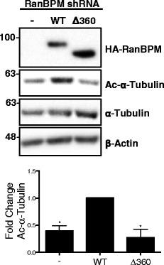
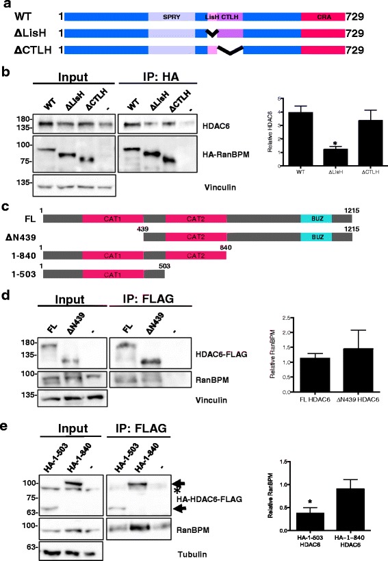
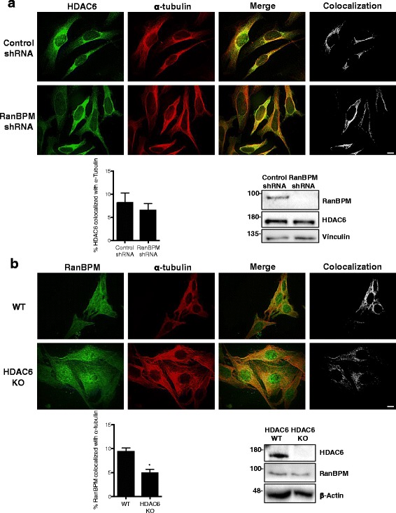
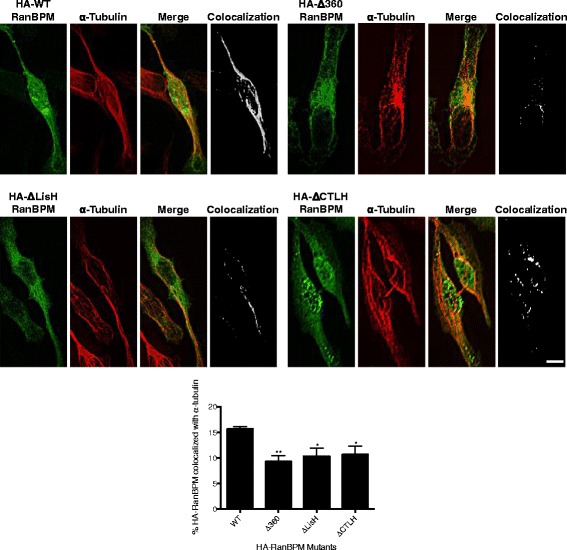
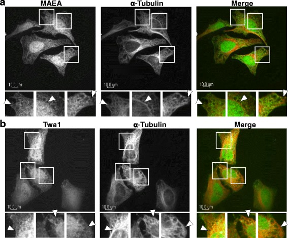
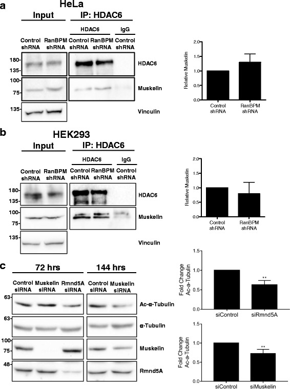
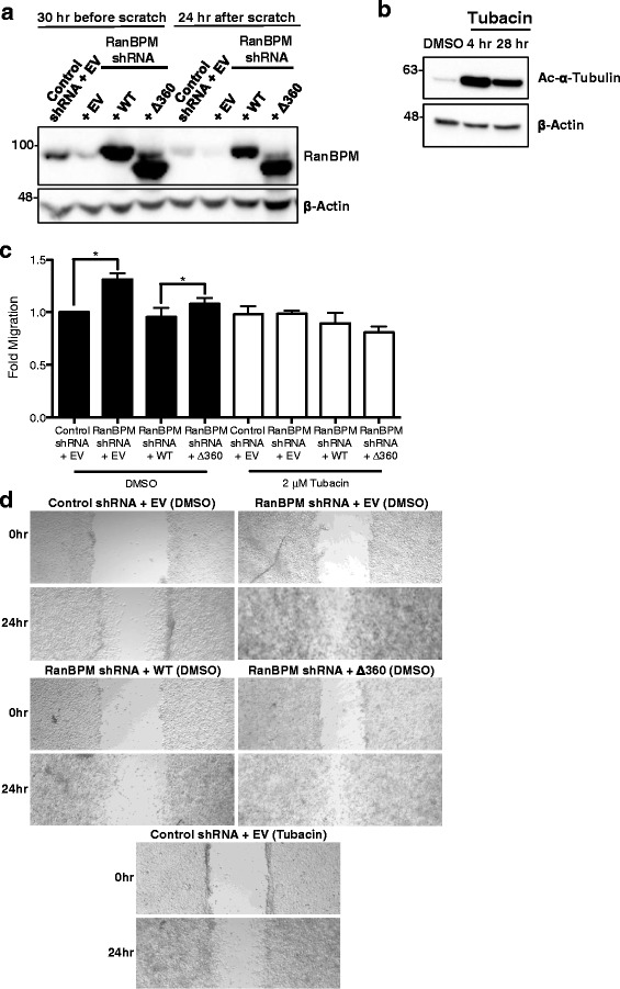
Results: We demonstrate that RanBPM-mediated inhibition of HDAC6 is dependent on its association with HDAC6. We show that, while HDAC6 does not require RanBPM to associate with microtubules, RanBPM association with microtubules requires HDAC6. Additionally, we show that Twa1 (Two-hybrid-associated protein 1 with RanBPM) and MAEA (Macrophage Erythroblast Attacher), two CTLH complex members, also associate with α-tubulin and that muskelin, another component of the CTLH complex, is able to associate with HDAC6. Downregulation of CTLH complex members muskelin and Rmnd5A (Required for meiotic nuclear division homolog A) resulted in decreased acetylation of HDAC6 substrate α-tubulin. Finally, we demonstrate that the increased cell migration resulting from downregulation of RanBPM is due to the relief in inhibition of HDAC6 α-tubulin deacetylase activity.

Conclusions: Our work shows that RanBPM, together with the CTLH complex, associates with HDAC6 and restricts cell migration through inhibition of HDAC6 activity. This study uncovers a novel function for the CTLH complex and suggests that it could have a tumour suppressive role in restricting HDAC6 oncogenic properties.

Keywords: CTLH complex; Cell migration; HDAC6; RanBPM; α-tubulin.


Figures

Fig. 1
Fig. 1
RanBPM association with HDAC6 is required for inhibition of HDAC6 deacetylase activity. Top, whole cell extracts from RanBPM shRNA Hela cells either left untransfected or transfected with HA-RanBPM-WT or HA-RanBPM-Δ360 were analyzed by western blot with the antibodies indicated. Bottom, quantification of relative amounts of acetylated α-tubulin was normalized to total α-tubulin levels. Relative amounts of acetylated α-tubulin in untransfected and HA-RanBPM-Δ360-transfected cells were normalized to WT. Results are averaged from three different experiments, with error bars indicating SEM. P < 0.05 (*)
Fig. 2
Fig. 2
Identification of the RanBPM and HDAC6 domains that mediate their association. a Schematic representation of HA-tagged RanBPM wildtype (WT) and deletion mutant constructs ΔLisH and ΔCTLH. b Left, whole cell extracts were prepared from RanBPM shRNA Hela cells untransfected (−) or transfected with HA-RanBPM-WT, HA-RanBPM-ΔLisH, or HA-RanBPM-ΔCTLH constructs. RanBPM was immunoprecipitated with an HA antibody and immunoprecipitates were analyzed by western blot with HDAC6 and RanBPM antibodies. Input, 5% input extract. Right, quantification of relative amounts of co-immunoprecipitated HDAC6 normalized to immunoprecipitated RanBPM. Results are averaged from three different experiments with error bars indicating SEM. P < 0.05 (*). c Schematic representation of HDAC6 full length (FL) and deletion mutant constructs. d Left, whole cell extracts were prepared from HDAC6 knockout MEFs untransfected (−) or transfected with full length (FL) or ΔN439 HDAC6 constructs. HDAC6 was immunoprecipitated with a FLAG antibody and immunoprecipitates were analyzed by western blot with RanBPM and HDAC6 antibodies. Input, 5% input extract. Right, quantification of relative amounts of co-immunoprecipitated RanBPM normalized to immunoprecipitated HDAC6. Results are averaged from three different experiments with error bars indicating SEM. P < 0.05 (*). e Left, whole cell extracts were prepared from HDAC6 knockout MEFs untransfected (−) or transfected with HA-1-503 or HA-1-840 HDAC6 constructs and immunoprecipitation and analysis was performed as described above except that HDAC6 mutant immunoprecipitation was verified using an HA antibody. Arrows indicate the position of the HA-HDAC6 mutants. The asterisk indicates residual RanBPM signal from previous hybridization. Right, quantification of relative amounts of co-immunoprecipitated RanBPM normalized to immunoprecipitated HDAC6. Results are averaged from three different experiments with error bars indicating SEM. P < 0.05 (*)
Fig. 3
Fig. 3
RanBPM requires HDAC6 for association with α-tubulin. a Control or RanBPM shRNA Hela cells were fixed and incubated with antibodies against HDAC6 (green) and α-tubulin (red). Colocalization of HDAC6 and α-tubulin was analyzed using Imaris software. Top, representative images where white signal represents the top 2% of HDAC6 and α-tubulin colocalization. Bottom left, quantification of the top 2% of pixels representing HDAC6 colocalized with α-tubulin. Data are representative of a minimum of 60 cells from three experiments. Error bars represent SEM. P < 0.05 (*). Bottom right, western blot analysis of extracts from Hela control and RanBPM shRNA cells, showing RanBPM and HDAC6 expression with respect to vinculin loading control. b Wildtype (WT) or HDAC6 knockout (KO) MEFs were fixed and incubated with antibodies against RanBPM (green) and α-tubulin (red). Colocalization of RanBPM and α-tubulin was analyzed using Imaris software. Top, representative images where white signal represents the top 2% of RanBPM and α-tubulin colocalization. Bottom left, quantification of the top 2% of pixels representing RanBPM colocalized with α-tubulin. Data are representative of a minimum of 60 cells from three experiments. Error bars represent SEM. P < 0.05 (*). Bottom right, western blot analysis of extracts from WT and HDAC6 KO MEFs, showing HDAC6 and RanBPM expression with respect to β-actin loading control
Fig. 4
Fig. 4
RanBPM requires the LisH and CTLH domains to associate with α-tubulin. Hela RanBPM shRNA cells were transfected with the indicated HA-RanBPM deletion constructs. Cells were fixed and incubated with antibodies against HA (green) and α-tubulin (red). Colocalization of HA-RanBPM and α-tubulin was analyzed using Imaris software. Top, representative images where white signal represents the top 2% of HA-RanBPM and α-tubulin colocalization. Scale bar: 10 μm. Bottom, Quantification of the top 2% of pixels representing HA-RanBPM colocalized with α-tubulin. Data are representative of a minimum of 10 transfected cells from three experiments. Error bars represent SEM. P < 0.05 (*)
Fig. 5
Fig. 5
CTLH components associate with microtubules. a Hela cells were fixed and incubated with antibodies against MAEA and α-tubulin. Shown are single plane confocal images. Insets are enlarged images of the boxed regions from the above panels and arrows indicate areas of colocalization. The right panels show merged images (MAEA, green; α-tubulin, red) Scale bar: 10 μm. b Hela cells were fixed and incubated with antibodies against Twa1 and α-tubulin and analysis was performed as described above (Twa1, green; α-tubulin, red)
Fig. 6
Fig. 6
Muskelin associates with HDAC6 and downregulation of Rmnd5A and muskelin affects α-tubulin acetylation. a HDAC6 associates with Muskelin independently of RanBPM. Left, whole cell extracts were prepared from control or RanBPM shRNA Hela cells and immunoprecipitated with either HDAC6 or IgG. Immunoprecipitates were analyzed by western blot with a muskelin antibody. HDAC6 immunoprecipitation was verified using an HDAC6 antibody. Input, 5% input extract. Right, quantification of relative amounts of co-immunoprecipitated muskelin normalized to immunoprecipitated HDAC6. Results are averaged from three different experiments with error bars indicating SEM. P < 0.05 (*). b Whole cell extracts were prepared from Control or RanBPM shRNA HEK cells and analyzed as described above. c Whole cell extracts were prepared from HeLa cells transfected with control, Rmnd5A, or Muskelin siRNA. Extracts were prepared either 72 or 144 h after transfection and analyzed by western blot with the indicated antibodies. Right, quantification of relative amounts of acetylated α-tubulin normalized to total α-tubulin levels. Results are averaged from at least three different experiments, with error bars indicating SD. P < 0.005 (**)
Fig. 7
Fig. 7
RanBPM inhibits HDAC6 mediated cell migration. a Control or RanBPM shRNA HEK293 cells transfected with either empty vector (EV), WT-RanBPM (WT) or Δ360 RanBPM (Δ360) were harvested 30 h before the scratch or 24 h after the scratch. Whole cell extracts were analyzed by western blot and hybridized with the antibodies indicated. b Control shRNA HEK293 cells were treated with either DMSO or 2 μM Tubacin for the indicated time points. Whole cell extracts were analyzed by western blot and hybridized with the antibodies indicated. c Confluent monolayers of control or RanBPM shRNA HEK293 cells were transfected with either EV, WT or Δ360 RanBPM and were cultured in the presence of 2 mM hydroxyurea for 24 h, and pretreated with either DMSO (black bars) or 2 μM Tubacin (white bars) for 4 h. Cells were then scratched and width of the wound was assessed at the time of the scratch and 24 h later. Fold migration was calculated by normalizing the average wound width at 24 h to the average wound width at 0 h and each sample was normalized to control shRNA EV transfected, DMSO treated samples. Results are averaged from three independent experiments, with error bars indicating SEM. P < 0.05 (*). d Representative images of Control or RanBPM shRNA HEK cells transfected and treated as above following the scratch (0 h) and 24 h later

Similar articles

Cited by

References

    1. Rhodes DA, de Bono B, Trowsdale J. Relationship between SPRY and B30.2 protein domains. Evolution of a component of immune defence? Immunology. 2005;116:411–417. - PMC - PubMed
    1. Atabakhsh E, Bryce DM, Lefebvre KJ, Schild-Poulter C. RanBPM has proapoptotic activities that regulate cell death pathways in response to DNA damage. Mol Cancer Res. 2009;7:1962–1972. doi: 10.1158/1541-7786.MCR-09-0098. - DOI - PubMed
    1. Emes RD, Ponting CP. A new sequence motif linking lissencephaly, Treacher Collins and oral-facial-digital type 1 syndromes, microtubule dynamics and cell migration. Hum Mol Genet. 2001;10:2813–2820. doi: 10.1093/hmg/10.24.2813. - DOI - PubMed
    1. Menon RP, Gibson TJ, Pastore A. The C terminus of fragile X mental retardation protein interacts with the multi-domain ran-binding protein in the microtubule-organising centre. J Mol Biol. 2004;343:43–53. doi: 10.1016/j.jmb.2004.08.024. - DOI - PubMed
    1. Kobayashi N, Yang J, Ueda A, Suzuki T, Tomaru K, Takeno M, et al. RanBPM, Muskelin, p48EMLP, p44CTLH, and the armadillo-repeat proteins ARMC8alpha and ARMC8beta are components of the CTLH complex. Gene. 2007;396:236–47. - PubMed

MeSH terms