

High glucose induces the aging of mesenchymal stem cells via Akt/mTOR signaling
- PMID: 28656269
- PMCID: PMC5562095
- DOI: 10.3892/mmr.2017.6832
High glucose induces the aging of mesenchymal stem cells via Akt/mTOR signaling
Abstract
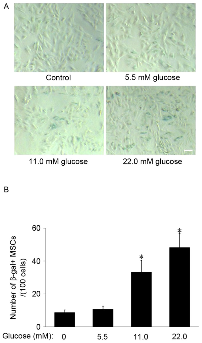
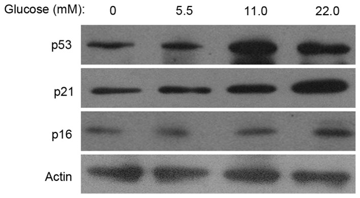
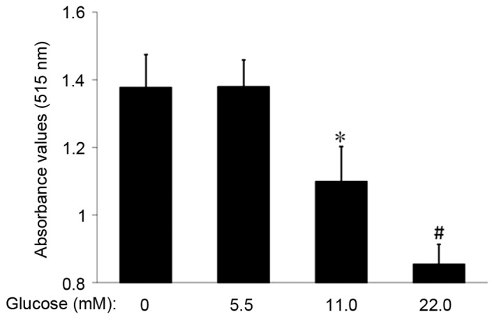
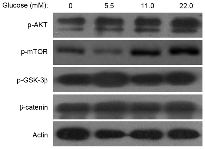
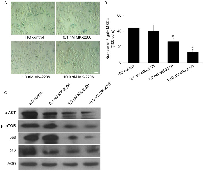
It has previously been demonstrated that glucose is important in the process of stem cell aging. However, the mechanisms of cell senescence induced by high glucose (HG) remain to be elucidated. The preliminary study indicated that D‑galactose induced mesenchymal stem cell (MSCs) aging. The present study demonstrated, following treatment with 11.0 or 22.0 mM HG for 14 days, that HG significantly promoted MSCs aging and the expression levels of phosphorylated (p-)phosphatidylinositol 3-kinase/protein kinase B (Akt) and p‑mammalian target of rapamycin signaling (mTOR) in the HG groups were increased compared with the control group. However, following Akt inhibition with 1.0 or 10.0 nM MK‑2206, which is an Akt‑specific small molecule inhibitor, the senescence‑cell value in the HG group was significantly decreased compared with the control group. These results indicated that HG induced MSCs senescence and this effect was primarily mediated via the Akt/mTOR signaling pathway.
Figures
Similar articles
-
Coenzyme Q10 inhibits the aging of mesenchymal stem cells induced by D-galactose through Akt/mTOR signaling.Oxid Med Cell Longev. 2015;2015:867293. doi: 10.1155/2015/867293. Epub 2015 Feb 18. Oxid Med Cell Longev. 2015. PMID: 25789082 Free PMC article.
-
Enhanced expression of glucose transporter-1 in vascular smooth muscle cells via the Akt/tuberous sclerosis complex subunit 2 (TSC2)/mammalian target of rapamycin (mTOR)/ribosomal S6 protein kinase (S6K) pathway in experimental renal failure.J Vasc Surg. 2013 Feb;57(2):475-85. doi: 10.1016/j.jvs.2012.07.037. Epub 2012 Dec 21. J Vasc Surg. 2013. PMID: 23265586
-
Mesenchymal stem cells ameliorate inflammatory cytokine-induced impairment of AT-II cells through a keratinocyte growth factor-dependent PI3K/Akt/mTOR signaling pathway.Mol Med Rep. 2016 May;13(5):3755-62. doi: 10.3892/mmr.2016.5004. Epub 2016 Mar 18. Mol Med Rep. 2016. PMID: 27035760 Free PMC article.
-
[DEVELOPMENT PROGRESS OF MESENCHYMAL STEM CELLS SENESCENCE].Zhongguo Xiu Fu Chong Jian Wai Ke Za Zhi. 2015 Jul;29(7):899-904. Zhongguo Xiu Fu Chong Jian Wai Ke Za Zhi. 2015. PMID: 26540988 Review. Chinese.
-
Targeting mTOR signaling by polyphenols: A new therapeutic target for ageing.Ageing Res Rev. 2016 Nov;31:55-66. doi: 10.1016/j.arr.2016.07.004. Epub 2016 Jul 21. Ageing Res Rev. 2016. PMID: 27453478 Review.
Cited by
-
Annexin A2 Improves the Osteogenic Differentiation of Mesenchymal Stem Cells Exposed to High-Glucose Conditions through Lessening the Senescence.Int J Mol Sci. 2022 Oct 19;23(20):12521. doi: 10.3390/ijms232012521. Int J Mol Sci. 2022. PMID: 36293376 Free PMC article.
-
Low-glucose culture environment can enhance the wound healing capability of diabetic adipose-derived stem cells.Stem Cell Res Ther. 2023 Sep 4;14(1):236. doi: 10.1186/s13287-023-03478-2. Stem Cell Res Ther. 2023. PMID: 37667384 Free PMC article.
-
Limited Potential or Unfavorable Manipulations? Strategies Toward Efficient Mesenchymal Stem/Stromal Cell Applications.Front Cell Dev Biol. 2020 May 19;8:316. doi: 10.3389/fcell.2020.00316. eCollection 2020. Front Cell Dev Biol. 2020. PMID: 32509777 Free PMC article. Review.
-
Cellular Senescence as the Pathogenic Hub of Diabetes-Related Wound Chronicity.Front Endocrinol (Lausanne). 2020 Sep 16;11:573032. doi: 10.3389/fendo.2020.573032. eCollection 2020. Front Endocrinol (Lausanne). 2020. PMID: 33042026 Free PMC article. Review.
-
The Regulation of the AMPK/mTOR Axis Mitigates Tendon Stem/Progenitor Cell Senescence and Delays Tendon Aging.Stem Cell Rev Rep. 2023 Jul;19(5):1492-1506. doi: 10.1007/s12015-023-10526-0. Epub 2023 Mar 14. Stem Cell Rev Rep. 2023. PMID: 36917311
References
MeSH terms
Substances
LinkOut - more resources
Full Text Sources
Other Literature Sources
Miscellaneous
