. 2017 Aug 10;91(17):e00720-17.
doi: 10.1128/JVI.00720-17. Print 2017 Sep 1.

Interplay of PA-X and NS1 Proteins in Replication and Pathogenesis of a Temperature-Sensitive 2009 Pandemic H1N1 Influenza A Virus

Affiliations

Interplay of PA-X and NS1 Proteins in Replication and Pathogenesis of a Temperature-Sensitive 2009 Pandemic H1N1 Influenza A Virus

Aitor Nogales et al. J Virol. .

Abstract

Influenza A viruses (IAVs) cause seasonal epidemics and occasional pandemics, representing a serious public health concern. It has been described that one mechanism used by some IAV strains to escape the host innate immune responses and modulate virus pathogenicity involves the ability of the PA-X and NS1 proteins to inhibit the host protein synthesis in infected cells. It was reported that for the 2009 pandemic H1N1 IAV (pH1N1) only the PA-X protein had this inhibiting capability, while the NS1 protein did not. In this work, we have evaluated, for the first time, the combined effect of PA-X- and NS1-mediated inhibition of general gene expression on virus pathogenesis, using a temperature-sensitive, live-attenuated 2009 pandemic H1N1 IAV (pH1N1 LAIV). We found that viruses containing PA-X and NS1 proteins that simultaneously have (PAWT+/NS1MUT+) or do not have (PAMUT-/NS1WT-) the ability to block host gene expression showed reduced pathogenicity in vivo However, a virus where the ability to inhibit host protein expression was switched between PA-X and NS1 (PAMUT-/NS1MUT+) presented pathogenicity similar to that of a virus containing both wild-type proteins (PAWT+/NS1WT-). Our findings suggest that inhibition of host protein expression is subject to a strict balance, which can determine the successful progression of IAV infection. Importantly, knowledge obtained from our studies could be used for the development of new and more effective vaccine approaches against IAV.IMPORTANCE Influenza A viruses (IAVs) are one of the most common causes of respiratory infections in humans, resulting in thousands of deaths annually. Furthermore, IAVs can cause unpredictable pandemics of great consequence when viruses not previously circulating in humans are introduced into humans. The defense machinery provided by the host innate immune system limits IAV replication; however, to counteract host antiviral activities, IAVs have developed different inhibition mechanisms, including prevention of host gene expression mediated by the viral PA-X and NS1 proteins. Here, we provide evidence demonstrating that optimal control of host protein synthesis by IAV PA-X and/or NS1 proteins is required for efficient IAV replication in the host. Moreover, we demonstrate the feasibility of genetically controlling the ability of IAV PA-X and NS1 proteins to inhibit host immune responses, providing an approach to develop more effective vaccines to combat disease caused by this important respiratory pathogen.

Keywords: host response; influenza; influenza vaccines; virus-host interactions.


Figures

FIG 1
FIG 1
Effect of temperature on the polymerase activity of the pH1N1 LAIV virus. (A) Schematic representation of PB2 and PB1 viral segments. pH1N1 PB2 and PB1 WT (gray, top) segments with the residues mutated to generate the LAIV virus (black, bottom) are indicated. Numbers on the right signify the amino acid (aa) length of the PB2 and PB1 proteins. (B) Viral replication and transcription. Human 293T cells were transiently cotransfected, using LPF2000, with the pH1N1 WT (gray bars) or LAIV (black bars) ambisense pDZ expression plasmids encoding the minimal requirements for viral genome replication and gene transcription (PB2, PB1, and PA) and NP, together with a vRNA-like expression plasmid encoding Gluc under the control of the human polymerase I promoter (hpPol-I Gluc) and the SV40-Cluc plasmid to normalize transfection efficiencies. At 6 h p.t., cells were placed at 33°C, 37°C, or 39°C, and viral replication and transcription were evaluated at 24 h by luminescence. Gluc (left) and Cluc (middle) activity are represented. Gluc activity was normalized to that of Cluc, and the data were represented as relative activity considering the activity of pH1N1 WT at each indicated temperature as 100% (right). Data represent the means and SDs of the results determined from triplicate wells. *, P < 0.05 (WT versus LAIV) using Student's t test (n = 3 per time point) from Microsoft Excel. (C) NP protein expression levels from cell lysates were evaluated by Western blotting using a specific monoclonal antibody against the viral NP. Sizes of molecular markers are noted on the left.
FIG 2
FIG 2
Characterization of the pH1N1 LAIV virus. (A) Viral growth kinetics. TCS of MDCK cells infected at a low MOI (0.001) with pH1N1 WT (gray bars) or LAIV (black bars) virus at 33°C, 37°C, and 39°C were analyzed at the indicated times p.i. (12, 24, 48, and 72 h) by immunofocus assay using an anti-NP MAb (HB-65). Data represent the means and SDs of the results determined from triplicate wells. The dashed line indicates the limit of detection (200 FFU/ml). *, P < 0.05 (WT versus LAIV) using Student's t test (n = 3 per time point) from Microsoft Excel. (B) Plaque assays. MDCK cells were infected with pH1N1 WT and LAIV viruses and incubated at 33°C, 37°C, and 39°C for 3 days. The plaques phenotype was assessed by immunostaining with an anti-NP MAb (HB-65).
FIG 3
FIG 3
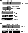
Schematic representation of WT and mutant PA and NS1 proteins. (A) PA (top) and PA-X (bottom) WT viral proteins (gray) and the different mutations (black) introduced into the frameshift motif (PAPMUT or PAMUT) to abolish PA-X expression are shown. Numbers on the right indicate the lengths of the PA and PA-X proteins. (B) NS1 WT (gray) and mutant (black) proteins containing amino acid substitutions allowing binding to CPSF30 and inhibition of host protein expression (NS1MUT). Numbers on the right signify the lengths of the WT and mutant NS1 proteins.
FIG 4
FIG 4
Ability of WT and mutant PA-X and NS1 proteins to block host protein expression. Human 293T cells were transiently cotransfected, using LPF2000, with expression plasmids encoding GFP and Gluc under the control of a polymerase II promoter (pCAGGS GFP and pCAGGS Gluc, respectively) together with pDZ plasmids encoding WT or mutant (MUT) PA or NS1 proteins or empty (E) plasmid as a control. (A, B, D, and E) At 24 h p.t., cells were analyzed by GFP expression under a fluorescence microscope (A and D) and by Gluc activity from TCS (B and E). Representative images are shown. Scale bar = 100 μm. Results represent the means and standard deviations of triplicate values. (C and F) Protein expression from cell lysates was evaluated by Western blotting using specific antibodies for the viral PA and NS1 proteins or actin as loading control. Molecular markers are indicated on the left.
FIG 5
FIG 5
Ability of PA-X and NS1 proteins to block host protein expression in combination. Human 293T cells were transiently cotransfected, using LPF2000, with pCAGGS GFP and pCAGGS Gluc together with the indicated combination of pDZ plasmids encoding WT or mutant (MUT) PA or NS1 proteins or empty (E) plasmid as a control. (A and B) At 24 h p.t., cells were analyzed for GFP expression under a fluorescence microscope (A) and for Gluc activity from TCS (B). Representative images are shown. Scale bar = 100 μm. Results represent the means and standard deviations of triplicate values. (C) Protein expression from cell lysates was evaluated by Western blotting using specific antibodies for the viral PA and NS1 proteins. Actin was used as a loading control. Molecular markers are noted on the left.
FIG 6
FIG 6
(A) Effects of WT and mutant PA alone or in combination with NS1 on polymerase viral replication and transcription. Human 293T cells were transiently cotransfected, using LPF2000, with the pH1N1 LAIV ambisense pDZ expression plasmids encoding the minimal requirements for viral genome replication and gene transcription (PB2, PB1, and PA) and NP, together with the vRNA-like expression plasmid hpPol-I Gluc and the SV40-Cluc plasmid to normalize transfection efficiencies. Viral replication and transcription were evaluated at 24 h by Gluc expression. Gluc activity was normalized to that of Cluc. Data for Gluc (top), Cluc (middle), and Gluc activity normalized to that of Cluc (bottom) are shown. Data represent the means and SDs of the results determined from triplicate wells. *, P < 0.05 using Student's t test from Microsoft Excel. (B) Analysis of protein expression by Western blotting. Protein expression levels from cell lysates were evaluated by Western blotting using specific antibodies for the viral PB2, PB1, PA, NP, and NS1 proteins. Actin was used as a loading control. Molecular markers are noted on the left.
FIG 7
FIG 7
Multicycle growth kinetics and plaque assay of pH1N1 LAIV viruses. (A and B) Viral growth kinetics. Canine MDCK (A) or human A549 (B) cells were infected (MOI of 0.001 for MDCK cells or 0.1 for A549 cells) with the indicated pH1N1 LAIV viruses and incubated at 33°C. TCS were collected at multiple times p.i., and viral titers were determined by immunofocus assay using an anti-NP MAb (HB-65). Data represent the means and SDs of the results determined from triplicate wells. The dashed line indicates the limit of detection (200 FFU/ml). (C) Plaque assays. MDCK cells were infected with the indicated pH1N1 LAIV viruses and incubated at 33°C for 3 days. Plaque phenotypes were assessed by immunostaining with the anti-NP monoclonal antibody HB-65. PAWT+/NS1WT, virus containing WT PA and NS1 proteins. PAMUT/NS1WT, virus containing a mutant PA (PAMUT) affecting its ability to inhibit host protein expression and a WT NS1. PAWT+/NS1MUT+, virus containing a WT PA and a mutant NS1 allowing inhibition of host protein expression. PAMUT/NS1MUT+, virus containing both PA and NS1 mutants. In the virus illustrations, WT and mutant segments are indicated with gray and black lines, respectively.
FIG 8
FIG 8
Induction of IFN by pH1N1 LAIV viruses. MDCK cells constitutively expressing GFP-CAT and Fluc reporter genes under the control of the IFN-β promoter (MDCK IFN-β-GFP/IFN-β-Fluc) were infected (MOI, 3) with the different pH1N1 LAIV viruses. Cells infected with an NS1 deficient PR8 virus (ΔNS1) were used as internal control. (A) Viral infections were evaluated by immunofluorescence using an anti NP MAb (HB-65). (B) At 16 h p.i., IFN-β promoter activation was determined by Fluc expression. (C) Supernatants from infected MDCK IFN-β GFP-CAT/Fluc cells were collected at the same time p.i. and, after UV virus inactivation, used to treat fresh MDCK cells for 24 h prior to infection with rNDV-GFP. GFP expression from infected cells was determined at 24 h p.i. using a microplate reader. *, P < 0.05 using Student's t test from Microsoft Excel.
FIG 9
FIG 9
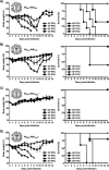
Virulence of pH1N1 LAIV viruses. Six- to 8-week-old female C57BL/6 mice (n = 5) were infected i.n. with the indicated PFU of pH1N1 LAIV PAWT+/NS1WT (A), PAMUT/NS1WT (B), PAWT+/NS1MUT+ (C), and PAMUT/NS1MUT+ (D) viruses and then monitored daily for 2 weeks for body weight loss (left) and survival (right). Mice that lost 25% of their initial body weight were sacrificed. Data represent the means and SDs of the results determined for individual mice (n = 5).
FIG 10
FIG 10
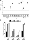
Replication of pH1N1 LAIV viruses in mouse lungs. Six- to eight-week-old female C57BL/6 mice (n = 9) were infected i.n. with 1 × 10³ PFU of the indicated pH1N1 LAIV viruses. (A) Viral replication in the lungs of infected mice was evaluated at 1, 3, and 5 days p.i. (dpi) (n = 3) by immunofocus assay using an anti-NP MAb (HB-65). Symbols represent data from individual mice. Bars represent the geometric means of lung viral titers. The dotted line represents the limit of detection of the assay (200 FFU/ml). *, P < 0.05 using Student's t test (n = 3 per time point) from Microsoft Excel. (B) mRNA expression levels of the viral M1 in lungs of infected mice were quantified by RT-qPCR analysis. Fold expression changes in each mouse group were calculated relative to the control group of mock (PBS)-infected mice. Data represent the averages and SD values for three mice in each group on the days indicated.
FIG 11
FIG 11
Induction of innate immune responses by pH1N1 LAIV viruses. mRNA expression levels of IFN-β, TNF-α and CCL2 in lungs of mice infected with 1 × 10³ PFU of the indicated pH1N1 LAIV viruses at 1, 3, and 5 dpi (n = 3) were quantified by RT-qPCR analysis. Fold expression changes in each mouse group were calculated relative to the control group of mock (PBS)-infected mice. Data represent the averages and SD values for three mice in each group on the days indicated.
FIG 12
FIG 12
Humoral responses to pH1N1 LAIV viral infections. Six- to 8-week-old female C57BL/6 mice (n = 5) were infected i.n. with 1 × 10² PFU of the indicated pH1N1 LAIV viruses. (A to C) At 21 days p.i., mice were bled and sera were collected and evaluated by ELISA for IgG antibodies against total influenza virus proteins using cell extracts of pH1N1 virus-infected MDCK cells (A) or recombinant pH1N1 HA (B) or NA (C) protein. OD, optical density. (D) HAI titers from mock (PBS)-infected or infected mouse sera were calculated.

Similar articles

Cited by

References

    1. Palese P, Shaw ML. 2007. Orthomyxoviridae: the viruses and their replication. In Knipe DM, Howley PM, Griffin DE, Lamb RA, Martin MA (ed), Fields virology, 5th ed Lippincott Williams and Wilkins, Philadelphia, PA.
    1. Resa-Infante P, Jorba N, Coloma R, Ortin J. 2011. The influenza virus RNA synthesis machine: advances in its structure and function. RNA Biol 8:207–215. doi:10.4161/rna.8.2.14513. - DOI - PMC - PubMed
    1. Wong SS, Webby RJ. 2013. Traditional and new influenza vaccines. Clin Microbiol Rev 26:476–492. doi:10.1128/CMR.00097-12. - DOI - PMC - PubMed
    1. Chan W, Zhou H, Kemble G, Jin H. 2008. The cold adapted and temperature sensitive influenza A/Ann Arbor/6/60 virus, the master donor virus for live attenuated influenza vaccines, has multiple defects in replication at the restrictive temperature. Virology 380:304–311. doi:10.1016/j.virol.2008.07.027. - DOI - PubMed
    1. Cox NJ, Kitame F, Kendal AP, Maassab HF, Naeve C. 1988. Identification of sequence changes in the cold-adapted, live attenuated influenza vaccine strain, A/Ann Arbor/6/60 (H2N2). Virology 167:554–567. - PubMed

Publication types

MeSH terms

Substances

LinkOut - more resources