. 2017 Jul;66(7):1890-1900.
doi: 10.2337/db16-1170. Epub 2017 Jun 12.

Kv2.1 Clustering Contributes to Insulin Exocytosis and Rescues Human β-Cell Dysfunction

Affiliations

Kv2.1 Clustering Contributes to Insulin Exocytosis and Rescues Human β-Cell Dysfunction

Jianyang Fu et al. Diabetes. 2017 Jul.

Abstract

Insulin exocytosis is regulated by ion channels that control excitability and Ca2+ influx. Channels also play an increasingly appreciated role in microdomain structure. In this study, we examine the mechanism by which the voltage-dependent K+ (Kv) channel Kv2.1 (KCNB1) facilitates depolarization-induced exocytosis in INS 832/13 cells and β-cells from human donors with and without type 2 diabetes (T2D). We find that Kv2.1, but not Kv2.2 (KCNB2), forms clusters of 6-12 tetrameric channels at the plasma membrane and facilitates insulin exocytosis. Knockdown of Kv2.1 expression reduces secretory granule targeting to the plasma membrane. Expression of the full-length channel (Kv2.1-wild-type) supports the glucose-dependent recruitment of secretory granules. However, a truncated channel (Kv2.1-ΔC318) that retains electrical function and syntaxin 1A binding, but lacks the ability to form clusters, does not enhance granule recruitment or exocytosis. Expression of KCNB1 appears reduced in T2D islets, and further knockdown of KCNB1 does not inhibit Kv current in T2D β-cells. Upregulation of Kv2.1-wild-type, but not Kv2.1-ΔC318, rescues the exocytotic phenotype in T2D β-cells and increases insulin secretion from T2D islets. Thus, the ability of Kv2.1 to directly facilitate insulin exocytosis depends on channel clustering. Loss of this structural role for the channel might contribute to impaired insulin secretion in diabetes.


Figures

Figure 1
Figure 1
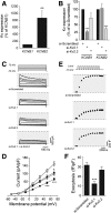
Kv2.1, but not Kv2.2, controls exocytosis in human β-cells. A: Expression of mRNA encoding Kv2.1 (KCNB1) and Kv2.2 (KCNB2) in islets from human donors assessed by quantitative PCR (n = 11 donors). B: Knockdown of KCNB1 and KCNB2 expression in human islet cells, assessed by quantitative PCR, following transfection with control siRNA duplexes (si-Scrambled) or siRNAs targeting Kv2.1 (si-Kv2.1) or Kv2.2 (si-Kv2.2) (n = 6 donors). Representative traces (C) and averaged current–voltage relationships (D) of Kv currents recorded from human β-cells following transfection with si-Scrambled (gray squares), si-Kv2.1 (black circles), si-Kv2.2 (gray circles), or both (black squares) (n = 20, 13, 21, and 17 cells from 4 donors, respectively). Representative capacitance traces (E) and averaged cumulative exocytotic responses (F) of human β-cells to a series of membrane depolarizations following transfection with si-Scrambled, si-Kv2.1, or si-Kv2.2 (n = 23, 32, and 31 cells from 5 donors, respectively). *P < 0.05; **P < 0.01; ***P < 0.001 compared with the Kv2.1 group (A) or scrambled control (B–F).
Figure 2
Figure 2
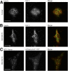
Kv2.1 forms clusters in insulin-secreting cells. Human β-cells (A) or INS 832/13 cells (B) were immunostained with anti-Kv2.1 (red) and anti-insulin (green) antibodies and visualized by TIRF microscopy (representative of 23 cells from 3 donors and 26 cells in 3 experiments, respectively). C: INS 832/13 cell expressing mCherry-Kv2.1-WT (red) and NPY-Venus (green; 33 cells in 3 experiments). Scale bars, 10 μm.
Figure 3
Figure 3
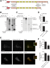
Superresolution imaging of Kv2.1 clusters. A: PALM images of HEK 293 and INS 832/13 cells expressing PAmCherry-Kv2.1-WT (red). Inset zoom-in area corresponds to the region of interest (ROI). Scale bars, 5 μm. B: PAmCherry-Kv2.1-WT molecule coordinate plots for ROIs in A, with cluster regions determined by spatial pattern analysis, highlighted in red in the background. The percentage of PAmCherry-Kv2.1-WT molecules found within clustered domains (C) and the average number of molecules per cluster (D) in HEK 293 cells (n = 22) and INS 832/13 cells (n = 29). E: Extracellular cross-linking with DTSSP followed by blotting of protein lysates with anti-Kv2.1 antibody demonstrates that the tagged (PAmCherry-Kv2.1-WT) channels form large-molecular-weight complexes at the cell surface, consistent with channel clustering. Breakdown of cross-links with 2-ME reveals the expression of channel monomers (representative of n = 6 experiments).
Figure 4
Figure 4
A clustering-deficient Kv2.1 reduces secretory granule recruitment to the plasma membrane. A: mCherry and Myc-tagged clustering-deficient Kv2.1 channels were generated by truncating the final 318 amino acids (mCherry/Myc-Kv2.1-ΔC318) of the rat sequence. B–D: When expressed in HEK 293 (B; n = 6 experiments) or INS 832/13 cells (C; n = 10 experiments), the Myc-Kv2.1-ΔC318 formed fewer high-molecular-weight clusters, which could be broken down to channel monomers by 2-ME. Although Myc-Kv2.1-ΔC318 retained some ability to form clusters in INS 832/13 cells, likely because of combination with native Kv2.1 (Supplementary Fig. 5), quantification (D; n = 6 and 10) revealed that the majority of the signal remains tetrameric (i.e., single-channel rather than cluster). Additional assessment of the clustering of these constructs is presented in Supplementary Fig. 4. E and F: When expressed in INS 832/13 cells and visualized by TIRF microscopy, mCherry-Kv2.1-ΔC318 forms fewer clusters than mCherry-Kv2.1-WT and results in fewer membrane-resident secretory granules, marked by NPY-Venus (n = 37; 62 cells from 5 experiments). Scale bars, 10 μm. ***P < 0.001 compared with mCherry-Kv2.1-WT.
Figure 5
Figure 5
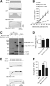
Clustering-deficient Kv2.1 retains electrical activity and syntaxin 1A binding, but does not facilitate exocytosis. Representative Kv currents (A) and quantified current-voltage relationships (B) from INS 832/13 cells expressing GFP alone, Myc-Kv2.1-WT, or Myc-Kv2.1-ΔC318 (n = 17, 19, and 18 cells, respectively). C and D: Coimmunoprecipitation (IP) of syntaxin 1A (Syn1A) pulled down both Myc-Kv2.1-WT and Myc-Kv2.1-ΔC318 equally well (n = 7 experiments). Representative capacitance traces (E) and cumulative exocytotic responses (F) of INS 832/13 cells expressing GFP alone, Myc-Kv2.1-WT, or Myc-Kv2.1-ΔC318 (n = 15, 22, and 17 cells, respectively). *P < 0.01 as indicated; ***P < 0.001 versus GFP control.
Figure 6
Figure 6
Kv2.1 and a role for the channel in granule recruitment. A: Knockdown of Kv2.1 protein in Ad-shKv2.1–infected INS 832/13 cells compared with control subjects (Ad-shScrambled; n = 4 experiments). B and C: Knockdown of Kv2.1 in INS 832/13 cells resulted in a decreased density of membrane-resident secretory granules marked with NPY-Venus and visualized by TIRF microscopy at both 1 and 16.7 mmol/L glucose. Representative images (B) and quantified data (C) are shown (n = 30, 30, 27, 30, 28, and 30 cells in 3 experiments). Syn1A, syntaxin 1A. Scale bar, 10 μm. **P < 0.01; ***P < 0.001 compared with 1 mmol/L glucose control or as indicated.
Figure 7
Figure 7
Kv2.1 clusters promote secretory granule recruitment. Representative images (A and B) and quantified data (C and D) of INS 832/13 cells expressing mCherry-Kv2.1-WT (red) or mCherry-Kv2.1-ΔC318 (red) and NPY-Venus (green) at 1 mmol/L glucose and after 15 or 30 min of 16.7 mmol/L glucose, visualized by TIRF microscopy. Glucose stimulation increased the density of membrane-resident secretory granules (C) in the mCherry-Kv2.1-WT group (black bars), but not the mCherry-Kv2.1-ΔC318 group (gray bars), the latter of which had fewer channel clusters (D). Scale bars, 10 μm. *P < 0.05; **P < 0.01 compared with the 1 mmol/L glucose condition, or as indicated.
Figure 8
Figure 8
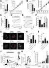
Upregulation of Kv2.1 in human T2D β-cells improves exocytotic function. Knockdown of Kv2.1 (A; KCNB1; n = 13 and 13 cells from 2 donors) or Kv2.2 (B; KCNB2; n = 19 and 23 cells from 3 donors) expression failed to reduce voltage-dependent K+ currents in β-cells from donors with T2D. C: Expression of Kv2.1 (KCNB1) mRNA in islets from ND donors or donors with T2D (n = 11 and 5 donors, respectively). Representative capacitance responses at 10 mmol/L glucose from untransfected (D) β-cells of ND donors and donors with T2D and of transfected β-cells (E) of donors with T2D expressing GFP alone or together with Kv2.1-WT. F: The cumulative exocytotic responses from D and E (n = 30 cells from 3 ND donors and n = 30, 32 and 34 cells from 3–5 donors with T2D). G: Images from live-cell TIRF of ND and T2D β-cells expressing mCherry, mCherry-Kv2.1-WT, or mCherry-Kv2.1-ΔC318 and the granule marker NPY-EGFP (greyscale). Red dots indicate exocytotic events occurring over 2 min upon increasing glucose from 2.8 to 5 mmol/L. Scale bars, 5 μm. Average frequency of exocytotic events in ND (H; n = 17, 20, and 21 cells from 2 donors) and T2D (I; n = 24, 34, and 30 cells from 2 donors with T2D) β-cells. J and K: Insulin secretory profiles of islets from a donor with T2D transduced with control adenovirus (Ad-GFP; circles) or an electrically silent full-length Kv2.1 (Ad-Kv2.1W365C/Y380T; squares). The first phase secretory response is shown on expanded time scale (K) (one donor; experiment run in duplicate). L: Averaged and individual (circles) area under the curve (AUC) values for baseline, first-phase, and second-phase responses of islets from donors with T2D transduced as in J and K (n = 3 donors in duplicate). *P < 0.05 versus ND or as indicated; **P < 0.01 versus mCherry; ***P < 0.001 as indicated.

Similar articles

Cited by

References

    1. Kahn SE, Cooper ME, Del Prato S. Pathophysiology and treatment of type 2 diabetes: perspectives on the past, present, and future. Lancet 2014;383:1068–1083 - PMC - PubMed
    1. Rorsman P, Braun M. Regulation of insulin secretion in human pancreatic islets. Annu Rev Physiol 2013;75:155–179 - PubMed
    1. MacDonald PE, Sewing S, Wang J, et al. . Inhibition of Kv2.1 voltage-dependent K+ channels in pancreatic beta-cells enhances glucose-dependent insulin secretion. J Biol Chem 2002;277:44938–44945 - PubMed
    1. Jacobson DA, Kuznetsov A, Lopez JP, Kash S, Ammälä CE, Philipson LH. Kv2.1 ablation alters glucose-induced islet electrical activity, enhancing insulin secretion. Cell Metab 2007;6:229–235 - PMC - PubMed
    1. Herrington J, Sanchez M, Wunderler D, et al. . Biophysical and pharmacological properties of the voltage-gated potassium current of human pancreatic beta-cells. J Physiol 2005;567:159–175 - PMC - PubMed