Potential therapeutic impact of CD13 expression in non-small cell lung cancer
- PMID: 28604784
- PMCID: PMC5467809
- DOI: 10.1371/journal.pone.0177146
Potential therapeutic impact of CD13 expression in non-small cell lung cancer
Erratum in
-
Correction: Potential therapeutic impact of CD13 expression in non-small cell lung cancer.PLoS One. 2017 Aug 9;12(8):e0183201. doi: 10.1371/journal.pone.0183201. eCollection 2017. PLoS One. 2017. PMID: 28793330 Free PMC article.
Abstract
Background: Aminopeptidase N (CD13) is a zinc-binding protease that has functional effects on both cancerogenesis and tumor angiogenesis. Since CD13 is an antigen suitable for molecular targeted therapies (e.g. tTF-NGR induced tumor vascular infarction), we evaluated its impact in NSCLC patients, and tested the effects of the CD13-targeted fusion protein tTF-NGR (truncated tissue factor (tTF) containing the NGR motif: asparagine-glycine-arginine) in vivo in nude mice.
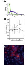
Methods: Expression of both CD13 and CD31 was studied in 270 NSCLC patients by immunohistochemistry. Clinical correlations and prognostic effects of the expression profiles were analyzed using univariate and multivariate analyses. In addition, a microarray-based analysis on the basis of the KM plotter database was performed. The in vivo effects of the CD13-targeted fusion protein tTF-NGR on tumor growth were tested in CD1 nude mice carrying A549 lung carcinoma xenotransplants.
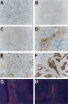
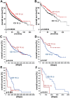
Results: CD13 expression in tumor endothelial and vessel associated stromal cells was found in 15% of the investigated samples, while expression in tumor cells was observed in 7%. Although no significant prognostic impact was observed in the full NSCLC study cohort, both univariate and multivariate models identified vascular CD13 protein expression to correlate with poor overall survival in stage III and pN2+ NSCLC patients. Microarray-based mRNA analysis for either adenocarcinomas or squamous cell carcinomas did not reveal any significant effect. However, the analysis of CD13 mRNA expression for all lung cancer histologies demonstrated a positive prognostic effect. In vivo, systemic application of CD13-targeted tissue factor tTF-NGR significantly reduced CD13+ A549 tumor growth in nude mice.
Conclusions: Our results contribute a data basis for prioritizing clinical testing of tTF-NGR and other antitumor molecules targeted by NGR-peptides in NSCLC. Because CD13 expression in NSCLC tissues was found only in a specific subset of NSCLC patients, rigorous pre-therapeutic testing will help to select patients for these studies.
Conflict of interest statement
Figures


Similar articles
-
CD13 as target for tissue factor induced tumor vascular infarction in small cell lung cancer.Lung Cancer. 2017 Nov;113:121-127. doi: 10.1016/j.lungcan.2017.09.013. Epub 2017 Sep 22. Lung Cancer. 2017. PMID: 29110838
-
Radiation synergizes with antitumor activity of CD13-targeted tissue factor in a HT1080 xenograft model of human soft tissue sarcoma.PLoS One. 2020 Feb 21;15(2):e0229271. doi: 10.1371/journal.pone.0229271. eCollection 2020. PLoS One. 2020. PMID: 32084238 Free PMC article.
-
Vascular infarction by subcutaneous application of tissue factor targeted to tumor vessels with NGR-peptides: activity and toxicity profile.Int J Oncol. 2010 Dec;37(6):1389-97. doi: 10.3892/ijo_00000790. Int J Oncol. 2010. PMID: 21042706
-
Targeting Tissue Factor to Tumor Vasculature to Induce Tumor Infarction.Cancers (Basel). 2021 Jun 7;13(11):2841. doi: 10.3390/cancers13112841. Cancers (Basel). 2021. PMID: 34200318 Free PMC article. Review.
-
NGR-Based Radiopharmaceuticals for Angiogenesis Imaging: A Preclinical Review.Int J Mol Sci. 2023 Aug 11;24(16):12675. doi: 10.3390/ijms241612675. Int J Mol Sci. 2023. PMID: 37628856 Free PMC article. Review.
Cited by
-
Cancer-specific glycosylation of CD13 impacts its detection and activity in preclinical cancer tissues.iScience. 2023 Oct 16;26(11):108219. doi: 10.1016/j.isci.2023.108219. eCollection 2023 Nov 17. iScience. 2023. PMID: 37942010 Free PMC article.
-
Animal Safety, Toxicology, and Pharmacokinetic Studies According to the ICH S9 Guideline for a Novel Fusion Protein tTF-NGR Targeting Procoagulatory Activity into Tumor Vasculature: Are Results Predictive for Humans?Cancers (Basel). 2020 Nov 26;12(12):3536. doi: 10.3390/cancers12123536. Cancers (Basel). 2020. PMID: 33256235 Free PMC article.
-
Expression of the plasma membrane citrate carrier (pmCiC) in human cancerous tissues-correlation with tumour aggressiveness.Front Cell Dev Biol. 2024 Jul 3;12:1308135. doi: 10.3389/fcell.2024.1308135. eCollection 2024. Front Cell Dev Biol. 2024. PMID: 39022761 Free PMC article.
-
Aminopeptidase N (CD13): Expression, Prognostic Impact, and Use as Therapeutic Target for Tissue Factor Induced Tumor Vascular Infarction in Soft Tissue Sarcoma.Transl Oncol. 2018 Dec;11(6):1271-1282. doi: 10.1016/j.tranon.2018.08.004. Epub 2018 Aug 17. Transl Oncol. 2018. PMID: 30125801 Free PMC article.
-
Future Options of Molecular-Targeted Therapy in Small Cell Lung Cancer.Cancers (Basel). 2019 May 17;11(5):690. doi: 10.3390/cancers11050690. Cancers (Basel). 2019. PMID: 31108964 Free PMC article. Review.
References
-
- Fitzmaurice C, Dicker D, Pain A, Hamavid H, Moradi-Lakeh M, MacIntyre MF, et al. The Global Burden of Cancer 2013. JAMA Oncol. 2015;1(4):505–527. doi: 10.1001/jamaoncol.2015.0735 - DOI - PMC - PubMed
-
- Jemal A, Bray F, Center MM, Ferlay J, Ward E, Forman D. Global cancer statistics. CA Cancer J Clin. 2011; 61(2):69–90. doi: 10.3322/caac.20107 - DOI - PubMed
-
- Antczak C, De Meester I, Bauvois B. Ectopeptidases in pathophysiology. Bioessays. 2001;23(3):251–260. doi: 10.1002/1521-1878(200103)23:3<251::AID-BIES1035>3.0.CO;2-O - DOI - PMC - PubMed
-
- Ashmun RA, Look AT. Metalloprotease activity of CD13/aminopeptidase N on the surface of human myeloid cells. Blood. 1990;75(2):462–469. - PubMed
Grants and funding
LinkOut - more resources
Full Text Sources
Other Literature Sources
Miscellaneous

