. 2017 Aug 10;130(6):817-828.
doi: 10.1182/blood-2016-12-755587. Epub 2017 Jun 2.

The disulfide isomerase ERp72 supports arterial thrombosis in mice

Affiliations

The disulfide isomerase ERp72 supports arterial thrombosis in mice

Junsong Zhou et al. Blood. .

Abstract

Several CGHC motif-containing disulfide isomerases support thrombosis. We here report that endoplasmic reticulum protein 72 (ERp72), with 3 CGHC redox-active sites (ao, a, and a'), supports thrombosis. We generated a new conditional knockout mouse model and found that Tie2-Cre/ERp72fl/fl mice with blood and endothelial cells lacking ERp72 had prolonged tail bleeding times and decreased platelet accumulation in laser-induced cremaster arteriole injury and FeCl3-induced mesenteric arterial injury. Fibrin deposition was decreased in the laser injury model. Both platelet and fibrin accumulation defects were fully rescued by infusion of recombinant ERp72 containing functional a and a' CGHC motifs (ERp72(oo-ss-ss)). Infusion of ERp72 containing inactivated a and a' CGHC motifs (ERp72(ss-oo-oo)) inhibited platelet accumulation and fibrin deposition in wild-type mice. Infusion of ERp72(oo-ss-ss) into β3-null mice increased fibrin deposition in the absence of platelets. ERp72-null platelets had defective aggregation, JON/A binding, P-selectin expression, and adenosine triphosphate (ATP) secretion. The aggregation and ATP secretion defects were fully rescued by ERp72(oo-ss-ss) but partially rescued by ERp72(ss-oo-ss) and ERp72(ss-ss-oo). Aggregation and ATP secretion of human platelets was potentiated by ERp72(oo-ss-ss) but inhibited by ERp72(ss-oo-ss) and ERp72(ss-ss-oo). These data suggest that both the a and a' active sites are required for platelet function. ERp72 bound poorly to β3-null mouse platelets, and the addition of ERp72(oo-ss-ss) to human platelets generated thiols in αIIbβ3, suggesting a direct interaction of ERp72 with αIIbβ3. Defective aggregation of ERp72-null platelets was recovered by ERp72, but not other thiol isomerases. In summary, ERp72 plays a critical role in platelet function and coagulation through the a and a' CGHC motifs.


Conflict of interest statement

Conflict-of-interest disclosure: The authors declare no competing financial interests.

Figures

Figure 1.
Figure 1.
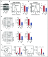
Intravascular ERp72 is required for hemostasis and platelet accumulation into a growing thrombus. (A-C) Characterization of Tie2-Cre/ERp72fl/fl mice. (A) Platelet mRNA expression was evaluated by RT-PCR to demonstrate the absence of ERp72 mRNA. The mRNA expressions of other PDIs serve as control. Western blots of platelet (B) and endothelial (C) lysates using a polyclonal rabbit anti–ERp72 antibody and antibodies against PDI, ERp57, ERp46, and ERp5. Shown are the PLCγ2 loading controls for ERp72. Separate loading controls were run for ERp57, ERp5, and ERp72 with similar amounts of protein found in each sample (not depicted). Blots are representative of 3 separate experiments. (D) Tail bleeding times; ***P < .001, Student t test. (E and F) Incorporation of platelets into growing thrombus in ERp72fl/fl mice and Tie2-Cre/ERp72fl/fl mice was detected by Alexa 488 anti-CD41 using FeCl3-induced mesenteric arterial injury. Mean artery diameters were 125.2 ± 2.6 μm in ERp72fl/fl mice and 121.3 ± 2.1 μm in Tie2-Cre/ERp72fl/fl mice (P = not significant). (E) Images at 7, 9, and 12 minutes. Dotted lines mark the vessel wall. Scale bar, 200 μm. Images are original magnification ×100. (F) Composite of fluorescence intensity (FI) per area analyzed (FI/μm2) in ERp72fl/fl (n = 20 from 8 mice) and Tie2-Cre/ERp72fl/fl (n = 17 from 8 mice) mice; mean ± standard error of the mean (SEM), *P < .05, ***P < .001, Student t test.
Figure 2.
Figure 2.
ERp72 is critical for platelet aggregation and interacts with β3 integrins. (A) Western blots of platelet lysates using a polyclonal rabbit anti–ERp72 antibody and antibodies against ERp57 and PDI. Shown are the PLCγ2 loading controls for ERp72. Blots are representative of 3 separate experiments, with separate loading controls run for ERp57 and PDI (not depicted). (B) ERp72-deficient platelets have defective thrombin (0.025 U/mL)–induced activation of αIIbβ3 (detected by the JON/A activation-dependent antibody). (C) P-selectin expression is decreased in thrombin-stimulated ERp72-null platelets. (B and C) Representative histogram (left) and combined results (right); mean ± SEM, n = 6 for each group, ***P < .001, Student t test. (D and E) Representative aggregation and ATP release tracings (left) and combined results (right) showing the defect in ERp72-deficient platelets using convulxin (15 ng/mL) or thrombin (0.025 U/mL); mean ± SEM, n = 3, *P < .05, **P < .01, Student t test. Aggregation and ATP secretion were monitored in the lumiaggregometer. (F and G) ERp72 interacts with β3 integrins on mouse platelets. Binding of Alexa Fluor 488–conjugated ERp72 to nonactivated (NA) and thrombin-activated (F) or Mn2+-treated (G) wild-type and β3-null mouse platelets. Cumulative data for ERp72 binding to thrombin-activated platelets (right panel of F) and to Mn2+-treated platelets (right panel of G); mean ± SEM, n = 3 for each group, ***P < .001, analysis of variance. Washed mouse platelets (3 × 10⁸/mL) were preincubated with Alexa Fluor 488 ERp72 (30 μg/mL) for 10 minutes at room temperature and then activated by thrombin (0.1 U/mL) (F) or treated with Mn2+ (12 mM) (G) for 5 minutes at room temperature. Surface binding of Alexa Fluor 488 ERp72 was detected by flow cytometry. ATP, adenosine triphosphate.
Figure 3.
Figure 3.
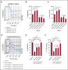
The a and a′ active sites of ERp72 are critical for platelet aggregation and ATP release. (A-C) Effect of preincubating human platelets (2 × 10⁸ platelets/mL) with single active site mutants of ERp72, ERp72(oo-ss-ss), ERp72(ss-oo-ss), and ERp72(ss-ss-oo). Submaximal aggregation (baseline) was stimulated with collagen (1 µg/mL). The concentrations for each type of protein are indicated. Representative aggregation tracings with ATP release for the 3 single active site mutants (A) and corresponding cumulative data for aggregation (B) and secretion (C) with wild-type ERp72(ss-ss-ss) and the double active site mutant ERp72(ss-oo-oo) results added to the cumulative data; mean ± SEM, n = 3, *P < .05, analysis of variance. (D-F) Correction of the aggregation and secretion defects of ERp72-null platelets (2 × 10⁸ platelets/mL) from Pf4-Cre/ERp72fl/fl mice compared with wild type (ERp72fl/fl) littermate mice by ERp72 mutants. The mutant proteins were added 5 minutes prior to the addition of thrombin (0.015 U/mL). Representative tracings (D) and cumulative data for aggregation (E) and secretion (F); mean ± SEM, n = 3, *P < .05, **P < .01, ***P < .001, analysis of variance. ERp72fl/fl, PDIfl/fl, and ERp57fl/fl represent the Cre-negative littermate controls. NS, not significant.
Figure 4.
Figure 4.
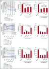
Preferential recovery of aggregation of ERp72-, PDI-, and ERp57-null platelets by the specific PDI that is deleted. (A) ERp72-null platelets were incubated with 150 nM PDI or ERp57 (as in Figure 3D). (B) PDI-null platelets were incubated with 100 nM ERp57, ERp72, or PDI. (C) ERp57-null platelets were incubated with 100 nM ERp57, ERp72, or PDI. (A-C) Platelet aggregation was stimulated using thrombin (0.015 U/mL) (right); cumulative data, mean ± SEM, n = 3, *P < .05, **P < .01, ***P < .001, analysis of variance. ERp72fl/fl, PDIfl/fl, and ERp57fl/fl represent the Cre-negative littermate controls. NS, not significant.
Figure 5.
Figure 5.
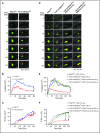
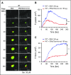
Intravascular ERp72 is required for platelet accumulation and fibrin formation in vivo. (A-C) Cremaster arteriole injury was induced in Pf4-Cre/ERp72fl/fl mice and their Cre-negative ERp72fl/fl littermate control mice. Platelets and fibrin accumulated at the site of injury were detected using anti-CD41 F(ab)2 fragments conjugated to Alexa Fluor 488 and anti–fibrin antibody conjugated to Alexa Fluor 647. (A) Representative fluorescence images in intravital microscopy for platelet accumulation (green) and fibrin deposition (red) at the indicated time points after injury . Original magnification ×64. Scale bar, 30 μm. The median integrated fluorescence intensities (FIs) of anti-CD41 (platelet, B) and anti–fibrin (fibrin, C) antibodies over 300 seconds. (D-F) Cremaster arteriole injury was induced in Tie2-Cre/ERp72fl/fl mice and Cre-negative ERp72fl/fl littermate control mice. Tie2-Cre/ERp72fl/fl mice were infused with ERp72(ss-ss-ss) or ERp72(oo-ss-ss) (150 μg/mouse). (D) Representative fluorescence images of platelet accumulation (green) and fibrin deposition (red). Original magnification ×64. Scale bar, 30 μm. The data of fluorescence intensities of anti–CD41 antibody (platelet, E) and anti–fibrin antibody (fibrin, F) were obtained from 30 thrombi in 3 mice. Fluorescence signal was not observed using fluorescently labeled control immunoglobulin G (not depicted). The area under the curve of fluorescence intensity over 300 seconds was analyzed using a Kruskal-Wallis test. Only significant differences are shown; *P < .05; ***P < .001. The data were obtained from 30 thrombi in 3 mice for each experimental condition.
Figure 6.
Figure 6.
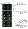
Recombinant ERp72(ss-oo-oo) protein inhibits platelet accumulation and fibrin deposition in wild-type mice. Wild-type (WT) C57BL/6 mice were infused with ERp72 (ss-oo-oo) or bovine serum albumin (BSA; control) (150 μg per mouse), followed by cremaster arteriole injury. Platelets and fibrin were detected using anti-CD41 F(ab)2 conjugated to Alexa Fluor 488 and anti–fibrin antibody conjugated to Alexa Fluor 647. (A) Representative fluorescence images of intravital microscopy for platelet accumulation (green) and fibrin deposition (red). Original magnification ×64. Scale bars, 30 μm. Median fluorescent intensities (FIs) of anti–CD41 antibodies (platelets) (B) and anti–fibrin antibody (C) with the area under curve were analyzed using a Mann-Whitney rank-sum test; **P < .01; ***P < .001. The data were obtained from 30 thrombi in 3 mice for each experimental condition.
Figure 7.
Figure 7.
Recombinant ERp72(oo-ss-ss) protein directly enhances fibrin deposition. (A) Cremaster arteriole injury was induced in wild-type mice after they received intravenous injection of anti–fibrin antibodies conjugated to Alexa Fluor 647, with anti-CD41 F(ab)2 conjugated to Alexa Fluor 488 or 3′-dihexyloxacarbocyanine iodide (DIOC6) (2.5 μL of a 100 μM solution per gram of body weight). Representative images for platelet accumulation visualized by these 2 methods (green) and fibrin formation (red). The total original magnification is ×64. Scale bar, 30 μm. (B) Wild-type mice and β3−/− mice received intravenous infusion of ERp72(oo-ss-ss) or BSA (control) (150 μg per mouse) as indicated, followed by cremaster arteriole injury. Platelets and fibrin formation were detected using DIOC6 and anti–fibrin antibody conjugated to Alexa Fluor 647. Median fluorescence intensities (FIs) of platelets (B) and fibrin (C) with the area under curve analyzed using a Kruskal-Wallis test. Only significant differences are shown; ***P < .001. The data were obtained from 40 thrombi in 5 mice for each experimental condition.

Similar articles

Cited by

References

    1. Hatahet F, Ruddock LW. Protein disulfide isomerase: a critical evaluation of its function in disulfide bond formation. Antioxid Redox Signal. 2009;11(11):2807-2850. - PubMed
    1. Holbrook LM, Watkins NA, Simmonds AD, Jones CI, Ouwehand WH, Gibbins JM. Platelets release novel thiol isomerase enzymes which are recruited to the cell surface following activation. Br J Haematol. 2010;148(4):627-637. - PubMed
    1. Wang L, Wu Y, Zhou J, et al. . Platelet-derived ERp57 mediates platelet incorporation into a growing thrombus by regulation of the αIIbβ3 integrin. Blood. 2013;122(22):3642-3650. - PMC - PubMed
    1. Kim K, Hahm E, Li J, et al. . Platelet protein disulfide isomerase is required for thrombus formation but not for hemostasis in mice. Blood. 2013;122(6):1052-1061. - PMC - PubMed
    1. Zhou J, Wu Y, Wang L, et al. . The disulfide isomerase ERp57 is required for fibrin deposition in vivo. J Thromb Haemost. 2014;12(11):1890-1897. - PMC - PubMed

Publication types

Substances