. 2017 May 30;8(3):e00713-17.
doi: 10.1128/mBio.00713-17.

Viral DNA Replication Orientation and hnRNPs Regulate Transcription of the Human Papillomavirus 18 Late Promoter

Affiliations

Viral DNA Replication Orientation and hnRNPs Regulate Transcription of the Human Papillomavirus 18 Late Promoter

Xiaohong Wang et al. mBio. .

Abstract

The life cycle of human papillomaviruses (HPVs) is tightly linked to keratinocyte differentiation. Although expression of viral early genes is initiated immediately upon virus infection of undifferentiated basal cells, viral DNA amplification and late gene expression occur only in the mid to upper strata of the keratinocytes undergoing terminal differentiation. In this report, we show that the relative activity of HPV18 TATA-less late promoter P811 depends on its orientation relative to that of the origin (Ori) of viral DNA replication and is sensitive to the eukaryotic DNA polymerase inhibitor aphidicolin. Additionally, transfected 70-nucleotide (nt)-long single-strand DNA oligonucleotides that are homologous to the region near Ori induce late promoter activity. We also found that promoter activation in raft cultures leads to production of the late promoter-associated, sense-strand transcription initiation RNAs (tiRNAs) and splice-site small RNAs (spliRNAs). Finally, a cis-acting AAGTATGCA core element that functions as a repressor to the promoter was identified. This element interacts with hnRNP D0B and hnRNP A/B factors. Point mutations in the core prevented binding of hnRNPs and increased the promoter activity. Confirming this result, knocking down the expression of both hnRNPs in keratinocytes led to increased promoter activity. Taking the data together, our study revealed the mechanism of how the HPV18 late promoter is regulated by DNA replication and host factors.IMPORTANCE It has been known for decades that the activity of viral late promoters is associated with viral DNA replication among almost all DNA viruses. However, the mechanism of how DNA replication activates the viral late promoter and what components of the replication machinery are involved remain largely unknown. In this study, we characterized the P811 promoter region of HPV18 and demonstrated that its activation depends on the orientation of DNA replication. Using single-stranded oligonucleotides targeting the replication fork on either leading or lagging strands, we showed that viral lagging-strand replication activates the promoter. We also identified a transcriptional repressor element located upstream of the promoter transcription start site which interacts with cellular proteins hnRNP D0B and hnRNP A/B and modulates the late promoter activity. This is the first report on how DNA replication activates a viral late promoter.

Keywords: DNA replication; HPV18; gene expression; human papillomaviruses; promoters; small RNAs; transcription; transcriptional regulation.


Figures

FIG 1
FIG 1
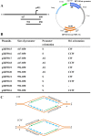
Construction of HPV18 late promoter- and replication origin-containing plasmids. (A) A line diagram of the HPV18 genome with the relative position of the P811 late promoter TSS (arrow) indicated. Below the genome are two versions (a longer version from nt 417 to nt 850 and a shorter version from nt 592 to nt 850) of the putative late-promoter region running over the TSS inserted at Asp718 and XhoI sites upstream of a firefly luciferase gene in either a sense (S) or an antisense (AS) orientation (see the map on the right). The minimal HPV18 replication origin (HPV18 Ori) from nt 7805 to nt 72 (124 bp) (44, 45) in the circular HPV18 genome was inserted in either a clockwise (CW) or a counterclockwise (CCW) orientation (relative to the plasmid map) into AgeI and NsiI sites downstream of the Luc ORF (see the map). The plasmid illustration is not drawn to scale. (B) Plasmid names and features are listed. (C) Diagrams of the replication forks derived from each replication Ori in the indicated orientation. Solid arrows, leading strand; dashed arrows, lagging strand; ATTAATACTTTTAA, a representative sequence from the HPV18 Ori; x and y, two DNA strands of the Ori in CW or CCW orientation relative to the reporter.
FIG 2
FIG 2
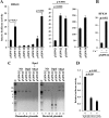
Characterization of the late-promoter region of HPV18. (A) The strong activity of a P811 promoter region is dependent on CCW orientation of the HPV18 Ori. HPV18-infected human foreskin keratinocytes (HFK18), differentiated by adding 2 mM calcium, were transfected for 48 h with each of the indicated plasmids. (A) Renilla luciferase plasmid pRL-TS was cotransfected as an internal control. The supernatant of the cell lysates from each transfection was examined for dual luciferase activities. Relative promoter activity levels were calculated by dividing the value representing the light unit readings from a testing promoter-firefly luciferase reporter by the value representing the light unit readings from the Renilla luciferase reporter (left panel). Plasmid pXHW61 directly derived from pXHW21 has the HPV18 Ori flipped into a CCW orientation (middle panel). Plasmid pXHW21-derived pXHW49 and pXHW22-derived pXHW50 have their corresponding promoter regions replaced by the SV40 early promoter derived from the pGL3 control vector (right panel). The data shown are means ± standard deviations (SD) of results from two to three independent experiments. P values were calculated using Student’s t test. (B) The P811 promoter activity depends on keratinocyte differentiation. HFK18 cells with (+) or without (−) 2.0 mM calcium treatment were transfected with pXHW22 for 48 h and then analyzed for their luciferase activity. The data shown are from results from one of two experiments, with means ± SD calculated from triplicate samples. (C) Opposite orientations of the HPV18 Ori do not affect plasmid DNA replication in HFK18 cells. HPV18-infected HFK cells, differentiated by 2 mM calcium, were transfected with pXHW21 (Ori in CW orientation) or pXHW22 (Ori in CCW orientation) for 48 h. Replicated plasmid DNA isolated from the cells and the original input bacterial plasmid DNA were compared for their sensitivity to DpnI (digesting only methylated bacterial plasmid DNA) and MboI (digesting only unmethylated bacterial and replicated plasmid DNA). Because the input bacterial DNA is methylated at the adenine of GATC sequences, it is sensitive to digestion by DpnI but resistant to digestion by MboI (right panel), and because human cells lack adenine methylase activity, the newly replicated DNA lacking adenine methylation is thus resistant to DpnI digestion but susceptible to MboI digestion (left panel). ND, no digestion with a restriction enzyme. The digested DNA samples were then resolved in a 1% agarose gel and imaged by ethidium bromide staining. Lanes 1 and 8 represent DNA markers (M). (D) Aphidicolin, a DNA polymerase inhibitor, blocks CCW orientation-dependent HPV18 late promoter activity. The sensitivity of Ori-directed DNA replication and HPV18 late promoter activity in plasmid pXHW22 to aphidicolin at different doses was analyzed in the HFK18 cells cotransfected with plasmids pXHW22 and pRL-SV40 (an internal control). The supernatant of the cell lysates was examined for dual luciferase activities, and the relative promoter activity levels were calculated as described for panel A.
FIG 3
FIG 3
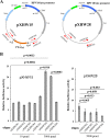
Strand-biased effect on HPV18 Ori-directed DNA replication and HPV18 late promoter activity revealed by a single-stranded, 70-nt-long DNA oligonucleotide. (A) The maps of plasmids pXHW15 (left panel) and pXHW28 (right panel) and the relative positions and orientations (arrow directions) of paired oXHW391/oXHW392 and oXHW393/oXHW394 oligonucleotides. (B) Effects of individual oligonucleotides (oligos) on promoter activity in pXHW15- and pXHW28-transfected HFK18 cells. HFK18 cells were cotransfected with pXHW15 or pXHW28 at the indicated doses of individual oligonucleotides, along with plasmid pRL-TS, and were cultured in a complete culture medium supplemented with 2.0 mM calcium. Dual luciferase activities were analyzed and calculated at 48 h after transfection.
FIG 4
FIG 4
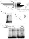
Identification of a transcriptional repressor element that affects P811 promoter activity and binds cellular proteins. (A) Schematic diagrams and their promoter activities for individual plasmids derived from pXHW16 with indicated deletions of the promoter region (nt 417 to nt 850 in the HPV18 genome) inserted upstream of a firefly luciferase (Luc) gene. The numbers above the lines represent nucleotide positions of the first and last nucleotides of the insertion in the HPV18 genome. The HPV18 late TSS at nt position 811 is indicated by an arrow. HFK18 cells in the presence of 2 mM calcium were cotransfected for 48 h with the individual plasmids along with Renilla luciferase plasmid pRL-TS. The supernatant of the cell lysates was examined for dual luciferase activities, and the relative promoter activity levels were calculated as described for Fig. 2A. (B) Diagrams of the nucleotide positions of four synthetic, double-stranded DNA oligonucleotide probes (a to d) used for electrophoretic mobility shift assays (EMSA). (C) Probe c, derived from nt 573 to nt 598 in the HPV18 genome, interacts with a cellular protein(s) from HFK18 cells. Probes were labeled with 32P, incubated with nuclear extract (NE) from HeLa or HFK18 cells, and then examined by EMSA. A Sp1 consensus oligonucleotide probe was used as a positive control. Protein-DNA complexes were resolved on a 4% native polyacrylamide gel. (D) Verification of the cellular proteins interacting with the repressor element by competitive gel shift assays. 32P-labeled probe c and a 32P-labeled Sp1 or TFIID (IID) oligonucleotide were incubated with NE prepared from HeLa or HFK18 cells in the presence or absence of an indicated cold competitor probe c, Sp1, TBP, or IID consensus oligonucleotide. Protein-DNA complexes were resolved on a 4% native polyacrylamide gel.
FIG 5
FIG 5
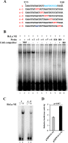
Mapping of a protein-binding core motif from the HPV18 late-promoter repressor element by 3-bp linker-scanning mutational analysis in EMSA. (A) Sequence of probe c and its substitutions with a 3-bp GTT linker. (B) Identification of an AAGTATGCA motif in the repressor element as a protein-binding core. Indicated probes were 32P-labeled double-stranded DNA oligonucleotide probe c and its derived variants in panel A used for EMSA with HeLa NE in the presence or absence of the indicated cold competitors. TBP and TFIID (IID) were included as controls. The protein-DNA complex was resolved on a 4% native polyacrylamide gel. (C) Protein binding profile of wild-type probe c and its c-4 mutant in EMSA. (D) Replacement of the probe c-corresponding sequence in plasmid pXHW16 with a c-4 sequence promotes HPV18 late promoter activity in HFK18 cells.
FIG 6
FIG 6
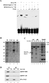
Identification of the cellular protein(s) bound to the repressor core element and its relation to HPV18 late promoter activity. (A) Expression of the repressor element-binding protein(s) in HFK18 cells depends on cell differentiation. 32P-labeled, double-stranded DNA probe c and its c-4 mutant probe used for EMSA were incubated with NE of HFK18 cells cultured under low- or high-calcium conditions. HeLa NE served as a control. (B) The repressor element-binding protein(s) (~45 kDa) is detectable by Southwestern blotting. Biotin-labeled dsDNA oligonucleotide probe c (wt) and its c-4 mutant (mt) were used for protein pulldown assays. The proteins pulled down from HeLa NE were separated by SDS-PAGE, transferred onto a nitrocellulose membrane, renatured, and probed by a 32P-labeled double-stranded DNA oligonucleotide probe (c). Ctrl, control. (C) Isolation of the repressor-binding protein(s) for LC-MS/MS analysis. Biotinylated probe c (wt) or its c-4 mutant (mt) was immobilized on magnetic streptavidin beads and incubated with HeLa NE. After extensively washing, bound proteins on the beads were eluted with a buffer containing 0.5 M or 1.0 M NaCl. Eluted fractions and the eluted beads were analyzed by SDS-PAGE and visualized by Coomassie blue staining. The specific repressor element-binding proteins (indicated with arrows) corresponding to probe c were excised for LC-MS/MS analysis. M (lane 5), Bio-Rad broad-range protein markers (7, 14, 21, 31, 45, 66, 97, 116, and 200 kDa). (D) Verification of repressor element-binding proteins hnRNP D0B and hnRNP A/B from the bound proteins on the beads by Western blotting using corresponding antibodies. Pol, polymerase.
FIG 7
FIG 7
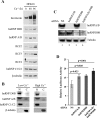
Expression and function of hnRNP D0B and hnRNP A/B in HFK18 keratinocytes. (A) Expression of hnRNP D0B, hnRNP A/B, RFC2, RFC3, and RFC4 in HFK18 cells under conditions of calcium-mediated differentiation. HFK18 cells were incubated with calcium-free EpiLife medium (no FBS) supplemented with 1× HKGS for 24 h, followed by induction of cell differentiation for 48 or 96 h with 3 mM Ca2+ EpiLife medium (5% FBS, 1× HKGS, 3 mM CaCl2) before total protein sample preparation was performed for Western blotting of individual proteins with corresponding antibodies. Involucrin served as a keratinocyte differentiation marker and β-tubulin and hnRNP C1/C2 as loading controls. (B) Nuclear and cytoplasmic distribution of hnRNP D0B and hnRNP A/B by cell fractionation of HFK18 cells under low- or high-calcium conditions. See details in the panel A legend. (C and D) Knockdown of hnRNP D0B and hnRNP A/B expression in HFK18 cells promotes HPV18 late promoter activity. The cells with efficient knockdown of the corresponding protein (C) were cotransfected by plasmid pXHW16 and pRL-TS and examined for HPV18 late promoter activity in the presence of 3 mM calcium (D). Nonspecific (NS) siRNA served as a control. hnRNP A/B and hnRNP D0B siRNA knockdown efficiency in HFK18 cells (C) was examined by Western blotting.

Similar articles

Cited by

References

    1. Walboomers JM, Jacobs MV, Manos MM, Bosch FX, Kummer JA, Shah KV, Snijders PJ, Peto J, Meijer CJ, Muñoz N. 1999. Human papillomavirus is a necessary cause of invasive cervical cancer worldwide. J Pathol 189:12–19. doi:10.1002/(SICI)1096-9896(199909)189:1<12::AID-PATH431>3.0.CO;2-F. - DOI - PubMed
    1. Muñoz N, Bosch FX, de Sanjosé S, Herrero R, Castellsagué X, Shah KV, Snijders PJ, Meijer CJ, International Agency for Research on Cancer Multicenter Cervical Cancer Study Group . 2003. Epidemiologic classification of human papillomavirus types associated with cervical cancer. N Engl J Med 348:518–527. doi:10.1056/NEJMoa021641. - DOI - PubMed
    1. Muñoz N, Castellsagué X, de González AB, Gissmann L. 2006. Chapter 1: HPV in the etiology of human cancer. Vaccine 24(Suppl 3):S1–S10. doi:10.1016/j.vaccine.2006.05.115. - DOI - PubMed
    1. Zheng ZM. 2014. Human papillomaviruses, p 87–112. In Yarchoan R. (), Cancers in people with HIV and AIDS. Springer, New York, NY.
    1. Barksdale SK, Baker CC. 1993. Differentiation-specific expression from the bovine papillomavirus type 1 P2443 and late promoters. J Virol 67:5605–5616. - PMC - PubMed

Publication types

MeSH terms

LinkOut - more resources