The Role and Mechanism of Erythrocyte Invasion by Francisella tularensis
- PMID: 28536678
- PMCID: PMC5423315
- DOI: 10.3389/fcimb.2017.00173
The Role and Mechanism of Erythrocyte Invasion by Francisella tularensis
Abstract
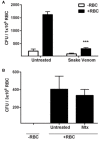
Francisella tularensis is an extremely virulent bacterium that can be transmitted naturally by blood sucking arthropods. During mammalian infection, F. tularensis infects numerous types of host cells, including erythrocytes. As erythrocytes do not undergo phagocytosis or endocytosis, it remains unknown how F. tularensis invades these cells. Furthermore, the consequence of inhabiting the intracellular space of red blood cells (RBCs) has not been determined. Here, we provide evidence indicating that residing within an erythrocyte enhances the ability of F. tularensis to colonize ticks following a blood meal. Erythrocyte residence protected F. tularensis from a low pH environment similar to that of gut cells of a feeding tick. Mechanistic studies revealed that the F. tularensis type VI secretion system (T6SS) was required for erythrocyte invasion as mutation of mglA (a transcriptional regulator of T6SS genes), dotU, or iglC (two genes encoding T6SS machinery) severely diminished bacterial entry into RBCs. Invasion was also inhibited upon treatment of erythrocytes with venom from the Blue-bellied black snake (Pseudechis guttatus), which aggregates spectrin in the cytoskeleton, but not inhibitors of actin polymerization and depolymerization. These data suggest that erythrocyte invasion by F. tularensis is dependent on spectrin utilization which is likely mediated by effectors delivered through the T6SS. Our results begin to elucidate the mechanism of a unique biological process facilitated by F. tularensis to invade erythrocytes, allowing for enhanced colonization of ticks.
Keywords: erythrocyte invasion; spectrin; tick borne disease; tularemia; type VI secretion system.
Figures


Similar articles
-
PdpC, a secreted effector protein of the type six secretion system, is required for erythrocyte invasion by Francisella tularensis LVS.Front Cell Infect Microbiol. 2022 Sep 27;12:979693. doi: 10.3389/fcimb.2022.979693. eCollection 2022. Front Cell Infect Microbiol. 2022. PMID: 36237421 Free PMC article.
-
OpiA, a Type Six Secretion System Substrate, Localizes to the Cell Pole and Plays a Role in Bacterial Growth and Viability in Francisella tularensis LVS.J Bacteriol. 2020 Jun 25;202(14):e00048-20. doi: 10.1128/JB.00048-20. Print 2020 Jun 25. J Bacteriol. 2020. PMID: 32366588 Free PMC article.
-
Invasion of erythrocytes by Francisella tularensis.J Infect Dis. 2011 Jul 1;204(1):51-9. doi: 10.1093/infdis/jir221. J Infect Dis. 2011. PMID: 21628658 Free PMC article.
-
Ticks and Tularemia: Do We Know What We Don't Know?Front Cell Infect Microbiol. 2019 May 8;9:146. doi: 10.3389/fcimb.2019.00146. eCollection 2019. Front Cell Infect Microbiol. 2019. PMID: 31139576 Free PMC article. Review.
-
The Francisella Type VI Secretion System.Front Cell Infect Microbiol. 2018 Apr 23;8:121. doi: 10.3389/fcimb.2018.00121. eCollection 2018. Front Cell Infect Microbiol. 2018. PMID: 29740542 Free PMC article. Review.
Cited by
-
The orange spotted cockroach (Blaptica dubia, Serville 1839) is a permissive experimental host for Francisella tularensis.Proc W Va Acad Sci. 2017;89(3):34-47. Epub 2017 Dec 4. Proc W Va Acad Sci. 2017. PMID: 29578544 Free PMC article.
-
Phenotypic and transcriptional characterization of F. tularensis LVS during transition into a viable but non-culturable state.Front Microbiol. 2024 Feb 6;15:1347488. doi: 10.3389/fmicb.2024.1347488. eCollection 2024. Front Microbiol. 2024. PMID: 38380104 Free PMC article.
-
A Francisella tularensis Chitinase Contributes to Bacterial Persistence and Replication in Two Major U.S. Tick Vectors.Pathogens. 2020 Dec 10;9(12):1037. doi: 10.3390/pathogens9121037. Pathogens. 2020. PMID: 33321814 Free PMC article.
-
Type VI Secretion System and Its Effectors PdpC, PdpD, and OpiA Contribute to Francisella Virulence in Galleria mellonella Larvae.Infect Immun. 2021 Jun 16;89(7):e0057920. doi: 10.1128/IAI.00579-20. Epub 2021 Jun 16. Infect Immun. 2021. PMID: 33875476 Free PMC article.
-
PdpC, a secreted effector protein of the type six secretion system, is required for erythrocyte invasion by Francisella tularensis LVS.Front Cell Infect Microbiol. 2022 Sep 27;12:979693. doi: 10.3389/fcimb.2022.979693. eCollection 2022. Front Cell Infect Microbiol. 2022. PMID: 36237421 Free PMC article.
References
-
- Boyum A. (1968). Separation of leukocytes from blood and bone marrow. Introduction. Scand. J. Clin. Lab. Invest. Suppl. 97:7. - PubMed
MeSH terms
Substances
Grants and funding
LinkOut - more resources
Full Text Sources
Other Literature Sources
Molecular Biology Databases
Research Materials

