. 2017 May 23;7(1):2255.
doi: 10.1038/s41598-017-01828-8.

IL-27R signaling controls myeloid cells accumulation and antigen-presentation in atherosclerosis

Affiliations

IL-27R signaling controls myeloid cells accumulation and antigen-presentation in atherosclerosis

Iuliia O Peshkova et al. Sci Rep. .

Abstract

Myeloid cells, key players in atherosclerosis, take up and present antigens, leading to systemic and local T cell activation. The recruitment and activation of immune cells to the aorta in atherosclerosis is regulated by adhesion molecules, chemokines and cytokines. IL-27R is an immunoregulatory signaling nod in autoimmune and infectious pathologies. IL-27R was shown to suppress T cells activation in atherosclerosis, however it's possible role in myeloid cell accumulation and activation is not understood. Here we demonstrate that Apoe -/- Il27ra -/- mice fed with "Western Diet" for 7 or 18 weeks developed significantly more atherosclerosis compared to Apoe -/- Il27ra +/- controls. Accelerated disease was driven by enhanced expression of adhesion molecules and chemokines causing the accumulation of immune cells. Myeloid cells produced more inflammatory cytokines and upregulated MHCII. Multiphoton microscopy revealed more efficient interactions between aortic myeloid cells and CD4+ T cells. Overall, we show that IL-27R signaling controls endothelial cells activation and myeloid cell recruitment at early and advanced stages of atherosclerosis. In the absence of IL-27R myeloid cells become hyperactivated, produce pro-inflammatory cytokines and act as more potent antigen presenting cells. Enhanced interactions between Il27ra -/- APC and CD4+ T cells in the aortic wall contribute to T cells re-activation and pro-atherogenic cytokine production.


Conflict of interest statement

The authors declare that they have no competing interests.

Figures
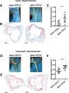
Figure 1
Figure 1
Accelerated atherosclerotic lesions development in Apoe / Il27ra /mice. Apoe / Il27ra +/ or Apoe / Il27ra / mice were fed with Western diet (WD) for 7 (A –C) or 18 weeks (D–F). Atherosclerotic lesions in aortic arch (A) and aortic root sections (B) of Apoe / Il27ra +/ and Apoe / Il27ra / mice fed with WD for 7 weeks. (C) Quantitative comparison of aortic lesion size in Apoe / Il27ra +/ (n = 8) and Apoe / Il27ra / (n = 8) mice feeding with WD for 7 weeks. Atherosclerotic lesions in aortic arch (D) and aortic root sections of (E) Apoe / Il27ra +/ and Apoe / Il27ra / mice fed with WD for 18 weeks. (F) Quantitative comparison of aortic lesion size in Apoe / Il27ra +/ (n = 10) and Apoe / Il27ra / (n = 9) mice feeding with WD for 18 weeks. Data are mean ± SEM from 4 independent experiments.
Figure 2
Figure 2
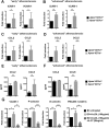
Upregulated expression of chemokines and adhesion molecules in aortas of Apoe / Il27ra /mice. (A,B) Relative gene expression of adhesion molecules ICAM-1 and VCAM-1 in aortas of Apoe / Il27ra / (n = 5) mice fed with WD for 7 weeks (A) or 18 weeks (B) were normalized to L-32 and fold induction was calculated based on the gene expression in aortas of control Apoe / Il27ra +/ (n = 5) mice. (C,D) Relative gene expression of CCL2 and CCL5 in aortas of Apoe / Il27ra / (n = 5) mice fed with WD for 7 weeks (C) or 18 weeks (D) were normalized to L-32 gene expression and fold induction was calculated based on the gene expression in aortas of control Apoe / Il27ra +/ (n = 5) mice. (E,F) CCL2 and CCL5 were measured by multiplex cytokines array in supernatants obtained from aortic cell suspensions of Apoe / Il27ra +/ (n = 5) or Apoe / Il27ra / (n = 5) mice fed with WD for 7 weeks (E) or 18 weeks (F) stimulated with anti-CD3/anti-CD28 for 48 hours. (G) Relative gene expression of vascular adhesion molecule VCAM-1, P-selectin, E-selectin and PECAM-1 in endothelial cells (mLEC cell line) treated with acLDL (100 μg/ml) and rIL-27 (25ng/ml) were normalized to L-32 and fold induction was calculated based on the gene expression in untreated endothelial cells. Data are mean ± SEM from at least 2 independent experiments.
Figure 3
Figure 3
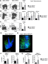
Enhanced accumulation of immune cells in aortas of Apoe / Il27ra / mice. Live CD45+ cells from aortas of Apoe / Il27ra +/ or Apoe / Il27ra / mice fed with WD for 7 weeks were stained for CD45+, CD11b+, CD11c+ and TCRβ+. Percentage (left) and cell number (right) (A–C) of live CD45+, CD11b+CD11c, CD11b+CD11c+ and CD11b- CD11c+ cells, TCRβ+ T cells in aortas of Apoe / Il27ra +/ or Apoe / Il27ra / mice fed with WD for 7 weeks was quantified by flow cytometry. Data are mean ± SEM from at least 3 independent experiments. Accumulation of CD11cYFP+ APC in aortas of Apoe / (D) and Apoe / Il27ra / (E) mice was analyzed by 2 photon microscopy. Green – CD11cYFP+ APC cells, blue – collagen detected by second harmonics generation. CD45.1+ monocytes from B6 mice were adoptively transferred to Apoe / Il27ra +/ (n = 5) or Apoe / Il27ra / (n = 6) mice fed with WD for 7 weeks. Monocyte recruitment to the aortas was assessed by flow cytometry 48 hours after cell transfer. Percentage and absolute number of recruited monocytes (F) and MHCII expression by recruited CD45.1 CD11b+ monocytes (G) in the aortic wall of Apoe / Il27ra / and Apoe / Il27ra +/ mice. Data are mean ± SEM from 2 independent experiments.
Figure 4
Figure 4
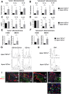
Enhanced activation of myeloid cells and T cells in Apoe / Il27ra / mice. (A,B) Relative gene expression of IL-6, IL-1α and IL-1β in aortas of Apoe / Il27ra / (n = 5) mice fed with WD for 7 weeks (A) or 18 weeks (B) were normalized to L-32 gene expression and then normalized to gene expression in aortas of control Apoe / Il27ra +/ (n = 5) mice. (C,D) IL-6, IL-1α and IL-1β were measured by bead array in supernatants of aortic cell suspension obtained from Apoe / Il27ra +/ (n = 5) or Apoe / Il27ra / (n = 5) mice fed with WD for 7 weeks (C) or 18 weeks (D), stimulated with anti-CD3/anti-CD28 for 48 hours. (E,F) Relative gene expression of F4/80 and MHCII in aortas of Apoe / Il27ra / (n = 5) mice fed with WD for 7 weeks (E) or 18 weeks (F) were normalized to L-32 gene expression and fold induction was calculated based on the gene expression in aortas of control Apoe / Il27ra +/ (n = 5) mice. (G) Expression of MHCII by CD11b+CD11c+ and CD11c+ cells in aorta of Apoe / Il27ra +/ or Apoe / Il27ra / mice fed with WD for 7 weeks. (H) Expression of CD69, a marker of T cell activation, by CD4+ T cells in aortas of Apoe / Il27ra +/ or Apoe / Il27ra / mice fed with WD for 18 weeks. Data are mean ± SEM from 3 independent experiments. (I) Localization of CD3+ T cells and MHCII+ cells in aortic roots of Apoe / Il27ra +/ or Apoe / Il27ra / mice fed with WD for 7 weeks (early lesions) or 18 weeks (advanced lesions) as demonstrated by confocal imaging. Arrows show co-localization of CD3+ T cells and MHCII+ cells.
Figure 5
Figure 5
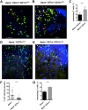
IL-27R deficiency promotes CD4+ T cells-APC interactions in the aortas. CD4+ T cells were labeled by administration of anti-CD4-PE antibody into live Apoe / and Apoe / Il27ra / mice after 5 weeks of WD feeding and localization of CD11cYFP+ APC and CD4-PE+ T cells were imaged by 2 photon and confocal microscopy in aortas of Apoe / Il27ra +/ CD11c YFP (A) and Apoe / Il27ra / CD11c YFP (B) mice. (C) Quantification of co-localizing cells in Apoe / Il27ra +/ CD11c YFP and Apoe / Il27ra / CD11c YFP aortas. (D,E) Two photon microscopy imaging revealed increased number of interacting cells in explanted aortas of Apoe / Il27ra / CD11c YFP mice (E) compared to control Apoe / CD11c YFP (D) fed with WD for 15 weeks. Isolated aortas were co-cultured with sorted from spleens CD4+ T cells obtained from atherosclerotic Apoe / Il27ra / mice. (F) Percent of CD4+ T cells interacting with CD11cYFP+ APC. (G) Reduced velocity of CD4+ T cells in Apoe / Il27ra / CD11c YFP aortas compared to Apoe / CD11c YFP aortas. Data are mean ± SEM from 4 independent experiments.
Figure 6
Figure 6
Increased expression of pro-inflammatory cytokines in aortas of Apoe / Il27ra / mice. (A,C) Relative gene expression of IFNγ, TNF-α and IL-17A in aortas of Apoe / Il27ra / (n = 5) mice fed with WD for 7 weeks (A) or 18 weeks (C) were normalized to L-32 gene expression and fold induction was calculated based on the gene expression in aortas of control Apoe / Il27ra +/ (n = 5). (B,D) Production of pro-inflammatory cytokines IFNγ, TNF-α and IL-17A in aortas of Apoe / Il27ra +/ or Apoe / Il27ra / mice at different stages of atherosclerosis was measured by multiplex cytokines array in supernatants of aortic cell suspension obtained from Apoe / Il27ra +/ (n = 5) or Apoe / Il27ra / (n = 5) mice fed with WD for 7 (B) or 18 weeks (D), stimulated with anti-CD3/anti-CD28 for 48 hours. Data are mean ± SEM from 3 independent experiments.

Similar articles

Cited by

References

    1. Robbins CS, et al. Local proliferation dominates lesional macrophage accumulation in atherosclerosis. Nat Med. 2013;19:1166–1172. doi: 10.1038/nm.3258. - DOI - PMC - PubMed
    1. Ensan S, et al. Self-renewing resident arterial macrophages arise from embryonic CX3CR1(+) precursors and circulating monocytes immediately after birth. Nat Immunol. 2016;17:159–168. doi: 10.1038/ni.3343. - DOI - PubMed
    1. Hansson GK, Hermansson A. The immune system in atherosclerosis. Nat Immunol. 2011;12:204–212. doi: 10.1038/ni.2001. - DOI - PubMed
    1. Galkina E, Ley K. Immune and inflammatory mechanisms of atherosclerosis (*) Annu Rev Immunol. 2009;27:165–197. doi: 10.1146/annurev.immunol.021908.132620. - DOI - PMC - PubMed
    1. Mallat Z, Taleb S, Ait-Oufella H, Tedgui A. The role of adaptive T cell immunity in atherosclerosis. J Lipid Res. 2009;50(Suppl):S364–369. - PMC - PubMed

Publication types

MeSH terms