

The catalytic function of cytochrome P450 is entwined with its membrane-bound nature
- PMID: 28529725
- PMCID: PMC5428493
- DOI: 10.12688/f1000research.11015.1
The catalytic function of cytochrome P450 is entwined with its membrane-bound nature
Abstract
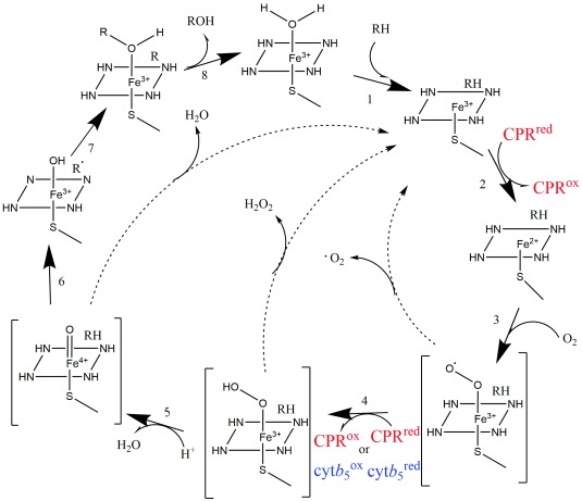
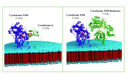
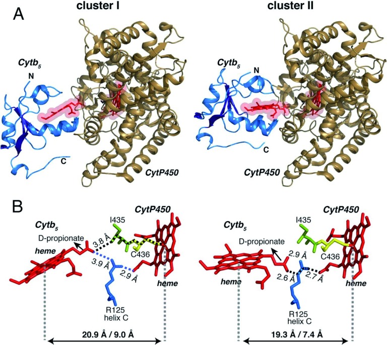
Cytochrome P450, a family of monooxygenase enzymes, is organized as a catalytic metabolon, which requires enzymatic partners as well as environmental factors that tune its complex dynamic. P450 and its reducing counterparts-cytochrome P450-reductase and cytochrome b 5 -are membrane-bound proteins located in the cytosolic side of the endoplasmic reticulum. They are believed to dynamically associate to form functional complexes. Increasing experimental evidence signifies the role(s) played by both protein-protein and protein-lipid interactions in P450 catalytic function and efficiency. However, the biophysical challenges posed by their membrane-bound nature have severely limited high-resolution understanding of the molecular interfaces of these interactions. In this article, we provide an overview of the current knowledge on cytochrome P450, highlighting the environmental factors that are entwined with its metabolic function. Recent advances in structural biophysics are also discussed, setting up the bases for a new paradigm in the study of this important class of membrane-bound enzymes.
Keywords: NADPH-P450 reductase; cytochrome P450; cytochrome b5; membrane; sold-state NMR; structure.
Conflict of interest statement
Competing interests: The authors declare that they have no competing interests.No competing interests were disclosed.No competing interests were disclosed.No competing interests were disclosed.No competing interests were disclosed.No competing interests were disclosed.
Figures
Similar articles
-
Picturing the Membrane-assisted Choreography of Cytochrome P450 with Lipid Nanodiscs.Chemphyschem. 2018 Oct 19;19(20):2603-2613. doi: 10.1002/cphc.201800444. Epub 2018 Jul 31. Chemphyschem. 2018. PMID: 29995333 Review.
-
P450BM-3; a tale of two domains--or is it three?Steroids. 1997 Jan;62(1):117-23. doi: 10.1016/s0039-128x(96)00169-9. Steroids. 1997. PMID: 9029725 Review.
-
Organization of multiple cytochrome P450s with NADPH-cytochrome P450 reductase in membranes.Pharmacol Ther. 2003 May;98(2):221-33. doi: 10.1016/s0163-7258(03)00031-7. Pharmacol Ther. 2003. PMID: 12725870 Review.
-
Quantification of interactions between cytochrome P450 2B4 and cytochrome b5 in a functional membrane complex.Neuro Endocrinol Lett. 2014;35 Suppl 2:114-22. Neuro Endocrinol Lett. 2014. PMID: 25638375
-
Dissociation Constants of Cytochrome P450 2C9/Cytochrome P450 Reductase Complexes in a Lipid Bilayer Membrane Depend on NADPH: A Single-Protein Tracking Study.J Am Chem Soc. 2017 Dec 13;139(49):17923-17934. doi: 10.1021/jacs.7b08750. Epub 2017 Dec 4. J Am Chem Soc. 2017. PMID: 29148818
Cited by
-
Layer-by-Layer Assembly of Nanosized Membrane Fractions for the Assessment of Cytochrome P450 Xenobiotic Metabolism.ACS Omega. 2018 Oct 3;3(10):12535-12544. doi: 10.1021/acsomega.8b01738. eCollection 2018 Oct 31. ACS Omega. 2018. PMID: 31457987 Free PMC article.
-
Structural Insights Uncover the Specific Phosphoinositide Recognition by the PH1 Domain of Arap3.Int J Mol Sci. 2023 Jan 6;24(2):1125. doi: 10.3390/ijms24021125. Int J Mol Sci. 2023. PMID: 36674645 Free PMC article.
-
Ligand Access Channels in Cytochrome P450 Enzymes: A Review.Int J Mol Sci. 2018 May 30;19(6):1617. doi: 10.3390/ijms19061617. Int J Mol Sci. 2018. PMID: 29848998 Free PMC article. Review.
-
NADPH diaphorase detects S-nitrosylated proteins in aldehyde-treated biological tissues.Sci Rep. 2020 Dec 3;10(1):21088. doi: 10.1038/s41598-020-78107-6. Sci Rep. 2020. PMID: 33273578 Free PMC article.
-
Interaction Modes of Microsomal Cytochrome P450s with Its Reductase and the Role of Substrate Binding.Int J Mol Sci. 2020 Sep 11;21(18):6669. doi: 10.3390/ijms21186669. Int J Mol Sci. 2020. PMID: 32933097 Free PMC article.
References
-
- De Montellano PR: Cytochrome P450: structure, mechanism, and biochemistry.Springer,2005. 10.1007/b139087 - DOI
-
- Bernhardt R: Mammalian and Bacterial Cytochromes P450 Involved in Steroid Hydroxylation: Regulation of Catalysis and Selectivity, and Potential Applications. Fifty Years of Cytochrome P450 Research. 2014;135–151. 10.1007/978-4-431-54992-5_8 - DOI
Publication types
Grants and funding
LinkOut - more resources
Full Text Sources
Other Literature Sources