. 2017 May 16:5:42.
doi: 10.1186/s40425-017-0246-1. eCollection 2017.

A tandem CD19/CD20 CAR lentiviral vector drives on-target and off-target antigen modulation in leukemia cell lines

Affiliations

A tandem CD19/CD20 CAR lentiviral vector drives on-target and off-target antigen modulation in leukemia cell lines

Dina Schneider et al. J Immunother Cancer. .

Abstract

Background: Clinical success with chimeric antigen receptor (CAR)- based immunotherapy for leukemia has been accompanied by the associated finding that antigen-escape variants of the disease are responsible for relapse. To target hematologic malignancies with a chimeric antigen receptor (CAR) that targets two antigens with a single vector, and thus potentially lessen the chance of leukemic escape mutations, a tandem-CAR approach was investigated.

Methods: Antigen binding domains from the FMC63 (anti-CD19) and Leu16 (anti-CD20) antibodies were linked in differing configurations to transmembrane and T cell signaling domains to create tandem-CARs. Expression on the surface of primary human T cells was induced by transduction with a single lentiviral vector (LV) encoding the tandem-CAR. Tandem-CARs were compared to single antigen targeting CARs in vitro and in vivo, and to an admixture of transduced cells expressing each CAR in vivo in immunodeficient (NSG) disease-bearing mice.

Results: Tandem constructs efficient killed the Raji leukemia cell line both in vitro and in vivo. Tandem CARs generated less cytokine than the CD20 CAR, but similar to CD19 CARs, on their own. In co-culture experiments at low effector to target ratios with both single- and tandem- CAR-T cells, a rapid down-modulation of full-length CD19 expression was seen on leukemia targets. There also was a partial down-modulation of CD22, and to a lesser degree, of CD20. Our data also highlight the extreme sensitivity of the NALM-6 cell line to general lymphocyte-mediated cytotoxicity. While single and tandem constructs were effective in vivo in a standard setting, in a high-disease burden setting, the tandem CAR proved both effective and less toxic than an admixture of transduced T cell populations expressing single CARs.

Conclusion: Tandem CARs are equally effective in standard disease models to single antigen specificity CARs, and may be both more effective and less toxic in a higher disease burden setting. This may be due to optimized cell killing with more moderate cytokine production. The rapid co-modulation of CD19, CD20, and CD22 may account for the ability to rapidly evolve escape mutants by selecting for leukemic clones that not require these target antigens for continued expansion.

Keywords: Adoptive immunotherapy; CAR T; CD19; CD20; CD22; Hematologic malignancy; Lentiviral vector; Tandem -targeting CAR; Tumor antigen escape.


Figures
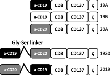
Fig. 1
Fig. 1
Construction of CARs targeting CD19 and CD20. Anti-CD19 and anti-CD20 single targeting CAR constructs were generated by linking single chain fragment variable region of monoclonal antibody FMC-63 (CD19) of Leu-16 (CD20) in frame to CD8 hinge and transmembrane domain, the 4-1BB (CD137) signaling domain and the CD3 zeta signaling domain. Constructs 19A and 19B differ only in the linker sequence connecting the heavy and the light chains of FMC63. Tandem targeting constructs 2019 and 1920 were generated in a similar manner to single targeting constructs, except that the single chain fragment variable regions of CD20 and CD19 were linked to each other sequentially by a flexible linker, followed by CD8, 4-1BB and CD3 zeta domains
Fig. 2
Fig. 2
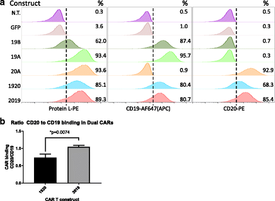
Surface expression of single and tandem-CAR T constructs on human primary T cells. a CAR T expression was determined by flow cytometry. T cells were activated with Transact CD3 CD28 reagent in the presence of IL-2, and transduced with LV as described in Methods. On culture day 10, viable transduced T cells (7-AAD negative) were assayed for CAR surface expression using one of three staining reagents: Protein L (column 1), CD19 Fc followed by anti-Fc-AF647 (column 2), or CD20-biotin followed by streptavidin-PE staining (column 3). The LV used in transduction is listed to the left of each row. Percentage of CAR T-positive populations in relation to non-transduced T cell control is noted in the right-hand corner of each histogram. GFP-transduced cells served as an additional negative control. Representative data of three separate donors is shown. b The ratio of CD19 and CD20 antigen binding by each tandem-CAR is expressed as the ratio of percent cells bound by CD20 biotin vs CD19 Fc. The average + SD of three separate experiments using three donors is shown, **p < 0.01
Fig. 3
Fig. 3
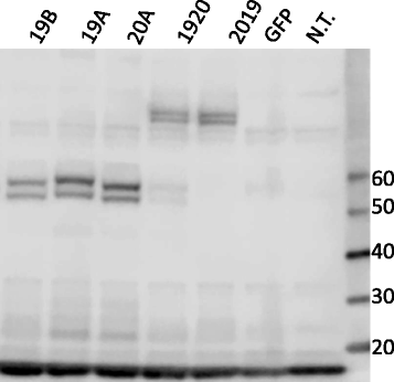
Western blot of CAR T proteins in primary human T cells. Cell lysates prepared from human primary T cells transduced with single (19B, 19A, 20A) or tandem-CAR19 and CAR20 (1920, 2019) constructs were resolved on a gradient (4–12%) SDS-PAGE gel and probed with an antibody against CD3-zeta. T cells from the same donor transduced with a lentiviral vector encoding green fluorescent protein (GFP), or non-transduced T cells (N.T.) served as controls. Molecular weight markers, in KDa, are listed to the right
Fig. 4
Fig. 4
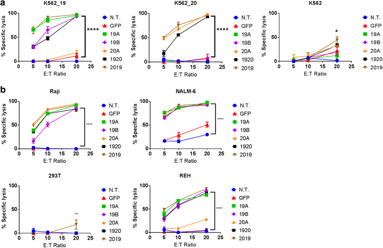
CAR T cytotoxicity in vitro. Luciferase-based cytotoxicity assays were performed using, a) K562, K562 CD19+, or K562 CD20+ cell lines, or b) leukemia or lymphoma cell lines (Raji, NALM6, REH), stably transduced with luciferase. CAR T cells and target tumor cells were co-incubated overnight at the listed effector to target (E:T) ratios, x-axis. Differences between groups were determined using 1-way ANOVA followed by Dunnett’s post-hoc test. Mean + SD, ****p < 0.0001, **p < 0.01 vs non-transduced control from the same donor (N.T.)
Fig. 5
Fig. 5
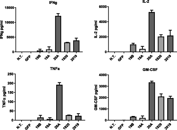
CAR T cytokine release in response to leukemia cell lines. Cytokine production by CAR-T, listed on the x-axis, upon overnight co-culture with the Raji leukemia line at an E:T ratio of 10:1, was measured using a flow-based bead array. Bars represent mean + SD of replicate samples. Data are representative of three independent experiments performed with CAR T cells from three separate donors
Fig. 6
Fig. 6
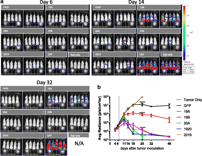
In vivo activity of CAR T constructs. NSG mice were injected i.v. with Raji-luciferase cells on Day 0, and treated with CAR T cells on day 7. a Bioluminescent images of the tumor burden in mice treated with singe and tandem-CAR T constructs on day 6, 14, or 32, post tumor engraftment are shown. Above each group is a listed the CAR-T used for treatment. b Time course of tumor growth based on mouse whole body bioluminescence. 10 mice per CAR T treatment group, and five mice per control group were studied. Mean signal per mouse ± SD is plotted. Statistical analysis for Day 25 (the last time point when subjects in the no treatment control group remained alive) is shown, using two-way ANOVA followed by Dunnett’s multiple comparisons test vs no treatment group. Mean + SD, ***P < 0.001
Fig. 7
Fig. 7
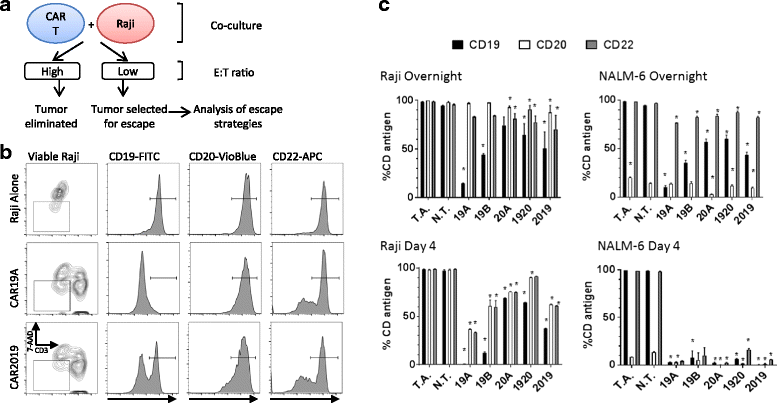
Single CAR19 construct strongly selects Raji tumor escape variants. a Diagram of the experimental design for tumor escape experiments. Raji and CAR T cells were co-cultured at E:T ratio of 1:1 either overnight or for 4 days. After overnight incubation and on day 4, cultures were harvested and viable Raji cells examined for CD19, CD20, and CD22 surface expression by flow cytometry. b Gating strategy for flow cytometric analysis used to analyze viable Raji cells (7AAD- and CD3-) from co-cultures is shown for representative treatment groups is shown in column 1. Columns 2, 3, and 4 show CD19, CD20, and CD22 expression levels, respectively, when Raji cells were co-cultured with no T cells (row 1), 19A CAR (row 2), or 2019 CAR (row 3). c Graphs of CD19, CD20 and CD22 surface expression (solid, open, gray, respectively) in surviving Raji and NALM-6 cells after overnight or 4 days of co-culture with CAR T cells, as listed on x-axis, as determined by flow cytometry. Bars represent group means + SD. Statistical analysis was performed by one-way ANOVA followed by Dunnett’s multiple comparisons test vs N.T. (non-transduced T cells) control from the same donor, *p < 0.05. T.A.- tumor alone control group
Fig. 8
Fig. 8
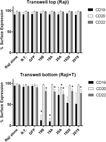
The down-modulation of CD19, CD20 and CD22 on Raji surface requires direct contact with CART cells. Multi-well plates with transwell inserts were used in this experiment. At the bottom of each well, 5 × 105 each of Raji and CAR T cells were combined, and in the transwell upper portion 2.5 × 105 Raji cells were cultured in the absence of T cells. After overnight incubation, cells from the upper transwell compartment and from the bottom compartment were harvested, and viable Raji cells were examined for CD19, CD20, and CD22 surface expression by flow cytometry (black, light grey, and dark grey bars, respectively. Surface expression for each marker with reference to the specific CAR T included in the lower compartment (x- axis) is shown. Bars depict mean + SD of three independent experiments performed using CAR T cells originating from three different donors. One way ANOVA, Dunnett’s multiple comparisons test *p < 0.05
Fig. 9
Fig. 9
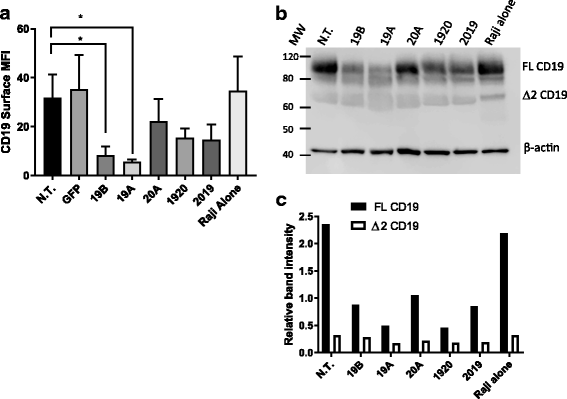
Down-modulation of CD19 full-length protein and CD19 splice variant by CAR19 constructs. Raji cells were co-incubated with CAR T cells at a 1:1 E:T ratio. After overnight incubation, T cells were removed from co-incubated cell populations using magnetic beads. CD19 expression on purified Raji populations was investigated by flow cytometry and Western blot. a Raji cell samples were stained with anti-CD19 antibody and acquired by flow cytometry. Median fluorescence intensity for Raji cells representing each treatment group is shown. Bars depict mean + SD of three independent experiments performed using CAR T cells originating from three different donors. One way ANOVA, Dunnett’s multiple comparisons test *p < 0.05. b Lysates of purified Raji cells from each of the co-incubated groups (CAR-T identity is listed above each line) were resolved on a 4–12% SDS polyacrylamide gel as described in the Methods, and probed with antibodies targeting the C-terminus of CD19 molecule, or β-actin (loading control). c The intensity of specific immunoreactive bands representing full-length CD19 protein (FL CD19), and the exon 2 spliced CD19 variant (Δ2CD19) was quantified using Image Studio software (LI-COR Biosciences). Relative band intensity of CD19 bands was calculated as signal CD19/signal β actin
Fig. 10
Fig. 10
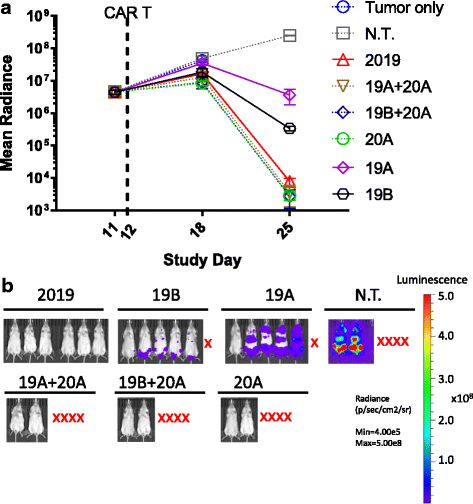
In vivo activity of CAR T cells in a high tumor burden model. NSG mice (n = 6) were injected i.v. with Raji-luciferase cells on Day 0, and treated with CAR T cells, as indicated in the figure, on day 12. a Disease burden is plotted as the average bioluminescent signal (mean radiance [p/s/cm2/sr]) ± SEM. Groups with less than half of the mice surviving to day 25 are plotted as dotted lines. Groups where more than half survived are plotted as solid lines. b Bioluminescent images of the tumor burden in mice treated with singe and tandem-CAR T constructs as indicated in the plot above on day 25 post tumor engraftment are shown. Red X indicates mice that did not survive to study day 25
Fig. 11
Fig. 11
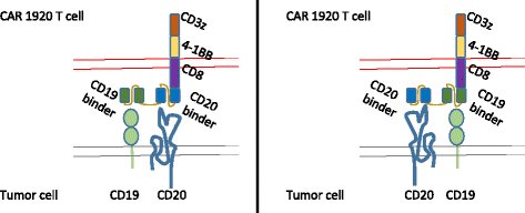
Schematic representation of Tandem-CAR T cells targeting CD19 and CD20 tumor antigens. The tandem-CAR 1920 (left) and 2019 (right) are comprised of tandem extracellular targeting domains linked in frame to CD8-derived hinge and transmembrane domains, followed by the 4-1BB costimulatory domain, and the CD3 zeta activation domain. Each CAR T construct is capable of activation via binding to either CD19 or CD20 tumor antigens, or both

Similar articles

Cited by

References

    1. Lee DW, et al. The future is now: chimeric antigen receptors as new targeted therapies for childhood cancer. Clin Cancer Res. 2012;18(10):2780–90. doi: 10.1158/1078-0432.CCR-11-1920. - DOI - PMC - PubMed
    1. Kochenderfer JN, et al. Construction and pre-clinical evaluation of an anti-CD19 chimeric antigen receptor. J Immunother. 2009;32(7):689. doi: 10.1097/CJI.0b013e3181ac6138. - DOI - PMC - PubMed
    1. Jensen M, et al. CD20 is a molecular target for scFvFc: zeta receptor redirected T cells: implications for cellular immunotherapy of CD20+ malignancy. Biol Blood Marrow Transplant. 1998;4(2):75–83. doi: 10.1053/bbmt.1998.v4.pm9763110. - DOI - PubMed
    1. Lee DW, et al. T cells expressing CD19 chimeric antigen receptors for acute lymphoblastic leukaemia in children and young adults: a phase 1 dose-escalation trial. Lancet. 2015;385(9967):517–28. doi: 10.1016/S0140-6736(14)61403-3. - DOI - PMC - PubMed
    1. Grupp SA, et al. Chimeric antigen receptor–modified T cells for acute lymphoid leukemia. N Engl J Med. 2013;368(16):1509–18. doi: 10.1056/NEJMoa1215134. - DOI - PMC - PubMed

Publication types

MeSH terms