. 2017 Jun 15;144(12):2294-2305.
doi: 10.1242/dev.146019. Epub 2017 May 15.

Fgf10 and Sox9 are essential for the establishment of distal progenitor cells during mouse salivary gland development

Affiliations

Fgf10 and Sox9 are essential for the establishment of distal progenitor cells during mouse salivary gland development

Lemonia Chatzeli et al. Development. .

Abstract

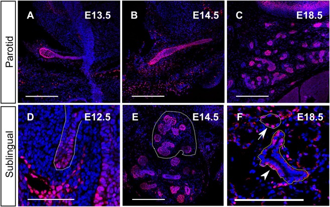
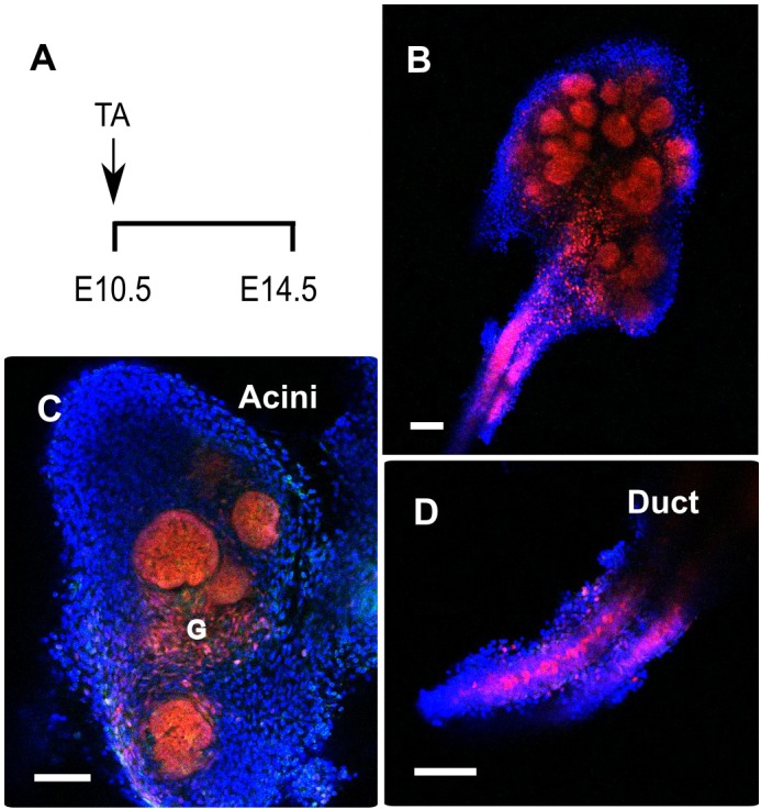
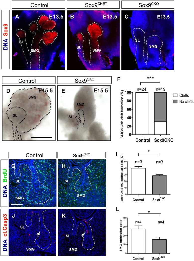
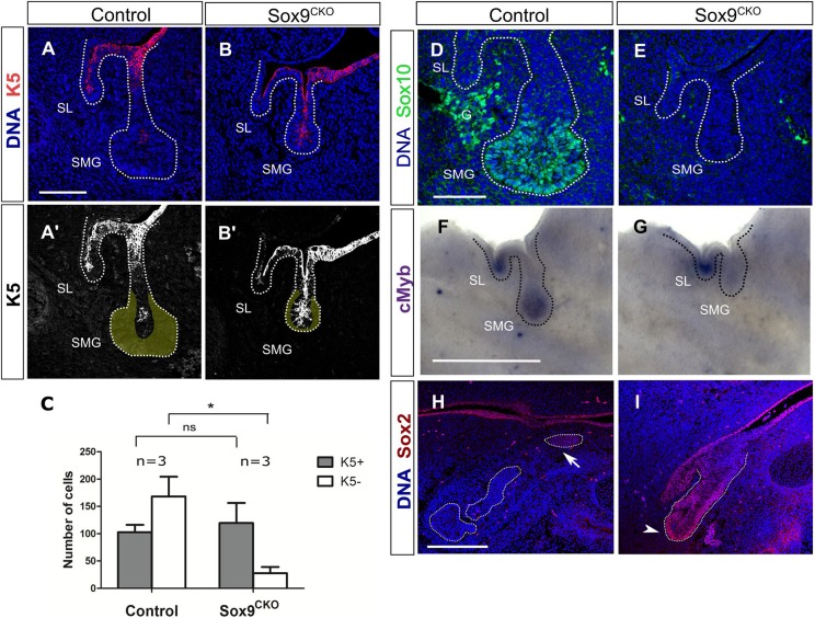
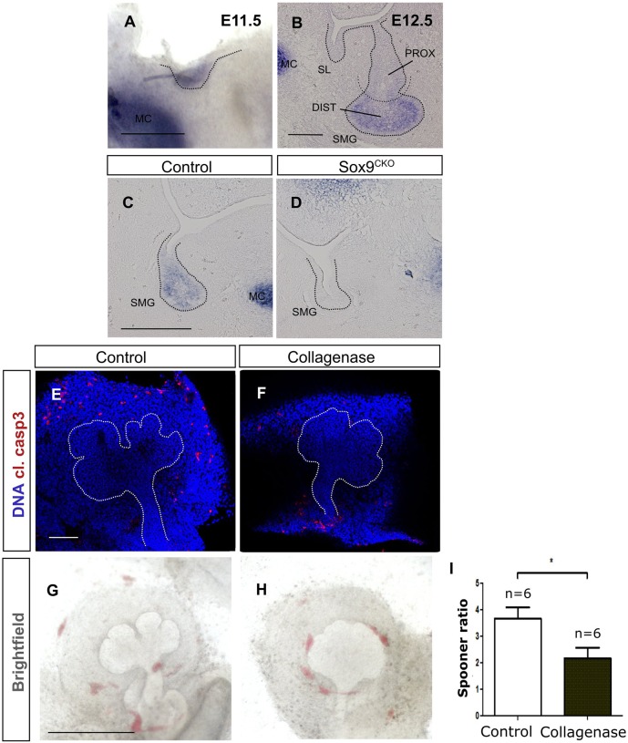
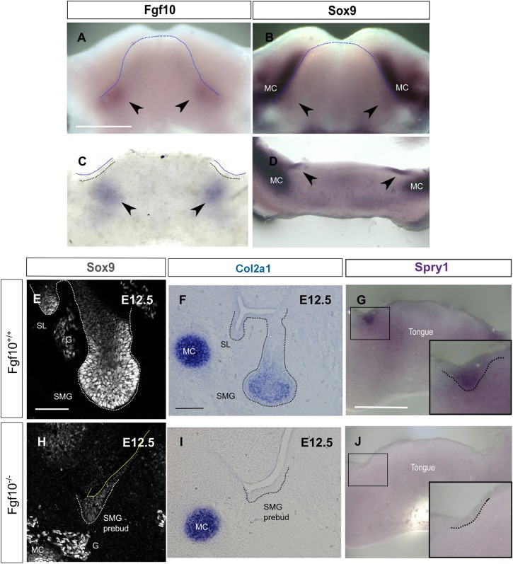
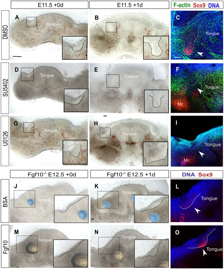
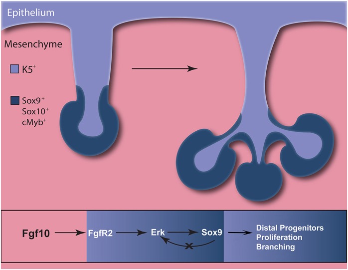
Salivary glands are formed by branching morphogenesis with epithelial progenitors forming a network of ducts and acini (secretory cells). During this process, epithelial progenitors specialise into distal (tips of the gland) and proximal (the stalk region) identities that produce the acini and higher order ducts, respectively. Little is known about the factors that regulate progenitor expansion and specialisation in the different parts of the gland. Here, we show that Sox9 is involved in establishing the identity of the distal compartment before the initiation of branching morphogenesis. Sox9 is expressed throughout the gland at the initiation stage before becoming restricted to the distal epithelium from the bud stage and throughout branching morphogenesis. Deletion of Sox9 in the epithelium results in loss of the distal epithelial progenitors, a reduction in proliferation and a subsequent failure in branching. We demonstrate that Sox9 is positively regulated by mesenchymal Fgf10, a process that requires active Erk signalling. These results provide new insights into the factors required for the expansion of salivary gland epithelial progenitors, which can be useful for organ regeneration therapy.

Keywords: Branching morphogenesis; Epithelial progenitors; Fgf signalling; Salivary glands; Sox9.


Conflict of interest statement

Competing interestsThe authors declare no competing or financial interests.

Figures

Fig. 1.
Fig. 1.
Sox9 is expressed throughout the development of the submandibular gland. (A-F) Sox9 immunofluorescence (red) at the placode [E11.0 (A), E11.5 (B)], initial bud [E12.5 (C)], pseudoglandular [E13.5 (D)], canalicular [E15.5 (E)] and adult (F) stages. DNA is shown in blue (DAPI), F-actin in yellow and Mist1 in green. Dotted lines in A-D delineate the salivary gland epithelium. Insets in F show magnifications of an acinus stained for Mist1 (green) and Sox9 (red). Arrowheads point to Sox9-positive cells within the striated duct. G, ganglion; MC, Meckel's cartilage; SL, sublingual gland; SMG, submandibular gland. Scale bars: 100 μm.
Fig. 2.
Fig. 2.
Sox9 expression in the parotid and sublingual gland is similar to the submandibular. (A-F) Sox9 immunofluorescence (red) in the parotid (A-C) and sublingual (D-F) glands at the bud stage (A,D) and at E14.5 (B,E) and E18.5 (C,F). Dotted white lines in D and E outline the sublingual glands. The white dotted line in F outlines an acinus and the yellow a duct. Arrow points to Sox9+ cells in the acinus and the arrowhead points to Sox9+ cells in the duct. DNA is shown in blue (DAPI). Scale bars: 250 μm (A-C,E,F); 50 μm (D).
Fig. 3.
Fig. 3.
Sox9-positive cells are progenitors of the entire submandibular gland epithelium. (A) Experimental strategy used to follow the progeny of Sox9-expressing cells with the Sox9-creERT2; R26-tdTomato line. Tamoxifen (TA) was given at E10.5 and embryos were collected at E14.5. (B-D) Tomato-labelled cells (red) were detected in the whole submandibular gland at E14.5, (B) in the acini (C) and in the duct (D). G, ganglion. Scale bars: 100 μm.
Fig. 4.
Fig. 4.
Sox9 is required for branching morphogenesis. (A-C) Sox9 immunofluorescence (red) in control (A), Sox9CHET (B) and Sox9CKO (C) at the pseudoglandular stage (E13.5). (D,E) Submandibular and sublingual glands dissected from control (D) and Sox9CKO (E) mice at E15.5. (F) Quantification of cleft formation in the control and Sox9CKO submandibular glands at the pseudoglandular stage (E13.5). ‘n’ equals the number of submandibular glands. ***P<0.0001. (G,H) BrdU immunofluorescence (green) in control (G) and Sox9CKO (H) submandibular glands at the bud stage (E12.5). (I) Quantification of the percentage of epithelial BrdU+ cells in the control and Sox9CKO submandibular glands at the bud stage (E12.5). (J,K) Cleaved caspase 3 immunofluorescence (red) in control and Sox9CKO submandibular glands at the bud stage (E12.5). Arrowheads indicate apoptotic cells at the stalk region. (L) Quantification of epithelial cell number in control and Sox9CKO submandibular glands at the bud stage (E12.5). Dotted lines in A-E,G,H,J,K delineate the salivary gland epithelium. Error bars in I and L represent s.e.m.; *P<0.05. DNA is shown in blue (DAPI) for A-C and G,H,J,K. SL, sublingual gland; SMG, submandibular gland. Scale bars: 200 μm (A-C); 500 μm (D,E); 100 μm (G,H,J,K).
Fig. 5.
Fig. 5.
Sox9 is required for parotid gland development. (A-C) Sox9 immunofluorescence (red) in control (A), Sox9CHET (B) and Sox9CKO (C) parotid glands at E15.5. Arrowheads indicate the position of the parotid gland. DNA is shown in blue (DAPI). Scale bar: 250 μm (A-C).
Fig. 6.
Fig. 6.
Sox9 is required for the specification of distal epithelial progenitors. (A-B′) Immunofluorescence for cytokeratin 5 (K5) (red) in control (A,A′) and Sox9CKO (B,B′) submandibular glands at the bud stage (E12.5). Yellow area in A′,B′ represents the K5 distal epithelial cells. (C) Total number of K5+ and K5 epithelial cells in control and Sox9CKO submandibular glands at the bud stage (E12.5). *P<0.01; ns, not significant. Error bars represent s.e.m. (D,E) Immunofluorescence for Sox10 (green) in control (D) and Sox9CKO (E) submandibular glands at the bud stage (E12.5). (F,G) In situ hybridisation for Myb in control (F) and Sox9CKO (G) submandibular glands at the bud stage (E12.5). (H,I) Immunofluorescence for Sox2 in control (H) and Sox9CKO (I) submandibular glands at the pseudoglandular stage (E13.5). Dotted lines (A-B',D-I) delineate the salivary gland epithelium. Arrow and arrowhead indicate the proximal and distal progenitors, respectively. DNA is shown in blue (DAPI) in A,B,D,E,H,I. G, ganglion; SL, sublingual gland; SMG, submandibular gland. Scale bars: 100 μm (A-B',H,I); 50 μm (D,E); 500 μm (F,G).
Fig. 7.
Fig. 7.
Type II collagen (Col2a1) is expressed in the distal progenitors and acts downstream of Sox9 possibly by contributing to branching. (A,B) In situ hybridisation for Col2a1 at the placode (A) and bud stage (B). (C,D) In situ hybridisation for Col2a1 at the bud stage (E12.5) in control (C) and Sox9CKO (D) submandibular glands. (E,F) Immunofluorescence for cleaved caspase 3 (red) in control (E) and collagenase-treated (F) submandibular gland explants. DNA is shown in blue (DAPI). (G,H) Brightfield images of control (G) and collagenase-treated (H) submandibular gland explants. (I) Spooner ratio of the number of buds produced in the control and collagenase-treated submandibular gland explants. *P<0.05. Dotted lines (A-H) delineate the salivary gland epithelium. Error bars represent s.e.m. DIST, distal; MC, Meckel's cartilage; PROX, proximal; SL, sublingual gland; SMG, submandibular gland. Scale bars: 250 μm (A); 50 μm (B); 100 μm (C-F); 500 μm (G,H).
Fig. 8.
Fig. 8.
Fgf10 maintains Sox9 expression during the initial stages of salivary gland development. (A-D) In situ hybridisation for Fgf10 (A,C) and Sox9 (B,D) on E11.0 mandibles (A,B) and frontal mandibular slices (C,D). Arrowheads indicate the site of expression in submandibular glands. (E,H) Immunofluorescence for Sox9 in Fgf10+/+ and Fgf10−/− submandibular glands at E12.5. (F-J) In situ hybridisation for Col2a1 (F,I) and Spry1 (G,J) in Fgf10+/+ (F,G) and Fgf10−/− (I,J) submandibular glands. Dotted lines delineate the tongue (A,B), the placode of the salivary glands (C,G,J) or the salivary gland epithelium (E,F,H,I). Boxes (G,J) indicate the placode of the developing submandibular glands, as magnified in insets. G, ganglion; MC, Meckel's cartilage; SL, sublingual gland; SMG, submandibular gland. Scale bars: 500 μm (A-D,G,J); 50 μm (E,H); 100 μm (F,I).
Fig. 9.
Fig. 9.
Fgf receptor signalling maintains Sox9 expression through the Erk pathway. (A,B,D,E,G,H) Brightfield images of wild-type mandibular slice cultures treated with DMSO (A,B), the Fgf receptor inhibitor SU5402 (D,E) or the Erk inhibitor U0126 (G,H). (C,F,I) Immunofluorescence for Sox9 (red) and F-actin (green) in DMSO- (C), SU5402- (F) and U0126- (I) treated mandibular slice cultures. (J,K,M,N) Brightfield images of Fgf10−/− mandibular slice cultures treated with BSA-treated beads (blue) (J,K) or Fgf10-treated beads (pale yellow) (M,N). (L,O) Immunofluorescence for Sox9 in Fgf10−/− mandibles treated with BSA-treated beads (L) or Fgf10-treated beads (O). DNA is shown in blue (DAPI) in C,F,I,L,O. Boxes indicate the placode of the developing submandibular glands. Insets show higher magnifications of the boxed areas. Dotted lines outline the epithelium of the placodes. Arrowheads indicate the submandibular glands. Mc, Meckel's cartilage. Scale bars: 200 μm (C,F,I,L,O); 500 µm (A,B,D,E,G,H,J,K,M,N).
Fig. 10.
Fig. 10.
Model of Fgf10 and Sox9 function during salivary gland budding and branching morphogenesis. Sox9 is required for branching initiation by promoting the formation of distal epithelial progenitors and their proliferation. Mesenchymal Fgf10 maintains epithelial Sox9 expression during salivary gland development by activating the Erk pathway through Fgfr2.

Similar articles

Cited by

References

    1. Abler L. L., Mansour S. L. and Sun X. (2009). Conditional gene inactivation reveals roles for Fgf10 and Fgfr2 in establishing a normal pattern of epithelial branching in the mouse lung. Dev. Dyn. 238, 1999-2013. 10.1002/dvdy.22032 - DOI - PMC - PubMed
    1. Affolter M., Bellusci S., Itoh N., Shilo B., Thiery J.-P. and Werb Z. (2003). Tube or not tube: remodeling epithelial tissues by branching morphogenesis. Dev. Cell 4, 11-18. 10.1016/S1534-5807(02)00410-0 - DOI - PubMed
    1. Arnold K., Sarkar A., Yram M. A., Polo J. M., Bronson R., Sengupta S., Seandel M., Geijsen N. and Hochedlinger K. (2011). Sox2(+) adult stem and progenitor cells are important for tissue regeneration and survival of mice. Cell Stem Cell 9, 317-329. 10.1016/j.stem.2011.09.001 - DOI - PMC - PubMed
    1. Aure M. H., Konieczny S. F. and Ovitt C. E. (2015). Salivary gland homeostasis is maintained through acinar cell self-duplication. Dev. Cell 33, 231-237. 10.1016/j.devcel.2015.02.013 - DOI - PMC - PubMed
    1. Bell D. M., Leung K. K. H., Wheatley S. C., Ng L. J., Zhou S., Ling K. W., Sham M. H., Koopman P., Tam P. P. and Cheah K. S. (1997). SOX9 directly regulates the type-II collagen gene. Nat. Genet. 16, 174-178. 10.1038/ng0697-174 - DOI - PubMed

Publication types

MeSH terms