. 2017 May 18;66(4):473-487.e9.
doi: 10.1016/j.molcel.2017.04.009. Epub 2017 May 11.

Mechanisms of Ubiquitin-Nucleosome Recognition and Regulation of 53BP1 Chromatin Recruitment by RNF168/169 and RAD18

Affiliations

Mechanisms of Ubiquitin-Nucleosome Recognition and Regulation of 53BP1 Chromatin Recruitment by RNF168/169 and RAD18

Qi Hu et al. Mol Cell. .

Abstract

The protein 53BP1 plays a central regulatory role in DNA double-strand break repair. 53BP1 relocates to chromatin by recognizing RNF168-mediated mono-ubiquitylation of histone H2A Lys15 in the nucleosome core particle dimethylated at histone H4 Lys20 (NCP-ubme). 53BP1 relocation is terminated by ubiquitin ligases RNF169 and RAD18 via unknown mechanisms. Using nuclear magnetic resonance (NMR) spectroscopy and biochemistry, we show that RNF169 bridges ubiquitin and histone surfaces, stabilizing a pre-existing ubiquitin orientation in NCP-ubme to form a high-affinity complex. This conformational selection mechanism contrasts with the low-affinity binding mode of 53BP1, and it ensures 53BP1 displacement by RNF169 from NCP-ubme. We also show that RAD18 binds tightly to NCP-ubme through a ubiquitin-binding domain that contacts ubiquitin and nucleosome surfaces accessed by 53BP1. Our work uncovers diverse ubiquitin recognition mechanisms in the nucleosome, explaining how RNF168, RNF169, and RAD18 regulate 53BP1 chromatin recruitment and how specificity can be achieved in the recognition of a ubiquitin-modified substrate.

Keywords: 53BP1; DNA repair; NMR spectroscopy; RAD18; RNF168; RNF169; biophysics; nucleosome; structural biology; ubiquitylation.


Figures

Figure 1
Figure 1. Specific interaction of RNF169, RNF168 and RAD18 with the nucleosome core particle ubiquitylated at histone H2A lysines 13 and 15
(A) Amino acid sequences of RNF168, RNF169 and RAD18 ubiquitylated nucleosome binding motifs. A stretch of 4 residues conserved in RNF169 and RAD18 in underlined. (B) Ni2+-NTA pull-down of unmodified (WT), RNF168-ubiquitylated (H2AK13/K15) and RING1B-BMI1-ubiquitylated (H2AK119) NCPs by His6-tagged RNF168, RNF169 and RAD18, immunoblotted (IB) for histone H2A and ubiquitin (ub). (C) Ni2+-NTA pull-down of NCP and NCPH2AK15ub (reconstituted with ubiquitin-fused H2A) by His6-tagged RNF168, RNF169 and GB1-RAD18, analyzed by SDS-PAGE. (D) ITC results (top, raw titration data; bottom, integrated heat measurements) for RNF169 interactions with the NCP enzymatically ubiquitylated at Lys13, Lys15 or both. n is the stoichiometry of binding. Kds are reported with s.d. determined by nonlinear least-squares analysis. (E) ITC results for RNF168 interactions with the NCP enzymatically ubiquitylated at Lys13, Lys15 or both. (F) ITC results for RNF169 interactions with H2A-H2B enzymatically ubiquitylated at Lys13, Lys15 or both. (G) ITC results for RNF168 interactions with H2A-H2B enzymatically ubiquitylated at Lys13, Lys15 or both.
Figure 2
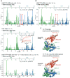
Figure 2. Methyl-TROSY spectra of H2AK15ub-H2B and NCPH2AK15ub
(A) Methyl-TROSY spectra of Ile, Val, Leu methyl-labeled H2A-H2B in the context of H2AK15ub-H2B, NCP and NCPH2AK15ub. From left to right: First panel corresponds to the enzymatically ubiquitylated long version of H2A-H2B used to reconstitute the NCP. Second panel corresponds to the enzymatically ubiquitylated short version of H2A-H2B used for NMR structure determination of H2AK15ub-H2B–RNF169. Third panel corresponds to the NCP reconstituted with the non-ubiquitylated long version of H2A-H2B. Fourth panel corresponds to ubiquitylated NCP reconstituted with the long version of H2AK15ub-H2B in the first panel. (B) Methyl-TROSY spectra of Ile, Val, Leu methyl-labeled ubiquitin in the context of H2AK15ub-H2B and NCPH2AK15ub described in A. In the right spectrum, the ubiquitin methyl signals that extensively broadened in the context of NCPH2AK15ub but not in H2AK15ub-H2B (left spectra) are highlighted with red circles. The corresponding residues (Val5, Val17 and Val26) are shown as gray spheres in the NCPH2AK15ub–RNF169 complex structure.
Figure 3
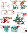
Figure 3. NMR characterization of RNF169 in complex with H2A-H2B and the nucleosome ubiquitylated at H2AK15
(A) Top: Regions of methyl-TROSY spectra of H2AK15ub-H2B selectively 1H-13C-labeled at methyl groups of Ile, Leu and Val residues of H2A-H2B or ubiquitin in an otherwise perdeuterated background, free (black) and bound to unlabeled RNF169 (red). Bottom: Regions of 1H-15N TROSY HSQC spectra of H2AK15ub-H2B prepared with 15N-labeled H2A-H2B and unlabeled ubiquitin, free (black) and bound to unlabeled RNF169 or RNF168 (red). Suffix A is for H2A and B for H2B. (B) Cartoon representation and NMR structure ensemble of H2AK15ub-H2B in complex with RNF169. (C) Cartoon representation of a region of the H2AK15ub-H2B–RNF169 complex highlighting the interaction of RNF169 α-helix and ubiquitin. Key residues are in stick representation. (D) Cartoon representation of a region of the H2AK15ub-H2B–RNF169 complex highlighting the interaction of RNF169 LRM with H2A-H2B. Key residues are in stick representation. H2A-H2B acidic patch area binding RNF169 Arg700 is circled in orange. (E) NMR/SAXS-based model of NCPH2AK15ub in complex with RNF169. Goodness of fit of the model to SAXS data recorded for the NCPH2AK15ub–RNF169 complex.
Figure 4
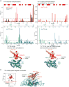
Figure 4. NMR of RNF169 and RNF168 interactions with H2AK15ub-H2B
(A) Top: Magnitude of 1H-15N chemical shift changes in H2A (blue) and H2B (green) in the context of H2AK15ub-H2B after adding 3-fold molar excess RNF169 (aa 653-708). The Kd determined using ITC is indicated. Isotope labeling schemes are specified in this and subsequent diagrams. Residues for which signals disappear due to exchange broadening are indicated with red elliptical disks on the x axis. The red rectangle highlights the main difference with the H2AK15ub-H2B–RNF168 interaction (see Figure 4B). Middle: Magnitude of 1H-15N chemical shift changes in H2A and H2B in the context of H2A-H2B after adding 10-fold molar excess RNF169 (aa 688-704). The Kd calculated from NMR chemical shift changes of selected H2A-H2B residues caused by interaction with RNF169C is indicated. Bottom: Magnitude of 1H-13C chemical shift changes in Ile, Val and Leu methyl groups of H2A and H2B in the context of H2AK15ub-H2B after adding 3-fold molar excess RNF169. (B) Magnitude of 1H-15N chemical shift changes in H2A (blue) and H2B (green) in the context of H2AK15ub-H2B after addition of 3-fold molar excess RNF168. The ITC-derived Kd is indicated. The red rectangle highlights the main difference with the H2AK15ub-H2B–RNF169 interaction (see Figure 4A). (C) Top: Cartoon representation of the H2AK15ub-H2B–RNF169 structure highlighting H2A and H2B residues (spheres) for which there are marked changes in 1H-15N chemical shifts (larger than the s.d. of the shift for all residues). Spheres are colored gray when interaction with RNF169 causes signal disappearance. Bottom: Like in Top but marked changes are in 1H-13C chemical shifts of Ile, Leu and Val methyl groups.
Figure 5
Figure 5. Ubiquitin adopts a preferred orientation relative to H2A-H2B
(A) Top: Magnitude of 1H-15N chemical shift changes in ubiquitin caused by ubiquitylation of H2A Lys15 in H2A-H2B (red) versus Lys164 in PCNA (black). Middle: Magnitude of 1H-15N chemical shift changes in H2A (blue) and H2B (green) caused by ubiquitylation of H2A Lys 15 in H2A-H2B. Bottom: Cartoon representation of the H2AK15ub-H2B–RNF169 structure highlighting residues (spheres) for which there are marked changes in 1H-15N chemical shifts (larger than the s.d. of the shift for all residues) in H2AK15ub-H2B caused by ubiquitylation of H2A Lys15. Note the shifts in helix α1 of H2B indicating a preferred orientation of ubiquitin in the absence of RNF169. (B) Top: Magnitude of 1H-13C chemical shift changes in Ile, Val and Leu methyl groups of ubiquitin caused by ubiquitylation of H2A Lys 15 in H2A-H2B. Middle: Magnitude of 1H-13C chemical shift changes in Ile, Val and Leu methyl groups of H2A (blue) and H2B (green) caused by ubiquitylation of H2A Lys 15 in H2A-H2B. Bottom: Cartoon representation of the H2AK15ub-H2B–RNF169 structure highlighting residues (spheres) for which there are marked changes in 1H-13C chemical shifts (larger than the s.d. of the shift for all residues) in H2AK15ub-H2B caused by ubiquitylation of H2A Lys15. Note the shifts in helix a1 of H2B indicating a preferred orientation of ubiquitin in the absence of RNF169. (C) Left: Comparison of 13C-methyl-lysine signals of ubiquitin in the free state (black) and in NCPH2AK15ub (red). Right: Cartoon representation of the NMR/SAXS-based model of NCPH2AK15ub–RNF169 in which ubiquitin Lys11 and Lys27 are highlighted in yellow and Lys6, Ly48 and Lys63 highlighted in gray. Note that there are two signals for Lys27.
Figure 6
Figure 6. Effects of RNF168, RNF169 and RAD18 on the association of 53BP1 with the nucleosome ubiquitylated at H2AK15 and dimethylated at H4K20
(A) Overlay of the cryo-EM structure of NCPH2AK15ubH4Kc20me2–53BP1 (aa 1611-1631) and NMR/SAXS-based model of NCPH2AK15ub–RNF169 (aa 653-708). (B) ITC results for the interactions of GST-53BP1 (aa 1484-1635) dimer, RNF168, RNF169 and RAD18 with NCPH2AK15ubH4Kc20me2. The GST-53BP1–NCPH2AK15ub interaction was also probed. n is the stoichiometry of binding. Kds are reported with s.d. determined by nonlinear least-squares analysis. (C) GST pull-down assays of NCPH2AK15ubH4Kc20me2 in the absence (lane 12) and presence of equimolar (lanes 1-3), 2-fold (lanes 4-6) and 4-fold (lanes 7-9) molar excess of RNF168, RNF169 and RAD18 with GST-53BP1 (aa 1484-1635), immunoblotted (IB) for GST, K15-ubiquitylated H2A (H2AK15ub) and ubiquitin (ub). Pull-downs of NCPH2AK15ubH4Kc20me2 with GST (lane 11) and GST-53BP1 (aa 1484-1635) T1609E/S1618E mutant (lane 13, red star) were done as negative controls. 53BP1 T1609E/S1618E is unable to bind NCPH2AK15ubH4Kc20me2 as reported (Lee et al., 2014; Orthwein et al., 2014). Input NCPH2AK15ubH4Kc20me2 is in lane 10.
Figure 7
Figure 7. Interaction of RAD18 with the nucleosome ubiquitylated at H2AK15
(A) ITC results for the interactions of RAD18 (aa 198-240; aa 198-227) with ubiquitin and with the NCP and H2A-H2B enzymatically ubiquitylated at H2AK15. n is the stoichiometry of binding. Kds are reported with s.d. determined by nonlinear least-squares analysis. (B) Regions of 1H-15N TROSY HSQC titration spectra of RAD18, H2A-H2B and H2AK15ub-H2B illustrating the interaction of RAD18 with H2A-H2B and ubiquitin in H2AK15ub-H2B. Different isotope labeling schemes (15N or 15N, 2H) were used as indicated. (C) Cartoon representation of the NMR-based model of NCPK15ub in complex with RAD18 (aa 198-240). The zinc atom is shown as a gray sphere. Side chains of RAD18 for which NMR signals were most affected (i.e. exchange broadened) by interaction with H2A-H2B are shown in yellow sticks. RAD18 R234 side chain interacting with H2A-H2B acidic patch is also shown.

Similar articles

Cited by

References

    1. Armache KJ, Garlick JD, Canzio D, Narlikar GJ, Kingston RE. Structural basis of silencing: Sir3 BAH domain in complex with a nucleosome at 3.0 Å resolution. Science. 2011;334:977–982. - PMC - PubMed
    1. Armstrong AA, Mohideen F, Lima CD. Recognition of SUMO-modified PCNA requires tandem receptor motifs in Srs2. Nature. 2012;483:59–63. - PMC - PubMed
    1. Barbera AJ, Chodaparambil JV, Kelley-Clarke B, Joukov V, Walter JC, Luger K, Kaye KM. The nucleosomal surface as a docking station for Kaposi's sarcoma herpesvirus LANA. Science. 2006;311:856–861. - PubMed
    1. Battiste JL, Wagner G. Utilization of site-directed spin labeling and high-resolution heteronuclear nuclear magnetic resonance for global fold determination of large proteins with limited nuclear overhauser effect data. Biochemistry. 2000;39:5355–5365. - PubMed
    1. Benirschke RC, Thompson JR, Nomine Y, Wasielewski E, Juranic N, Macura S, Hatakeyama S, Nakayama KI, Botuyan MV, Mer G. Molecular basis for the association of human E4B U box ubiquitin ligase with E2-conjugating enzymes UbcH5c and Ubc4. Structure. 2010;18:955–965. - PMC - PubMed

MeSH terms