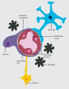
Development and Function of the Blood-Brain Barrier in the Context of Metabolic Control
- PMID: 28484368
- PMCID: PMC5399017
- DOI: 10.3389/fnins.2017.00224
Development and Function of the Blood-Brain Barrier in the Context of Metabolic Control
Abstract
Under physiological conditions, the brain consumes over 20% of the whole body energy supply. The blood-brain barrier (BBB) allows dynamic interactions between blood capillaries and the neuronal network in order to provide an adequate control of molecules that are transported in and out of the brain. Alterations in the BBB structure and function affecting brain accessibility to nutrients and exit of toxins are found in a number of diseases, which in turn may disturb brain function and nutrient signaling. In this review we explore the major advances obtained in the understanding of the BBB development and how its structure impacts on function. Furthermore, we focus on the particularities of the barrier permeability in the hypothalamus, its role in metabolic control and the potential impact of hypothalamic BBB abnormities in metabolic related diseases.
Keywords: blood-brain barrier; development; hypothalamus; inflammation; neurovascular unit; obesity.
Figures


Similar articles
-
Maternal obesity damages the median eminence blood-brain barrier structure and function in the progeny: the beneficial impact of cross-fostering by lean mothers.Am J Physiol Endocrinol Metab. 2023 Feb 1;324(2):E154-E166. doi: 10.1152/ajpendo.00268.2022. Epub 2023 Jan 4. Am J Physiol Endocrinol Metab. 2023. PMID: 36598900
-
Communication from the periphery to the hypothalamus through the blood-brain barrier: An in vitro platform.Int J Pharm. 2016 Feb 29;499(1-2):119-130. doi: 10.1016/j.ijpharm.2015.12.058. Epub 2015 Dec 28. Int J Pharm. 2016. PMID: 26732523
-
Neuronal and vascular interactions.Annu Rev Neurosci. 2015 Jul 8;38:25-46. doi: 10.1146/annurev-neuro-071714-033835. Epub 2015 Mar 12. Annu Rev Neurosci. 2015. PMID: 25782970 Free PMC article. Review.
-
Blood-brain Barrier and Neurovascular Unit Dysfunction in Parkinson's Disease: From Clinical Insights to Pathogenic Mechanisms and Novel Therapeutic Approaches.CNS Neurol Disord Drug Targets. 2024;23(3):315-330. doi: 10.2174/1871527322666230330093829. CNS Neurol Disord Drug Targets. 2024. PMID: 36999187 Review.
-
Structural pathways for macromolecular and cellular transport across the blood-brain barrier during inflammatory conditions. Review.Histol Histopathol. 2004 Apr;19(2):535-64. doi: 10.14670/HH-19.535. Histol Histopathol. 2004. PMID: 15024715 Review.
Cited by
-
The Effect of Short-Term Exposure to Cadmium on the Expression of Vascular Endothelial Barrier Antigen in the Developing Rat Forebrain and Cerebellum: A Computerized Quantitative Immunofluorescent Study.Cureus. 2022 Apr 5;14(4):e23848. doi: 10.7759/cureus.23848. eCollection 2022 Apr. Cureus. 2022. PMID: 35402117 Free PMC article.
-
A New Perspective on the Pathophysiology of Idiopathic Intracranial Hypertension: Role of the Glia-Neuro-Vascular Interface.Front Mol Neurosci. 2022 Jul 12;15:900057. doi: 10.3389/fnmol.2022.900057. eCollection 2022. Front Mol Neurosci. 2022. PMID: 35903170 Free PMC article.
-
Targeting fatty acid synthase in preclinical models of TNBC brain metastases synergizes with SN-38 and impairs invasion.NPJ Breast Cancer. 2024 Jun 10;10(1):43. doi: 10.1038/s41523-024-00656-0. NPJ Breast Cancer. 2024. PMID: 38858374 Free PMC article.
-
Hypothalamic Microglial Heterogeneity and Signature under High Fat Diet-Induced Inflammation.Int J Mol Sci. 2021 Feb 24;22(5):2256. doi: 10.3390/ijms22052256. Int J Mol Sci. 2021. PMID: 33668314 Free PMC article. Review.
-
Stress and the gut-brain axis: an inflammatory perspective.Front Mol Neurosci. 2024 Jul 18;17:1415567. doi: 10.3389/fnmol.2024.1415567. eCollection 2024. Front Mol Neurosci. 2024. PMID: 39092201 Free PMC article. Review.
References
Publication types
LinkOut - more resources
Full Text Sources
Other Literature Sources

