. 2017 May 5;12(5):e0177300.
doi: 10.1371/journal.pone.0177300. eCollection 2017.

Control of the induction of type I interferon by Peste des petits ruminants virus

Affiliations

Control of the induction of type I interferon by Peste des petits ruminants virus

Beatriz Sanz Bernardo et al. PLoS One. .

Abstract

Peste des petits ruminants virus (PPRV) is a morbillivirus that produces clinical disease in goats and sheep. We have studied the induction of interferon-β (IFN-β) following infection of cultured cells with wild-type and vaccine strains of PPRV, and the effects of such infection with PPRV on the induction of IFN-β through both MDA-5 and RIG-I mediated pathways. Using both reporter assays and direct measurement of IFN-β mRNA, we have found that PPRV infection induces IFN-β only weakly and transiently, and the virus can actively block the induction of IFN-β. We have also generated mutant PPRV that lack expression of either of the viral accessory proteins (V&C) to characterize the role of these proteins in IFN-β induction during virus infection. Both PPRV_ΔV and PPRV_ΔC were defective in growth in cell culture, although in different ways. While the PPRV V protein bound to MDA-5 and, to a lesser extent, RIG-I, and over-expression of the V protein inhibited both IFN-β induction pathways, PPRV lacking V protein expression can still block IFN-β induction. In contrast, PPRV C bound to neither MDA-5 nor RIG-I, but PPRV lacking C protein expression lost the ability to block both MDA-5 and RIG-I mediated activation of IFN-β. These results shed new light on the inhibition of the induction of IFN-β by PPRV.


Conflict of interest statement

Competing Interests: The authors have declared that no competing interests exist.

Figures

Fig 1
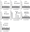
Fig 1. IFN-β induction during the infection with different strains of PPRV.
VHS cells (A-E) or G4 cells (F) were transfected with pIFN-β-luc (350 ng) and pJAT-lacZ (200 ng). At least 18 hours post transfection, cells were infected with (A-D) the indicated strain of PPRV at a MOI = 1 TCID50/cell or (E) with the Cantell strain of Sendai virus (50 HA units/well); in each case parallel samples were left uninfected. Duplicate samples were taken at each time point and cell extracts were prepared and assayed for luciferase and β-galactosidase activity as described in Methods. The luciferase reading was expressed relative to the β-galactosidase activity (Relative Light Units (RLU)). Induction of the IFN-β promoter was expressed as the ratio of RLUs in infected cells relative to that in uninfected cells (set as 1 for each time point). (G) G4 cells were infected with PPRV as in (F) and triplicate samples of cells harvested at the indicated times and RNA extracted. IFN-β mRNA was measured by RT-qPCR as described in Methods and normalized by setting the value for uninfected cells to 1. Error bars represent standard error of the mean (SEM). The ANOVA test and Tukey pairwise comparisons test were used to compare differences between the means (* = p < 0.05).
Fig 2
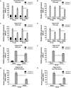
Fig 2. PPRV actively blocks MDA-5 and RIG-I mediated induction of the IFN-β promoter.
VHS cells (A-F) were transfected with pIFN-β-luc (350 ng) and pJAT-lacZ (200 ng). Additionally, for the poly(I:C)-mediated induction of IFN-β (A, C, E), 100 ng of MDA-5 plasmid was added to the transfection mix. Cells were infected with the indicated strain of PPRV at MOI = 3 for the indicated time, or left uninfected, before transfection of poly(I:C) (A, C, E, G) or infection with SeV-DI (B, D, F, H). Cell extracts were prepared from triplicate samples and were assayed for luciferase and β-galactosidase activity (A-F) or for IFN-β mRNA (G, H) as described in Methods. Results were normalised by setting the RLU, or the level of IFN-ß mRNA, in treated uninfected cells as 100. Error bars represent the SEM. The ANOVA test and Tukey pairwise comparisons test were used to compare differences between the means (* = p < 0.05).
Fig 3
Fig 3. The V protein of PPRV blocks the induction of IFN-β by intracellular poly(I:C).
(A) VHS cells were transfected with pIFN-β-luc (350 ng), pJAT-lacZ (200 ng), pEF-MDA-5 (100 ng) and an empty plasmid (500 ng) or expression plasmid encoding PIV5 V (500 ng), RPV V (300 ng) or PPRV V (500 ng). The total amount of DNA was kept constant in all samples by adding empty plasmid as required. At least twenty four hours post transfection, cells were transfected with 2 μg of poly(I:C) or left untreated; 7 hours later the cells were lysed and the cells extracts were assayed for luciferase and β-galactosidase activities. Samples were normalised by setting RLUs in poly(I:C) transfected cells without V protein to 100. Error bars represent the standard error of the mean. ANOVA and Tukey pairwise comparison test were used to determine the statistical significance of differences in means (* = p < 0.05). (B) Cell extracts from parallel transfected wells were run on 10% SDS-PAGE gels and Western blotted with anti-V5 or anti-PCNA (loading control).
Fig 4
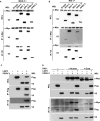
Fig 4. The V protein of PPRV binds to MDA-5, RIG-I and LGP2.
HEK-293FT cells were transfected with plasmids encoding (A) c-Myc-MDA-5 (1400 ng) and either an empty plasmid (1400 ng) or a plasmid encoding one of the viral accessory proteins PIV5 V (1400 ng), RPV V (100 ng), RPV C (400 ng), PPRV V (300 ng) or PPRV C (100 ng); (B) c-Myc-RIG-I (1400 ng) and the same set of empty and expression plasmids as in (A); (C) an expression plasmid encoding Flag-goat LGP2 (500 ng) and an empty plasmid (300 ng) or an expression plasmid encoding the PPRV V protein (300 ng); (D) expression plasmids encoding c-Myc-human RIG-I (1200 ng), c-Myc-goat RIG-I (2600 ng), Flag-goat LGP2 (500 ng), PPRV V (200 ng) and/or empty plasmid as indicated. The total amount of DNA was kept constant in all samples by adding empty plasmid as required. (A-D) Forty eight hours post transfection, cells were lysed and cell extracts were immune-extracted using antibodies against c-Myc and V5 (A, B) or against Flag and V5 (C) or V5 only (D). The whole cell lysate (WCL) and the immunoprecipitates (IP) were loaded onto 10% SDS-PAGE gel and Western blotted (WB) using antibodies against c-Myc, V5 or Flag as indicated.
Fig 5
Fig 5. Effect of V and C proteins on the induction of IFN-β by Sendai virus.
(A) VHS cells were transfected with pIFN-β-luc (350 ng), pJAT-lacZ (200 ng) and either an empty plasmid (300 ng) or an expression plasmid encoding PIV5 V (300 ng), RPV V (300 ng), RPV C (300 ng), PPRV V (300 ng) or PPRV C (300 ng). At least twenty four hours post transfection, cells were infected with 50 HA units of SeV-DI or left uninfected; 7 hours later the cells were lysed and the cells extracts were assayed for luciferase and β-galactosidase activity. RLU were normalised so that the induction seen in cells transfected with empty vector and infected with SeV-DI was set at 100. Error bars represent the standard error of the mean. ANOVA and Tukey pairwise comparison test were performed to determine under which conditions the SeV-DI-treated cells had lower induction than the control (* = p < 0.05). (B) Cell extracts from parallel transfected wells were run on 10% SDS-PAGE gels and Western blotted with anti-V5 or anti-PCNA (loading control). (C) VHS cells were transfected with the reporter plasmids as above, plus empty plasmid or the expression plasmid encoding PPRV V (500 ng), with increasing amounts of the expression plasmid encoding LGP2 (0 ng, 2 ng and 10 ng). The total amount of DNA was kept constant in all samples by adding empty plasmid as required. Twenty four hours post transfection, cells were infected with 50 HA units of SeV-DI or left uninfected. After 7 hours the cells were lysed and the cell extracts were assayed for luciferase and β-galactosidase activity and RLU values calculated and normalised as described above. (C, Inset) The effect of the expression of the V protein at each level of LGP2 is illustrated by replotting the data shown in (C). The normalized RLU for of each amount of LGP2-transfected/SeV-DI infected cells was used as a reference and set at 100. Error bars represent the standard error of the mean (SEM).
Fig 6
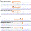
Fig 6. Confirmation of the presence of inserted mutations in the P gene of rescued viruses.
(A, B) Viral RNA was extracted from VDS cells infected with PPRV rNigeria/75/1, rNigeria/75/1_ΔC or rNigeria/75/1_ΔV and the P gene of each construct amplified by RT-PCR as described in Methods. (A) Sequence of the P gene from base 723 to 771 showing the V editing site. The specific bases in the editing site mutated to prevent editing are marked (*). (B) Sequence of the P gene from base 79 to 135 showing the start codon for the C protein (underlined) and the mutations introduced into the C ORF to introduce the STOP codons (TAA) (dotted underline) in rNigeria/75/1_ΔC.
Fig 7
Fig 7. Growth of and viral protein expression by rPPRV in VHS and G4 cells.
(A, B) Multi step growth curve of rNigeria/75/1_WT and the mutant viruses rNigeria/75/1_ΔC and rNigeria/75/1_ΔV. VHS cells (A) or G4 (B) cells were infected at a MOI = 0.01. Approximately two hours post infection the virus inoculum was removed and replaced with new medium. At the indicated times, samples of infected cells were frozen along with their medium. The virus titre (TCID50) was determined in VDS cells, allowing up to 10 days for signs of cytopathic effect (CPE) or GFP expression. The graph shows the titres for individual samples with the mean of the values marked with a line. The experiment was done twice in duplicate each titrated separately. (C, D) VHS cells (C) or G4 cells (D) were infected with rNigeria/75/1, rNigeria/75/1_ΔC or rNigeria/75/1_ΔV at a MOI = 3 or mock infected. At the indicated times post infection, the cells were lysed and the cell extracts were run on 8% SDS-PAGE gels and Western blotted with a mouse anti-RPV P antibody which cross-reacts with other morbillivirus P proteins, with mouse anti-PPRV N antibody or with rabbit anti-GFP antibody. The positions on the blot of the virally expressed proteins P, N and GFP are indicated by closed arrowheads, and the position of the main proteolytic fragment of N is indicated by the open arrowheads.
Fig 8
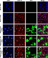
Fig 8. Effect of rPPRV on STAT1 activation.
VHS cells were infected with PPRV rNigeria/75/1, rNigeria/75/1_ΔC, rNigeria/75/1_ΔV (MOI ~ 0.5) or left uninfected. At 40 hpi, the cells were treated with 1,000 IU of IFN-α for 30 minutes or left untreated. STAT1P was detected by mouse anti-STAT1P and PPRV was detected using rabbit anti-GFP antibody. Primary antibodies were detected by Alexa Fluor 488 anti-rabbit IgG (green) and Alexa Fluor 568 anti-mouse IgG (red). Nuclei were stained with DAPI. Arrowheads point to the nuclei of infected cells.
Fig 9
Fig 9. IFN-β induction during the infection of VHS cells or G4 cells with PPRV lacking V or C expression.
VHS cells (A, D) or G4 cells (B, E) were transfected with reporter plasmids as described in for Fig 1. At least 18 hours post transfection, cells were infected with rNigeria/75/1_ΔV, rNigeria/75/1_ΔC (MOI = 1) or left uninfected. At each indicated time after infection, samples of cells were lysed and the cell extracts were assayed for luciferase and β-galactosidase activity. RLUs were normalised over time and between experiments by setting the value for uninfected cells to one. (C, F) G4 cells were infected with rNigeria/75/1_ΔV, rNigeria/75/1_ΔC (MOI = 1) or left uninfected and the cells lysed at the specified time points to extract total RNA. RT-qPCR was performed as described in Methods and the normalized relative quantities (NRQ) of the IFN-β mRNA calculated relative to the geometric mean of the amount of SDHA and GAPDH mRNA. The graphs show the relative NRQ of infected cells compared to uninfected cells, set to 1 at each time point. The error bars represent the standard error of the mean (SEM). The ANOVA test and Tukey pairwise comparison test were used to determine the significance of differences between the means (* = p < 0.05).
Fig 10
Fig 10. The V protein of PPRV is not essential in the context of virus infection to block the induction of IFN-β by poly(I:C) or Sendai virus.
(A, B) G4 cells were left uninfected or infected with rNigeria/75/1_ΔV (MOI = 3) for 16 hours before (A) transfection with 1 μg of poly(I:C) or (B) infection with 50 HA units of SeV-DI. At 5 hours after poly(I:C) transfection or 7 hours after SeV-DI infection the cells were lysed to extract total RNA. RT-qPCR was performed as described in Methods and the normalized relative quantities (NRQ) of the IFN-β mRNA calculated relative to the geometric means of the SDHA and the GAPDH mRNA. The NRQ was normalised between experiments by setting treated-uninfected cells to 100. The error bars represent the standard error of the mean (SEM). The ANOVA test and Tukey pairwise comparison test were used to determine the significance of differences between the means (* p = < 0.05). (C) G4 cells from parallel infected cells on coverslips were fixed in 3% PFA, permeabilized using 0.2% Triton and blocked with 0.2% gelatine in PBS. PPRV was detected using mouse anti N antibody followed by Alexa Fluor 488anti-mouse (green). Nuclei were stained by DAPI. Images were collected using the 63x magnification lens.
Fig 11
Fig 11. The C protein is necessary during PPRV infection for maximal inhibition of induction of IFN-β by either poly(I:C) and Sendai virus.
(A, B) G4 cells were left uninfected or infected with rNigeria/75/1_ΔC (MOI = 3) for 16 or 36 hours before (A) transfection with 1 μg of poly(I:C) or (B) infection with 50 HA units of SeV-DI. At 5 hours after poly(I:C) transfection or 7 hours after SeV-DI infection cells were lysed to extract total RNA. RT-qPCR was performed as described in Methods and the normalized relative quantities (NRQ) of the IFN-β mRNA calculated relative to the geometric means of the SDHA and the GAPDH mRNA. The NRQ was normalised between experiments by setting treated-uninfected cells to 100. (C) VHS cells were transfected with the reporter plasmids as described for Fig 1. At least 18 hours post transfection, cells were infected with rNigeria/75/1 (WT), with rNigeria/75/1_ΔC (ΔC) (MOI = 3) or left uninfected. At 16 or 36 hpi cells were infected with 50 HA units of SeV-DI or left uninfected. At 7 hours after SeV-DI infection the cells were lysed and the cell extracts were assayed for luciferase and β-galactosidase activity. RLUs were normalised between experiments by setting treated-uninfected cells to 100. (A-C) The error bars represent the standard error of the mean (SEM). The ANOVA test and Tukey pairwise comparison test were used to analyse differences between the means (* = p < 0.05). (D) VHS cell extracts from parallel infected/uninfected cells treated as in (C) were run on 10% SDS-PAGE gels and Western blotted with mouse anti-PPRV N antibody or with mouse anti-PCNA (loading control).

Similar articles

Cited by

References

    1. Baron MD, Diallo A, Lancelot R, Libeau G. Peste des Petits Ruminants Virus. Advances in virus research. 2016;95:1–42. doi: 10.1016/bs.aivir.2016.02.001 - DOI - PubMed
    1. Shaila MS, Shamaki D, Forsyth MA, Diallo A, Goatley L, Kitching RP, et al. Geographic distribution and epidemiology of peste des petits ruminants virus. Virus research. 1996;43(2):149–53. - PubMed
    1. Dhar P, Sreenivasa BP, Barrett T, Corteyn M, Singh RP, Bandyopadhyay SK. Recent epidemiology of peste des petits ruminants virus (PPRV). Veterinary microbiology. 2002;88(2):153–9. - PubMed
    1. Kwiatek O, Ali YH, Saeed IK, Khalafalla AI, Mohamed OI, Obeida AA, et al. Asian lineage of peste des petits ruminants virus, Africa. Emerging infectious diseases. 2011;17(7):1223–31. PubMed Central PMCID: PMC3381390. doi: 10.3201/eid1707.101216 - DOI - PMC - PubMed
    1. Brown CC, Mariner JC, Olander HJ. An immunohistochemical study of the pneumonia caused by peste des petits ruminants virus. Veterinary pathology. 1991;28(2):166–70. doi: 10.1177/030098589102800209 - DOI - PubMed

MeSH terms

Substances

LinkOut - more resources