Enhanced neuroinvasion by smaller, soluble prions
- PMID: 28431576
- PMCID: PMC5399838
- DOI: 10.1186/s40478-017-0430-z
Enhanced neuroinvasion by smaller, soluble prions
Abstract
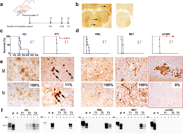
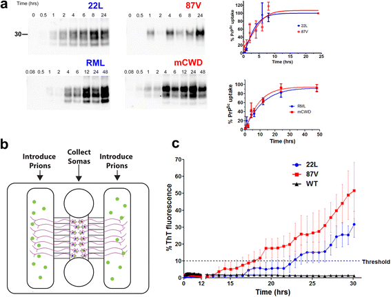
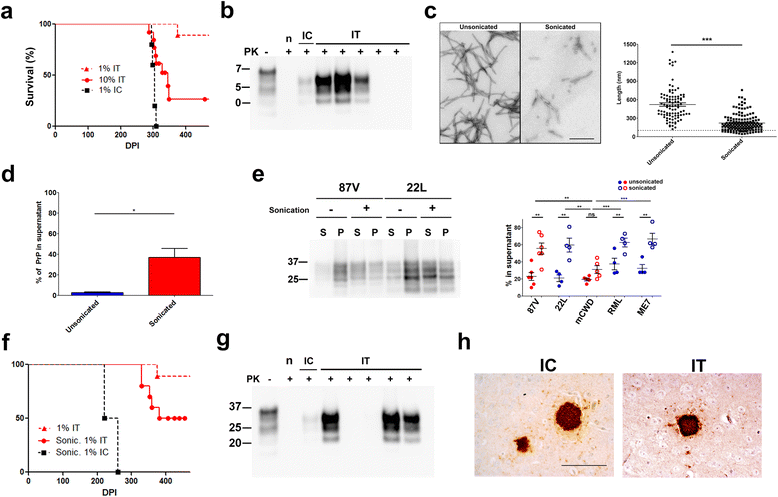
Infectious prion aggregates can propagate from extraneural sites into the brain with remarkable efficiency, likely transported via peripheral nerves. Yet not all prions spread into the brain, and the physical properties of a prion that is capable of transit within neurons remain unclear. We hypothesized that small, diffusible aggregates spread into the CNS via peripheral nerves. Here we used a structurally diverse panel of prion strains to analyze how the prion conformation impacts transit into the brain. Two prion strains form fibrils visible ultrastructurally in the brain in situ, whereas three strains form diffuse, subfibrillar prion deposits and no visible fibrils. The subfibrillar strains had significantly higher levels of soluble prion aggregates than the fibrillar strains. Primary neurons internalized both the subfibrillar and fibril-forming prion strains by macropinocytosis, and both strain types were transported from the axon terminal to the cell body in vitro. However in mice, only the predominantly soluble, subfibrillar prions, and not the fibrillar prions, were efficiently transported from the tongue to the brain. Sonicating a fibrillar prion strain increased the solubility and enabled prions to spread into the brain in mice, as evident by a 40% increase in the attack rate, indicating that an increase in smaller particles enhances prion neuroinvasion. Our data suggest that the small, highly soluble prion particles have a higher capacity for transport via nerves. These findings help explain how prions that predominantly assemble into subfibrillar states can more effectively traverse into and out of the CNS, and suggest that promoting fibril assembly may slow the neuron-to-neuron spread of protein aggregates.
Keywords: Amyloid; Axonal transport; Fibrils; Neurodegeneration; Prion disease; Prion strains.
Figures


Similar articles
-
Prion protein glycans reduce intracerebral fibril formation and spongiosis in prion disease.J Clin Invest. 2020 Mar 2;130(3):1350-1362. doi: 10.1172/JCI131564. J Clin Invest. 2020. PMID: 31985492 Free PMC article.
-
Generation of novel neuroinvasive prions following intravenous challenge.Brain Pathol. 2018 Nov;28(6):999-1011. doi: 10.1111/bpa.12598. Epub 2018 Jul 5. Brain Pathol. 2018. PMID: 29505163 Free PMC article.
-
Shortening heparan sulfate chains prolongs survival and reduces parenchymal plaques in prion disease caused by mobile, ADAM10-cleaved prions.Acta Neuropathol. 2020 Mar;139(3):527-546. doi: 10.1007/s00401-019-02085-x. Epub 2019 Oct 31. Acta Neuropathol. 2020. PMID: 31673874 Free PMC article.
-
Brain targeting through the autonomous nervous system: lessons from prion diseases.Trends Mol Med. 2004 Mar;10(3):107-12. doi: 10.1016/j.molmed.2004.01.008. Trends Mol Med. 2004. PMID: 15106608 Review.
-
Prion-like transmission of pathogenic protein aggregates in genetic models of neurodegenerative disease.Curr Opin Genet Dev. 2017 Jun;44:149-155. doi: 10.1016/j.gde.2017.03.011. Epub 2017 Apr 22. Curr Opin Genet Dev. 2017. PMID: 28441621 Review.
Cited by
-
Early stage prion assembly involves two subpopulations with different quaternary structures and a secondary templating pathway.Commun Biol. 2019 Oct 4;2:363. doi: 10.1038/s42003-019-0608-y. eCollection 2019. Commun Biol. 2019. PMID: 31602412 Free PMC article.
-
Prion protein glycans reduce intracerebral fibril formation and spongiosis in prion disease.J Clin Invest. 2020 Mar 2;130(3):1350-1362. doi: 10.1172/JCI131564. J Clin Invest. 2020. PMID: 31985492 Free PMC article.
-
Underglycosylated prion protein modulates plaque formation in the brain.J Clin Invest. 2020 Mar 2;130(3):1087-1089. doi: 10.1172/JCI134842. J Clin Invest. 2020. PMID: 31985491 Free PMC article.
-
Host prion protein expression levels impact prion tropism for the spleen.PLoS Pathog. 2020 Jul 23;16(7):e1008283. doi: 10.1371/journal.ppat.1008283. eCollection 2020 Jul. PLoS Pathog. 2020. PMID: 32702070 Free PMC article.
-
Extraneural infection route restricts prion conformational variability and attenuates the impact of quaternary structure on infectivity.PLoS Pathog. 2024 Jul 8;20(7):e1012370. doi: 10.1371/journal.ppat.1012370. eCollection 2024 Jul. PLoS Pathog. 2024. PMID: 38976748 Free PMC article.
References
Publication types
MeSH terms
Substances
Grants and funding
LinkOut - more resources
Full Text Sources
Other Literature Sources

