. 2017 May 9;8(19):31993-32008.
doi: 10.18632/oncotarget.16669.

miR-29a regulates the proliferation and differentiation of retinal progenitors by targeting Rbm8a

Affiliations

miR-29a regulates the proliferation and differentiation of retinal progenitors by targeting Rbm8a

Yi Zhang et al. Oncotarget. .

Abstract

During development, tight regulation of the expansion of retinal progenitor cells (RPCs) and their differentiation into neuronal and glial cells is important for retinal formation and function. Our study demonstrated that microRNA (miR)-29a modulated the proliferation and differentiation of RPCs by suppressing RBM8A (one of the factors in the exon junction complex). Particularly, overexpression of miR-29a reduced RPC proliferation but accelerated RPC differentiation. By contrast, reduction of endogenous miR-29a elicited the opposite effects. Overexpression of miR-29a repressed the translation of Rbm8a, thus negatively regulating RPC proliferation and promoting the neuronal and glial differentiation of RPCs, and knockdown of endogenous Rbm8a phenocopied the observed effects of miR-29a overexpression. Furthermore, a luciferase reporter assay showed that miR-29a directly interacted with the Rbm8a mRNA 3'UTR, which indicated that Rbm8a is the direct target of miR-29a. To further verify the result, co-overexpression of the Rbm8a 3' UTR-wt (plasmids into which the Rbm8a 3' UTR sequence had been introduced) and miR-29a in RPCs rescued the phenotype associated with miR-29a overexpression, reversing the promotion of differentiation and inhibition of proliferation. These results show a novel mechanism by which miR-29a regulates the proliferation and differentiation of RPCs through Rbm8a.

Keywords: RBM8A; differentiation; microRNA (miR)-29a; proliferation; retinal progenitor cells.


Conflict of interest statement

CONFLICTS OF INTEREST

The authors declare that they have no competing interest.

Figures
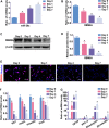
Figure 1
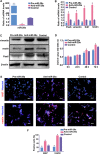
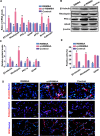
Figure 1. The endogenous expression levels of miR-29a and RBM8A during RPC differentiation
(A, B) The qPCR results showed that the expression level of miR-29a gradually increased, whereas RBM8A expression exhibited the opposite trend during the seven-day differentiation of RPCs. (C, D, E) Western blotting and immunostaining results revealed that the protein level of RBM8A decreased over the same period. (F, G) qPCR showed that the expression of proliferative marker (Ki67), and retinal progenitor markers (nestin, Pax6 and vimentin) was gradually decreased, but the expression of RPC differentiation markers (Rhodopsin, a marker for photoreceptors; β3-tubulin, a pan-neuronal marker; PKC-α, a marker for bipolar neurons; and GFAP, a glial cell marker) had the reversed trend during RPC differentiation. Western bands were scanned and normalized to β-actin. Day 0 represents the undifferentiated RPC state and was used as a normalizer. Scale bars: 100 μm. Data are averages of three independent experiments. Error bars indicate the standard error of the mean. *P ≤ 0.05 (one-way ANOVA).
Figure 2
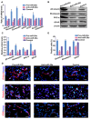
Figure 2. miR-29a inhibits RPC proliferation
(A) The qPCR results revealed that the expression of miR-29a was sharply upregulated by transfection of RPCs with the miR-29a mimic and significantly downregulated by the miR-29a inhibitor treatment. (B) According to the qPCR analysis, the expression of Ki67 (a cell proliferation marker) and vimentin, Pax6 and nestin (retinal progenitor markers) decreased in the miR-29a mimic-treated RPCs and increased when the RPCs were treated with the miR-29a inhibitor. (C) The results of the western blotting analysis were consistent with the qPCR analysis. Western blotting bands were scanned and normalized to β-actin. (D) The proliferation ability of the RPCs was assessed via a CCK-8 analysis. The proliferation ability of the RPCs markedly increased when treated with the miR-29a inhibitor and decreased when treated with the miR-29a mimic under proliferation conditions. (E, F) Immunostaining with antibodies against Ki67 and nestin revealed the effects on RPC proliferation, and was consistent with the results shown above. These data are averages of three independent experiments. Scale bars: 100 μm. Data are averages of three independent experiments. Error bars indicate the standard error of the mean. *P ≤ 0.05 (Student's t-test).
Figure 3
Figure 3. miR-29a promotes RPC differentiation
(AC) The expression levels of RPC differentiation-related markers, including PKC-α, Rhodopsin, β3-tubulin, Brn3a, calbindin and GFAP, were elevated by the miR-29a mimic but repressed by the miR-29a inhibitor according to qPCR and western blotting analyses. However, the expression of AP2α had no obvious change. Western blotting bands were scanned and normalized to β-actin. (D, E) The percentages of Rhodopsin-, β3-tubulin-, and GFAP-positive cells were evaluated to investigate RPC differentiation. The proportion of Rhodopsin- and β3-tubulin-positive cells was significantly augmented, and that of GFAP-positive cells slightly increased in the miR-29a mimic-treated RPC cultures. By contrast, the ratios of Rhodopsin, β3-tubulin and GFAP markedly decreased when treated with the miR-29a inhibitor. Retinal neuronal cell markers: PKC-α, Rhodopsin, β3-tubulin; glial cell marker: GFAP. Scale bars: 100 μm (for GFAP), 50 μm (for Rhodopsin and β3-tubulin). Data are averages of three independent experiments. Error bars indicate the standard error of the mean. *P ≤ 0.05 (Student's t-test).
Figure 4
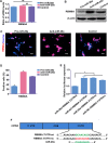
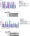
Figure 4. Rbm8a is a target gene of miR-29a in RPCs
(A) qPCR showed that neither exogenous miR-29a nor the miR-29a inhibitor had effects on Rbm8a mRNA levels. (BD) Western blotting and immunostaining analysis indicated that the RBM8A protein expression level was inhibited by the overexpression of miR-29a, but the downregulation of miR-29a via transfection with the miR-29a inhibitor promoted RBM8A protein expression. Western blotting bands were scanned and normalized to β-actin. Scale bars: 50 mm. (E) The dual luciferase reporter system indicated that the co-transfection of miR-29a and its wild type 3′-UTR binding site, termed Rbm8a 3′-UTRwt, dramatically reduced luciferase activity, whereas miR-29a had no effect on the mutated 3′-UTR binding region (Rbm8a 3′-UTRmu). The firefly luciferase activity data were all normalized to Renilla luciferase activity as a control. (F) Position 475-482 of the 3′-UTR of Rbm8a mRNA or a mutated sequence was designed and inserted into pGL3-control plasmids. Data are averages of three independent experiments. Error bars indicate the standard error of the mean. *P ≤ 0.05 (Student's t-test and one-way ANOVA ).
Figure 5
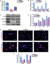
Figure 5. RBM8A enhances RPC proliferation
(A) The qPCR results revealed that the expression level of Rbm8a decreased sharply with siRbm8a treatment but increased significantly in the Rbm8a clone-treated RPC cultures compared with the control. (B) The qPCR results showed that the expression levels of nestin, Pax6, vimentin and Ki67 decreased in siRbm8a -treated RPC cultures and increased in cells treated with the Rbm8a clone compared with control cells. (C) The protein expression levels of nestin, Pax6 and vimentin exhibited similar trends to the qPCR results. Western blotting bands were scanned and normalized to β-actin. (D) The proliferation ability of the RPCs transfected with siRbm8a or the Rbm8a clone was assessed via CCK-8 analysis. The expansion capacity of the cells markedly improved in the Rbm8a clone-treated cultures, whereas a weakened proliferation capacity was detected for the siRbm8a-treated cells. (E, F) The percentages of Ki67- and nestin-positive cells decreased in the siRbm8a-treated RPCs and increased when treated with the Rbm8a clone. Scale bars: 100 μm. Data are averages of three independent experiments. Error bars indicate the standard error of the mean. *P ≤ 0.05 (Student's t-test).
Figure 6
Figure 6. RBM8A reduces RPC differentiation
(AC) qPCR and western blotting analysis revealed large increases in the expression levels of Rhodopsin, β3-tubulin, PKC-α, GFAP, Brn3a, and calbindin in the RPC cultures, but their expression levels were significantly decreased in the Rbm8a-treated groups. The expression level of AP2α had no marked change. Western blotting bands were scanned and normalized to β-actin. (D, E) Immunostaining with antibodies against Rhodopsin, β3-tubulin and GFAP revealed that the proportions of Rhodopsin-, and β3-tubulin-immunoreactive cells were higher in the siRbm8a -treated cultures than in the control cells, but these positive percentages decreased upon Rbm8a clone treatment. Scale bars: 100 μm (for GFAP), 50 μm (for Rhodopsin and β3-tubulin). Data are averages of three independent experiments. Error bars indicate the standard error of the mean. *P ≤ 0.05 (Student's t-test).
Figure 7
Figure 7. Rbm8a 3′-UTR overexpression rescues the observed effects of miR-29a overexpression on RPC proliferation and differentiation
(A, B) qPCR and western blotting analysis indicated that Rbm8a 3′-UTRwt antagonizes the effects of miR-29a on RPC proliferation. However, the co-transfection of Rbm8a 3′-UTRmu with miR-29a induced no obvious change in RPC proliferation in comparison with transfection with only miR-29a. (C, D) Rbm8a 3′-UTRwt overexpression rescued the miR-29a-mediated increase in RPC differentiation-related markers, including Rhodopsin, β3-tubulin, PKC-α and GFAP. Data are averages of three independent experiments. Western blotting bands were scanned and normalized to β-actin. Error bars indicate the standard error of the mean. *P ≤ 0.05 (one-way ANOVA).
Figure 8
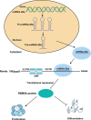
Figure 8. A model of the relationship between miR-29a and RBM8A in regulating RPCs proliferation and differentiation
miR-29a inhibited RBM8A expression by binding to the Rbm8a 3′-UTR and, thus, negatively regulated RPC proliferation and positively regulated RPC differentiation.

Similar articles

Cited by

References

    1. Gu P, Harwood LJ, Zhang X, Wylie M, Curry WJ, Cogliati T. Isolation of retinal progenitor and stem cells from the porcine eye. Mol Vis. 2007;13:1045–57. - PMC - PubMed
    1. Xia J, Liu H, Fan X, Hu Y, Zhang Y, Wang Z, Zhou X, Luo M, Gu P. An in vitro comparison of two different subpopulations of retinal progenitor cells for self-renewal and multipotentiality. Brain Res. 2012;1433:38–46. doi: 10.1016/j.brainres.2011.11.054. - DOI - PubMed
    1. Yang P, Seiler MJ, Aramant RB, Whittemore SR. In vitro isolation and expansion of human retinal progenitor cells. Exp Neurol. 2002;177:326–31. doi: 10.1006/exnr.2002.7955. - DOI - PubMed
    1. Fischer AJ, Reh TA. Identification of a proliferating marginal zone of retinal progenitors in postnatal chickens. Dev Biol. 2000;220:197–210. doi: 10.1006/dbio.2000.9640. - DOI - PubMed
    1. Tropepe V, Coles BL, Chiasson BJ, Horsford DJ, Elia AJ, McInnes RR, van der Kooy D. Retinal stem cells in the adult mammalian eye. Science. 2000;287:2032–36. doi: 10.1126/science.287.5460.2032. - DOI - PubMed

LinkOut - more resources