

Finite element analysis of sagittal balance in different morphotype: Forces and resulting strain in pelvis and spine
- PMID: 28377644
- PMCID: PMC5369862
- DOI: 10.1016/j.jor.2017.03.007
Finite element analysis of sagittal balance in different morphotype: Forces and resulting strain in pelvis and spine
Abstract
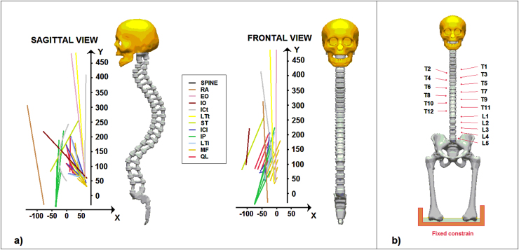
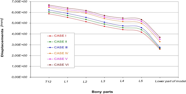
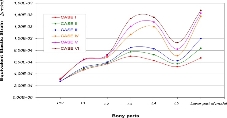
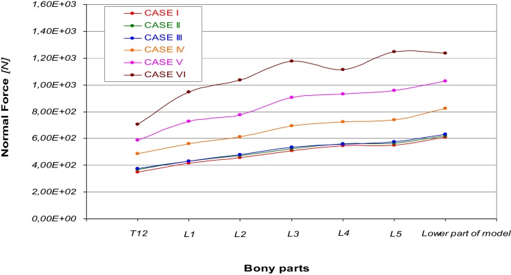
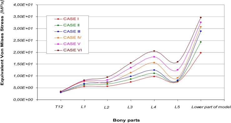
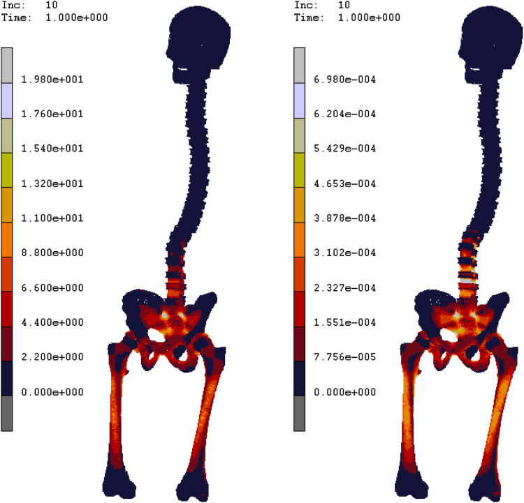
In humans, vertical posture acquisition caused several changes in bones and muscles which can be assumed as verticalization. Pelvis, femur, and vertebral column gain an extension position which decreases muscular work by paravertebral muscles in the latter. It's widely known that six different morphological categories exist; each category differs from the others by pelvic parameters and vertebral column curvatures. Both values depend on the Pelvic Incidence, calculated as the angle between the axes passing through the rotation centre of the two femur heads and the vertical axis passing through the superior plate of the sacrum. The aim of this study is to evaluate the distribution of stress and the resulting strain along the axial skeleton using finite element analysis. The use of this computational method allows performing different analyses investigating how different bony geometries and skeletal structures can behavior under specific loading conditions. A computerized tomography (CT) of artificial bones, carried on at 1.5 mm of distance along sagittal, coronal and axial planes with the knee at 0° flexion (accuracy 0.5 mm), was used to obtain geometrical data of the model developed. Lines were imported into a commercial code (Hypermesh by Altair®) in order to interpolate main surfaces and create the solid version of the model. In particular six different models were created according Roussoly's classification, by arranging geometrical position of the skeletal components. Loading conditions were obtained by applying muscular forces components to T1 till to L5, according to a reference model (Daniel M. 2011), and a fixed constrain was imposed on the lower part of the femurs. Materials were assumed as elastic with an Elastic modulus of 15 GPa, a Shear Modulus of 7 GPa for bony parts, and an Elastic modulus of 6 MPa, a Shear Modulus of 3 MPa for cartilaginous parts. Six different simulations have been carried out in order to evaluate the mechanical behavior of the human vertebral column arranged according to the Russoly's classification; results confirm higher solicitations obtained varying configurations from case I to case VI. In particular way, first three cases seem to supply the different loading configurations spreading stresses in almost all the bony parts of the column, while the remaining others three cases produce an higher concentration of stress around the lower part of spine (L3, L4, L5). Results confirm a good agreement with those present in literature (Winkle et al., 1999), an equivalent Von Mises average stress was of 0,55 MPa was found on the intervertebral disks with the higher values reached on the lower part of the column. A comparison of results obtained for Case I with literature (Galbusera et al., and El Rich et al., 2004), shows a good agreement in terms of normal compressive force, while more evident differences with Galbusera's results can be found for shear force and sagittal moment. The results underline a relationship between PI increase, and accordingly of PT and LL, and the distribution of load forces. Load forcesi is exerted mainly on distal vertebrae, especially on L4 and L5.
Keywords: Biomehcanics; Finite Element Analysis; Lumbar Spine; Sagittal Alignment; Sagittal Balance.
Figures
Similar articles
-
Biomechanical Effect of L4 -L5 Intervertebral Disc Degeneration on the Lower Lumbar Spine: A Finite Element Study.Orthop Surg. 2020 Jun;12(3):917-930. doi: 10.1111/os.12703. Epub 2020 May 31. Orthop Surg. 2020. PMID: 32476282 Free PMC article.
-
Does the anterior column realignment technique influences the stresses on posterior instrumentation in sagittal imbalance correction? A biomechanical, finite-element analysis of L5-S1 ALIF and L3-4 lateral ACR.Spine Deform. 2023 Jan;11(1):41-47. doi: 10.1007/s43390-022-00567-9. Epub 2022 Aug 23. Spine Deform. 2023. PMID: 35999490
-
Influence of spine morphology on intervertebral disc loads and stresses in asymptomatic adults: implications for the ideal spine.Spine J. 2005 May-Jun;5(3):297-309. doi: 10.1016/j.spinee.2004.10.050. Spine J. 2005. PMID: 15863086
-
[Correlation of lumbar disc degeneration and spinal-pelvic sagittal balance].Zhonghua Yi Xue Za Zhi. 2013 Apr 16;93(15):1123-8. Zhonghua Yi Xue Za Zhi. 2013. PMID: 23902878 Chinese.
-
Biomechanical analysis of the spino-pelvic organization and adaptation in pathology.Eur Spine J. 2011 Sep;20 Suppl 5(Suppl 5):609-18. doi: 10.1007/s00586-011-1928-x. Epub 2011 Aug 2. Eur Spine J. 2011. PMID: 21809016 Free PMC article. Review.
Cited by
-
High values of pelvic incidence: A possible risk factor for zigoapophyseal facet arthrosis in young.J Orthop. 2018 Feb 21;15(2):333-336. doi: 10.1016/j.jor.2018.02.011. eCollection 2018 Jun. J Orthop. 2018. PMID: 29881147 Free PMC article.
-
Ligament reconstruction for distal radioulnar joint instability with the biomechanical analysis: A case report.Medicine (Baltimore). 2024 Oct 11;103(41):e40057. doi: 10.1097/MD.0000000000040057. Medicine (Baltimore). 2024. PMID: 39465789 Free PMC article.
-
Stress shielding analysis on easy step staple prosthesis for calcaneus fractures.J Orthop. 2019 Sep 12;18:132-137. doi: 10.1016/j.jor.2019.09.008. eCollection 2020 Mar-Apr. J Orthop. 2019. PMID: 32021019 Free PMC article.
-
Flatfoot and normal foot a comparative analysis of the stress shielding.J Orthop. 2018 Aug 16;15(3):820-825. doi: 10.1016/j.jor.2018.08.002. eCollection 2018 Sep. J Orthop. 2018. PMID: 30140126 Free PMC article.
-
Statistical investigation about spinal clinical asymmetry in a school population.J Orthop. 2020 Aug 18;22:336-340. doi: 10.1016/j.jor.2020.08.011. eCollection 2020 Nov-Dec. J Orthop. 2020. PMID: 32904173 Free PMC article.
References
-
- Kardong . 6th ed. McGrawh-Hill; New York: 2012. Vertebrates: comparative anatomy, function, evolution.
-
- 4th ed. Elsevier.; 2016. Benzel’s spine surgery.
-
- Hutchinson JR, http://link.springer.com/article/10.1007%2Fs00114-008-0488-3. - PubMed
Publication types
LinkOut - more resources
Full Text Sources
Other Literature Sources
Research Materials
Miscellaneous
