Review
. 2017 Apr 4;7(2):35.
doi: 10.3390/biom7020035.

tRNA Modifications: Impact on Structure and Thermal Adaptation

Affiliations
Review

tRNA Modifications: Impact on Structure and Thermal Adaptation

Christian Lorenz et al. Biomolecules. .

Abstract

Transfer RNAs (tRNAs) are central players in translation, functioning as adapter molecules between the informational level of nucleic acids and the functional level of proteins. They show a highly conserved secondary and tertiary structure and the highest density of post-transcriptional modifications among all RNAs. These modifications concentrate in two hotspots-the anticodon loop and the tRNA core region, where the D- and T-loop interact with each other, stabilizing the overall structure of the molecule. These modifications can cause large rearrangements as well as local fine-tuning in the 3D structure of a tRNA. The highly conserved tRNA shape is crucial for the interaction with a variety of proteins and other RNA molecules, but also needs a certain flexibility for a correct interplay. In this context, it was shown that tRNA modifications are important for temperature adaptation in thermophilic as well as psychrophilic organisms, as they modulate rigidity and flexibility of the transcripts, respectively. Here, we give an overview on the impact of modifications on tRNA structure and their importance in thermal adaptation.

Keywords: archaeosine; dihydrouridine; dimethylguanosine; lysidine; methyladenosine; methylguanosine; post-transcriptional modifications; pseudouridine; tRNA; tRNA structure.


Conflict of interest statement

The authors declare no conflict of interest. The funding sponsors had no role in the design of the study; in the collection, analyses, or interpretation of data; in the writing of the manuscript, and in the decision to publish the results.

Figures
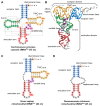
Figure 1
Figure 1
Variability of transfer RNA (tRNA) structures. (A) The canonical cloverleaf secondary structure of cytosolic tRNAPhe from S. cerevisiae is shown with acceptor stem (blue), D-arm (green), anticodon arm (red), variable loop (purple) and TΨC-arm (yellow). The anticodon is labeled in grey, the discriminator base in orange and post-transcriptional modifications in red. Grey dashed lines indicate tertiary interactions based on structural data [11]. Base numbering corresponds to Sprinzl et al. [25] and length of the RNA is indicated in parenthesis; (B) The L-shaped tertiary structure of the cytosolic tRNAPhe from S. cerevisiae. Protein Data Bank entry (PDB): 1EHZ [11]. The acceptor domain is composed of stacked T-arm and acceptor stem, whereas D- and anticodon arm form the anticodon domain. The region where both domains come together and interact with each other via tertiary base pairing is also called elbow region; (C) Secondary structure of human mitochondrial tRNASer1, which lacks the whole D-arm [26]; (D) Secondary structure of the mitochondrial tRNAArg from the nematode Romanomermis culicivorax, which lacks both D- and T-arm. Instead, we find a so-called replacement loop. It represents the shortest tRNA found in vivo [21].
Figure 2
Figure 2
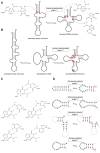
Variability of tRNA modifications. The upper part of the image illustrates the systematic abbreviation of RNA modifications with N2,N2,2′-O-trimethylguanosine (m22Gm) as an example and also shows the atom numbering in the purine and pyrimidine rings as well as in the ribose. An abbreviation in front of the base letter describes a base modification, whereas letters after the base stand for ribose alterations. Superscripted numbers specify the position at the base and subscripted numbers indicate the frequency of identical modification at the same position. Abbreviations are as follows: ac—acetyl, acp—aminocarboxypropyl, chm—carboxyhydroxymethyl, cmo—oxyacetic acid, cmnm—carboxymethylaminomethyl, f—formyl, g—glycinyl, gal—galactosyl, hn—hydroxynorvalylcarbamoyl, ho—hydroxy, i—isopentenyl, inm—isopentenylaminomethyl, io—cis-hydroxyisopentenyl, m—methyl, man—mannosyl, mchm—carboxyhydroxymethyl methyl ester, mcm—methoxycarbonylmethyl, mcmo—oxyacetic acid methyl ester, mnm—methylaminomethyl, mo—methoxy, ncm—carbamoylmethyl, nm—aminomethyl, r(p) —5-O-phosphono-b-d-ribofuranosyl, s—thio, se—seleno, t—threonylcarbamoyl, tm—taurinomethyl. The Venn diagram summarizes data collected from the RNA modification database and contains the 93 post-transcriptional modifications that are found in tRNAs [5]. Some examples mentioned throughout the text are shown with their chemical structure.
Figure 3
Figure 3
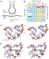
Post-transcriptional modifications in tRNA. (A) The colored tRNA structure shows the modification frequency of each base. The modification data were taken from the tRNAmodviz database [27] and plotted on the crystal structure of tRNAPhe from S. cerevisiae [11]. Blue colored bases are rarely modified; red colored bases are modification hotspots. tRNAs possess two regions with high modification levels—the anticodon loop (especially positions 34 and 37) and the core or elbow region, where D- and T-loop bases interact with each other and stabilize the tertiary fold. For some important positions, the chemical structure of the most frequent modification at this position is shown; (B) Three dimensional structure of pseudouridine at position 55 of tRNAPhe from S. cerevisiae. The additional H-bond donor at N1 interacts with the 5′-adjacent phosphates via a coordinated water molecule. The hydrogen bound to N1 was not resolved in the crystal structure. The ribose shows a stabilizing C3′-endo conformation. PDB: 1EHZ [11]; (C) Three dimensional structure of D16 in the D-arm of tRNAiMet from Schizosaccharomyces pombe. The C5-C6 bond of dihydrouridine is reduced, which leads to a non-planar structure of the base. The ribose takes the less stable C2′ -endo conformation. PDB: 2MN0 [41].
Figure 4
Figure 4
Modifications direct a tRNA secondary structure. (A) The in vitro transcript of human tRNALys shows an extended hairpin structure due to missing modifications. The introduction of N1-methyladenosine (m1A) at position 9 interrupts a base pair with U64 and forces the canonical folding of the tRNA. U64 now pairs with A50 [51,52]; (B) The unmodified transcript of human mitochondrial tRNAAsp shows an equilibrium between various secondary structures. It is suggested that the natural occurring modification m1A9, m2G10 and Ψ27 are important for correct folding. Queuosine at position 34 in the anticodon loop does likely not contribute to the secondary structure [55]. (A,B) adapted with permission from Motorin and Helm [40]. Copyright (2010) American Chemical Society; (C) The two methyl groups of m22G at the exocyclic N2 of guanosine create a steric hindrance and preclude base pairing with cytosine. However, the methylations do not alter the base pairing with A or U as the amino group at N2 is not involved in this interplay. An interaction of m22G with C is possible via a bridging water which leads to a greater distance between the two bases [56]; (D) Various examples show the impact of m22G for a correct tRNA folding. In Pyrococcus abyssi tRNAPro contains m22G at position 10, which wobble pairs with U25. A lack of this modification can lead to dramatic misfolding of the D-arm due to an extended D-stem [57]. tRNAAsp of the same organism also contains m22G10. However, this modification prevents a wrong base pairing with C27, which would lead to an enlarged D-loop [57]. In tRNALys of Haloferax volcanii, m22G can also direct the folding of the anticodon stem by interrupting C25-G45 and G26-C44 base pair which leads to an elongated anticodon and shortened D-stem [56]. A similar example is human cytosolic tRNAAsn. Here, m22G is found at position 26 and prohibits base pairing with C11 [56].
Figure 5
Figure 5
Modifications in decoding and anticodon loop structure. (A) The interaction of the anticodon bases (34–36) of a tRNA with the corresponding bases of the mRNA codons (3, 2, 1). A wobble interaction is possible between codon base 3 and anticodon base 34. The latter is frequently modified and directs the wobble interactions with the third codon base; (B) The standard genetic code is illustrated as a simple decoding table, 2-fold degenerate codon boxes are colored yellow, 4-fold degenerate boxes are blue. Start and stop codons are colored green and red, respectively; (C) Stereo image of the well-structured anticodon loop of tRNALys from E. coli. Modifications mnm5s2U34 and t6A37 prevent wrong base pairing inside the 7-nucleotide loop and promote the formation of the conserved U-turn motif. The stacked anticodon bases are located on the same side of the loop. PDB: 1FL8 [91]; (D) Stereo image of a collapsed and unmodified anticodon loop of tRNATyr from Bacillus subtilis. Here, bases 32 and 38 as well as 33 and 37 interact with each other and the U-turn motif is missing. The anticodon bases are not ordered and on opposite sides of the loop. PDB: 2LAC [89].

Similar articles

Cited by

References

    1. Hopper A.K. Transfer RNA post-transcriptional processing, turnover, and subcellular dynamics in the yeast Saccharomyces cerevisiae. Genetics. 2013;194:43–67. doi: 10.1534/genetics.112.147470. - DOI - PMC - PubMed
    1. Reinhold-Hurek B., Shub D.A. Self-splicing introns in tRNA genes of widely divergent bacteria. Nature. 1992;357:173–176. doi: 10.1038/357173a0. - DOI - PubMed
    1. Yoshihisa T. Handling tRNA introns, archaeal way and eukaryotic way. Front. Genet. 2014;5:213. doi: 10.3389/fgene.2014.00213. - DOI - PMC - PubMed
    1. Betat H., Rammelt C., Mörl M. tRNA nucleotidyltransferases: Ancient catalysts with an unusual mechanism of polymerization. Cell. Mol. Life Sci. 2010;67:1447–1463. doi: 10.1007/s00018-010-0271-4. - DOI - PMC - PubMed
    1. Cantara W.A., Crain P.F., Rozenski J., McCloskey J.A., Harris K.A., Zhang X., Vendeix F.A.P., Fabris D., Agris P.F. The RNA Modification Database, RNAMDB: 2011 update. Nucleic Acids Res. 2011;39:D195–D201. doi: 10.1093/nar/gkq1028. - DOI - PMC - PubMed

Publication types

LinkOut - more resources