. 2017 Apr;9(4):1153-1185.
doi: 10.18632/aging.101218.

The age- and sex-specific decline of the 20s proteasome and the Nrf2/CncC signal transduction pathway in adaption and resistance to oxidative stress in Drosophila melanogaster

Affiliations

The age- and sex-specific decline of the 20s proteasome and the Nrf2/CncC signal transduction pathway in adaption and resistance to oxidative stress in Drosophila melanogaster

Laura C D Pomatto et al. Aging (Albany NY). 2017 Apr.

Abstract

Hallmarks of aging include loss of protein homeostasis and dysregulation of stress-adaptive pathways. Loss of adaptive homeostasis, increases accumulation of DNA, protein, and lipid damage. During acute stress, the Cnc-C (Drosophila Nrf2 orthologue) transcriptionally-regulated 20S proteasome degrades damaged proteins in an ATP-independent manner. Exposure to very low, non-toxic, signaling concentrations of the redox-signaling agent hydrogen peroxide (H2O2) cause adaptive increases in the de novo expression and proteolytic activity/capacity of the 20S proteasome in female D. melanogaster (fruit-flies). Female 20S proteasome induction was accompanied by increased tolerance to a subsequent normally toxic but sub-lethal amount of H2O2, and blocking adaptive increases in proteasome expression also prevented full adaptation. We find, however, that this adaptive response is both sex- and age-dependent. Both increased proteasome expression and activity, and increased oxidative-stress resistance, in female flies, were lost with age. In contrast, male flies exhibited no H2O2 adaptation, irrespective of age. Furthermore, aging caused a generalized increase in basal 20S proteasome expression, but proteolytic activity and adaptation were both compromised. Finally, continual knockdown of Keep1 (the cytosolic inhibitor of Cnc-C) in adults resulted in older flies with greater stress resistance than their age-matched controls, but who still exhibited an age-associated loss of adaptive homeostasis.

Keywords: 20S proteasome; Nrf2; adaptive homeostasis; oxidative stress; protein aggregation; protein oxidation.


Conflict of interest statement

CONFLICTS OF INTEREST

The authors declare they have no conflicts of interest.

Figures

Figure 1
Figure 1. Hydrogen peroxide resistance declines with age
(A-D) Males and females of the Actin-GS-255B strain crossed to the w[1118] strain were aged to 3 days or 60 days and then transferred to vials containing kimwipes soaked in 1.0M to 8.0M H2O2 dissolved in 5% sucrose solution, and flies were scored as dead when completely immobile, as described previously [43]. (A) 3 day old females. (B) 3 day old males. (C) 60 day old females. (D) 60 day old males.
Figure 2
Figure 2. Adaptation to hydrogen peroxide declines with age in females
Progeny of the Actin-GS-255B strain crossed to the w[1118] strain were aged to 3 days and 60 days prior to H2O2 pretreatment. Following recovery, flies were fed H2O2 challenge dose: 4.4M in 3 day old males and females, 2M in 60 day old females, and 1M in 60 day old males. (A) 3 day old females showed increased survival following pretreatment. (B) 3 day old males showed no change in survival following pretreatment. (C) With age, 60 day old females no longer show increased survival. (D) 60 day old males show no change in adaptation following pretreatment. Statistical difference in survival (p < 0.05) was calculated using the Log-Rank test. Statistical summary is located in Supplementary Table S1.
Figure 3
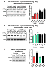
Figure 3. Adaptive de novo expression of the 20S proteasome diminishes with age in a sex-dependent manner
(A,B) Virgin females of the Actin-GS-255B strain were crossed to males of the w[1118] strain and progeny were assayed for 20Sα expression without H2O2 pretreatment, or after pretreatment with [10μM or 100μM] H2O2. (A) 3 day and 60 day aged females. (B) 3 day and 60 day aged males. All samples were compared to the 3 day old 0μM H2O2 controls (C) Basal expression of the 20S proteasome α subunits was measured between 3 day old females, 60 day old females, 3 day old males, and 60 day old males, with samples normalized to the 3 day old female. Western blots were performed in triplicate, normalized to Actin-HRP, and quantified using ImageJ. Error bars denote standard error of the mean (S.E.M) values. * P <0.05 and ** P < 0.01, relative to control using one-way ANOVA. Asterisks color indicates the age of the sample, young female (pink *) and aged female (black *). All statistical significance was calculated relative to the young controls.
Figure 4
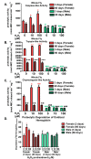
Figure 4. Adaptive proteolytic capacity of the 20S proteasome diminishes with age in a sex-dependent manner
Virgin females of the Actin-GS-255B strain were crossed to males of the w[1118] strain and progeny were assayed for the proteolytic activity of the three catalytic subunits of the 20S proteasome in 3 day-old (red or blue) and 60 day-old flies (checked pattern). Caspase-like activity in (A) Females and males. Trypsin-like activity in (B) Females and males. Chymotrypsin-like activity in (C) Females and males. (D) Proteolytic degradation of oxidized [3H] hemoglobin in flies pretreated with hydrogen peroxide at 3 days and 60 days. Statistical significance for proteolysis of oxidized substrate was compared to young control females. Error bars denote standard error of the mean (S.E.M) values. * P <0.05, ** P < 0.01, *** P < 0.001 relative to control using one-way ANOVA. Asterisk color corresponds to young females (pink *), aged females (black *), young males (green #), and aged males (black #). Statistical significance was calculated relative to the young control females (A-D).
Figure 5
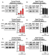
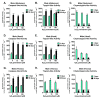
Figure 5. The adaptive expression of the 20S proteasome is age and tissue-dependent in females
Body segments collected from females of the Actin-GS-255B strain crossed to the w[1118] strain were used as controls, or were pretreated with either 10μM or 100μM hydrogen peroxide. (A,B) 20Sα expression in female abdominal tissue following pretreatment. (A) 3 day old. (B) 60 day old. (C,D) 20Sα expression in female head following pretreatment. (C) 3 day old. (D) 60 day old. (E,F) 20Sα expression in female thorax following pretreatment. (E) 3 day old. (F) 60 day old. Western blots were performed in triplicate, normalized to Actin-HRP, and quantified using ImageJ. The bar charts represent the quantification. Error bars denote standard error of the mean (S.E.M) values. * P <0.05, ** P <0.01, *** P < 0.001, relative to the female control using one-way ANOVA. Statistical significance is indicated in young females with pink asterisks (pink *).
Figure 6
Figure 6. Tissue-specific differences of the adaptive proteolytic capacity, and age-dependent changes in basal activity, of the 20S proteasome in females
Body segments collected from female progeny of the Actin-GS-255B strain crossed to the w[1118] strain were used as controls, or were pretreated with either 10μM or 100μM hydrogen peroxide. Individual proteolytic capacity of the 20S proteasome (caspase/peptidyl glutamyl-peptide hydrolyzing-like activity, trypsin-like, and chymotrypsin-like activity) was measured in the abdomen, head, and thorax. (A-C) Abdomen isolated from 3 day old (pink) and 60 day old (black) females following hydrogen peroxide pretreatment. (A) Caspase-like activity. (B) Trypsin-like activity. (C) Chymotrypsin-like activity. (D-F) Head isolated from 3 day old (pink) and 60 day old (black) females following hydrogen peroxide pretreatment. (D) Caspase-like activity. (E) Trypsin-like activity. (F) Chymotrypsin-like activity. (G-I) Thorax isolated from 3 day old (pink) and 60 day old (black) females following hydrogen peroxide pretreatment. (G) Caspase-like activity. (H) Trypsin-like activity. (I) Chymotrypsin-like activity. Error bars indicate the standard error of the mean (S.E.M) values. * P <0.05, ** P <0.01, *** P < 0.001, relative to the young female control using one-way ANOVA. Statistical significance is shown with pink asterisks (pink *) for young females and black asterisks (black *) in aged females.
Figure 7
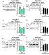
Figure 7. Males show no tissue-specific or age-related adaptive changes in 20S proteasome protein levels
Body segments were collected from males of the Actin-GS-255B strain crossed to the w[1118] strain that were used as controls, or that were pretreated with either 10μM or 100μM hydrogen peroxide. (A,B) 20Sα expression in male abdominal tissue following pretreatment. (A) 3 day old. (B) 60 day old. (C,D) 20Sα expression in male head following pretreatment. (C) 3 day old. (D) 60 day old. (E,F) 20Sα expression in male thorax following pretreatment. (E) 3 day old. (F) 60 day old. Western blots were performed in triplicate, normalized to Actin-HRP, and quantified using ImageJ. The bar charts represent the quantification. Error bars denote standard error of the mean (S.E.M) values, relative to the male control using one-way ANOVA.
Figure 8
Figure 8. Males show no tissue-specific differences in the adaptive proteolytic capacity of the 20S proteasome, but do exhibit age-dependent changes in proteasomal basal activity
Body segments were collected from male progeny of the Actin-GS-255B strain crossed to the w[1118] strain that were used as controls, or that were pretreated with either 10μM or 100μM hydrogen peroxide. Individual proteolytic capacity of the 20S proteasome (caspase/peptidyl glutamyl-peptide hydrolyzing-like activity, trypsin-like, and chymotrypsin-like activity) was measured in the abdomen, head, and thorax. (A-C) Abdomen isolated from 3 day old (green) and 60 day old (black) males following hydrogen peroxide pretreatment. (A) Caspase-like activity. (B) Trypsin-like activity. (C) Chymotrypsin-like activity. (D-F) Head isolated from 3 day old (green) and 60 day old (black) males following hydrogen peroxide pretreatment. (D) Caspase-like activity. (E) Trypsin-like activity. (F) Chymotrypsin-like activity. (G-I) Thorax isolated from 3 day old (green) and 60 day old (black) males following hydrogen peroxide pretreatment. (G) Caspase-like activity. (H) Trypsin-like activity. (I) Chymotrypsin-like activity.
Figure 9
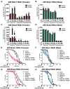
Figure 9. Adaptation is dependent upon the 20S proteasome
Progeny of the Actin-GS-255B strain crossed to the β1 or β2 RNAi strains were aged for 5 days in the absence or presence of RU486 prior to H2O2 pretreatment. (A-D). The purpose of the experiment was not to completely knockdown the entire pool of 20S proteasome, but only to block the transcription/translation-dependent adaptive increase in proteasome expression following hydrogen peroxide pretreatment. Thus, we used RNAi conditions that blocked increased proteasome expression, without depressing basal proteasome protein levels. Using this approach, we found at least a 50% decrease in mRNA in RNAi strains, and within proteasome western blots and activity, we found blockage of the adaptive increase. After pretreatment, proteolytic capacity of the individual subunits of the 20S proteasome (trypsin-like, caspase/peptidyl glutamyl-peptide hydrolyzing-like activity, and chymotrypsin-like activity) were measured in whole fly lysate. (A-B) Proteolytic capacity in β1 RNAi flies in the absence (black) “control” or presence (pink in females or blue in males, denoted with “+RU486”) of RU486. (A) Females. (B) Males. (C-D) Proteolytic capacity in β2 RNAi flies in the absence (black) “control” or presence (pink in females or blue in males, denoted with “+RU486”) of RU486. (C) Females. (D) Males. (E,G) Females of the β1 and β2 RNAi strains raised in the absence of RU486 were either not pretreated “control” (black circle) or were pretreated with either 10μM H2O2 (grey squares) or 100μM H2O2 (grey circles) for 8 hours, followed by a 16-hour recovery prior to H2O2 [4.4M] challenge. Females of the β1 and β2 RNAi strains raised in the presence of RU486 were either not pretreated “+RU486” (pink triangle) or were pretreated with either 10μM H2O2 (pink diamonds) or 100μM H2O2 (pink squares) for 8 hours, followed by a 16-hour recovery prior to H2O2 [4.4M] challenge. (F,H) Males of the β1 and β2 RNAi strains raised in the absence of RU486 were either not pretreated “control” (black circle) or were pretreated with either 10μM H2O2 (grey circles) or 100μM H2O2 (grey triangles) for 8 hours, followed by a 16-hour recovery prior to H2O2 [4.4M] challenge. Males of the β1 and β2 RNAi strains raised in the presence of RU486 were either not pretreated “+RU486” (green circle) or were pretreated with either 10μM H2O2 (blue square) or 100μM H2O2 (blue circle) for 8 hours, followed by a 16-hour recovery prior to H2O2 [4.4M] challenge. Statistical difference in survival (p < 0.05) was calculated using the Log-Rank test. Statistical summary is located in Supplementary Table S2.
Figure 10
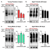
Figure 10. Decline in 20S proteasome induction is accompanied by an accumulation of oxidized proteins
Carbonyl content was detected with a DNP antibody in progeny of the Actin-GS-255B strain crossed to w[1118] strain following [0, 10, or 100μM] H2O2 pre-treatment for 8 hours, followed by a 16-hour recovery to allow for adaption before challenged with H2O2 [4.4M] for an additional 24 hours. (A) Carbonyl content showed significant decrease following H2O2 pretreatment in 3 day old females. (B) Carbonyl content showed no significant change in 60 day old females, irrespective H2O2 pretreatment. (C) Carbonyl content was measured in 3 day old males that were pre-treated with H2O2. (D) 60 day old males showed no change in carbonyl content upon H2O2 pre-treatment and subsequent recovery. Western blots were performed in triplicate and carbonyl content was normalized to Actin-HRP. Error bars denote standard error of the mean (S.E.M) values. * P <0.05, ** P <0.01, *** P < 0.001, relative to the young control using one-way ANOVA. Statistical significance is shown with asterisks (pink *) in young females.
Figure 11
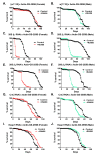
Figure 11. Loss of proteasomal subunits or regulators impacts lifespan
(A,B) To control for the effect of RU486 on males and females, lifespan of progeny from the Actin-GS-255B strain crossed to w[1118] strain raised in the absence/control (black line) or presence of RU486, pink line for females and blue line for males. (A) Females. (B) Males. (C-F) Effect of removal of proteasome subunits on life span. The Actin-GS-255B strain was crossed to the β1 RNAi or β2 RNAi strains and the progeny were assayed for life span in the absence/control (black line) or presence (pink line in females and blue line in males) of RU486, as indicated. (C) β1 RNAi females. (D) β1 RNAi males. (E) β2 RNAi females. (F) β2 RNAi males. (G,H) Effect of removal of the Cap-n-collar (CncC)/Nrf2 orthologue upon lifespan. The Actin-GS-255B strain was crossed to the CncC RNAi strain. Male and female lifespan was measured in the absence/control (black line) or presence (pink line for females and blue line for males) of RU486. (G) CncC RNAi female. (H) CncC RNAi male. (I,J) Effect of removal of Keap1 upon lifespan. The Actin-GS-255B strain was crossed to the Keap1 RNAi strain. Males and female lifespan was assessed in the absence/control (black line) and presence (pink line for females and blue line for males) of RU486. (I) Keap1 RNAi female. (J) Keap1 RNAi male. Statistical difference in survival (p < 0.05) was calculated using the Log-Rank test. Statistical summary is located in Supplementary Table S3.
Figure 12
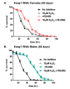
Figure 12. Hydrogen peroxide stress resistance improves with continual knockdown of Keap1 in aged (60 days) flies
In panels A and B, male and female progeny of the Actin-GS-255B strain crossed to Keap1 RNAi strain were collected and aged to 60 days. (A) Females raised in the absence of RU486 were either pretreated with 10μM H2O2 (gray line) “10μM H2O2” or not pre-treated with H2O2 (black line) “No Additions”. Females raised in the presence of RU486 were either pretreated with 10μM H2O2 (red line) “10μM H2O2 (+RU486)” or not pretreated with H2O2 (pink line) “+RU486”. (B) Males raised in the absence of RU486 were either pretreated with 10μM H2O2 (gray line) “10μM H2O2” or not pre-treated with H2O2 (black line) “No Additions”. Males raised in the presence of RU486 were either pretreated with 10μM H2O2 (blue line) “10μM H2O2 (+RU486)” or not pretreated with H2O2 (green line) “+RU486”. Statistical difference in survival (p < 0.05) was calculated using the Log-Rank test. Statistical summary is located in Supplementary Table S4.

Similar articles

Cited by

References

    1. Dröge W. Free radicals in the physiological control of cell function. Physiol Rev. 2002;82:47–95. doi: 10.1152/physrev.00018.2001. - DOI - PubMed
    1. Squier TC. Oxidative stress and protein aggregation during biological aging. Exp Gerontol. 2001;36:1539–50. doi: 10.1016/S0531-5565(01)00139-5. - DOI - PubMed
    1. Tanase M, Urbanska AM, Zolla V, Clement CC, Huang L, Morozova K, Follo C, Goldberg M, Roda B, Reschiglian P, Santambrogio L. Role of Carbonyl Modifications on Aging-Associated Protein Aggregation. Sci Rep. 2016;6:19311. doi: 10.1038/srep19311. - DOI - PMC - PubMed
    1. Davies KJ. Degradation of oxidized proteins by the 20S proteasome. Biochimie. 2001;83:301–10. doi: 10.1016/S0300-9084(01)01250-0. - DOI - PubMed
    1. Grune T, Reinheckel T, Davies KJ. Degradation of oxidized proteins in mammalian cells. FASEB J. 1997;11:526–34. doi: 10.1002/biof.5520060210. - DOI - PubMed

MeSH terms

LinkOut - more resources