Amyloid-β-Induced Changes in Molecular Clock Properties and Cellular Bioenergetics
- PMID: 28367108
- PMCID: PMC5355433
- DOI: 10.3389/fnins.2017.00124
Amyloid-β-Induced Changes in Molecular Clock Properties and Cellular Bioenergetics
Abstract
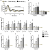
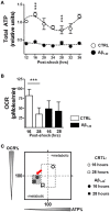
Ageing is an inevitable biological process that results in a progressive structural and functional decline, as well as biochemical alterations that altogether lead to reduced ability to adapt to environmental changes. As clock oscillations and clock-controlled rhythms are not resilient to the aging process, aging of the circadian system may also increase susceptibility to age-related pathologies such as Alzheimer's disease (AD). Besides the amyloid-beta protein (Aβ)-induced metabolic decline and neuronal toxicity in AD, numerous studies have demonstrated that the disruption of sleep and circadian rhythms is one of the common and earliest signs of the disease. In this study, we addressed the questions of whether Aβ contributes to an abnormal molecular circadian clock leading to a bioenergetic imbalance. For this purpose, we used different oscillator cellular models: human skin fibroblasts, human glioma cells, as well as mouse primary cortical and hippocampal neurons. We first evaluated the circadian period length, a molecular clock property, in the presence of different Aβ species. We report here that physiologically relevant Aβ1-42 concentrations ranging from 10 to 500 nM induced an increase of the period length in human skin fibroblasts, human A172 glioma cells as well as in mouse primary neurons whereas the reverse control peptide Aβ42-1, which is devoid of toxic action, did not influence the circadian period length within the same concentration range. To better understand the underlying mechanisms that are involved in the Aβ-related alterations of the circadian clock, we examined the cellular metabolic state in the human primary skin fibroblast model. Notably, under normal conditions, ATP levels displayed circadian oscillations, which correspond to the respective circadian pattern of mitochondrial respiration. In contrast, Aβ1-42 treatment provoked a strong dampening in the metabolic oscillations of ATP levels as well as mitochondrial respiration and in addition, induced an increased oxidized state. Overall, we gain here new insights into the deleterious cycle involved in Aβ-induced decay of the circadian rhythms leading to metabolic deficits, which may contribute to the failure in mitochondrial energy metabolism associated with the pathogenesis of AD.
Keywords: Alzheimer's disease; amyloid-β; bioenergetic balance; energetic state; mitochondria.
Figures


Similar articles
-
Manipulations of amyloid precursor protein cleavage disrupt the circadian clock in aging Drosophila.Neurobiol Dis. 2015 May;77:117-26. doi: 10.1016/j.nbd.2015.02.012. Epub 2015 Mar 10. Neurobiol Dis. 2015. PMID: 25766673 Free PMC article.
-
Alzheimer's amyloid-β peptide disturbs P2X7 receptor-mediated circadian oscillations of intracellular calcium.Folia Neuropathol. 2016;54(4):360-368. doi: 10.5114/fn.2016.64813. Folia Neuropathol. 2016. PMID: 28139817
-
The central molecular clock is robust in the face of behavioural arrhythmia in a Drosophila model of Alzheimer's disease.Dis Model Mech. 2014 Apr;7(4):445-58. doi: 10.1242/dmm.014134. Epub 2014 Feb 26. Dis Model Mech. 2014. PMID: 24574361 Free PMC article.
-
Alzheimer's disease.Subcell Biochem. 2012;65:329-52. doi: 10.1007/978-94-007-5416-4_14. Subcell Biochem. 2012. PMID: 23225010 Review.
-
Circadian rhythms in mitochondrial respiration.J Mol Endocrinol. 2018 Apr;60(3):R115-R130. doi: 10.1530/JME-17-0196. Epub 2018 Jan 29. J Mol Endocrinol. 2018. PMID: 29378772 Free PMC article. Review.
Cited by
-
Honeybush Extracts (Cyclopia spp.) Rescue Mitochondrial Functions and Bioenergetics against Oxidative Injury.Oxid Med Cell Longev. 2020 Aug 7;2020:1948602. doi: 10.1155/2020/1948602. eCollection 2020. Oxid Med Cell Longev. 2020. PMID: 32831989 Free PMC article.
-
The McGill Transgenic Rat Model of Alzheimer's Disease Displays Cognitive and Motor Impairments, Changes in Anxiety and Social Behavior, and Altered Circadian Activity.Front Aging Neurosci. 2018 Aug 28;10:250. doi: 10.3389/fnagi.2018.00250. eCollection 2018. Front Aging Neurosci. 2018. PMID: 30210330 Free PMC article.
-
Decreased expression of the clock gene Bmal1 is involved in the pathogenesis of temporal lobe epilepsy.Mol Brain. 2021 Jul 14;14(1):113. doi: 10.1186/s13041-021-00824-4. Mol Brain. 2021. PMID: 34261484 Free PMC article.
-
Associations of rest-activity patterns with amyloid burden, medial temporal lobe atrophy, and cognitive impairment.EBioMedicine. 2020 Aug;58:102881. doi: 10.1016/j.ebiom.2020.102881. Epub 2020 Jul 28. EBioMedicine. 2020. PMID: 32736306 Free PMC article.
-
Association between circadian rhythms and neurodegenerative diseases.Lancet Neurol. 2019 Mar;18(3):307-318. doi: 10.1016/S1474-4422(18)30461-7. Epub 2019 Feb 12. Lancet Neurol. 2019. PMID: 30784558 Free PMC article. Review.
References
LinkOut - more resources
Full Text Sources
Other Literature Sources
Research Materials

