

A Ratiometric Sensor for Imaging Insulin Secretion in Single β Cells
- PMID: 28366620
- PMCID: PMC5404835
- DOI: 10.1016/j.chembiol.2017.03.001
A Ratiometric Sensor for Imaging Insulin Secretion in Single β Cells
Abstract
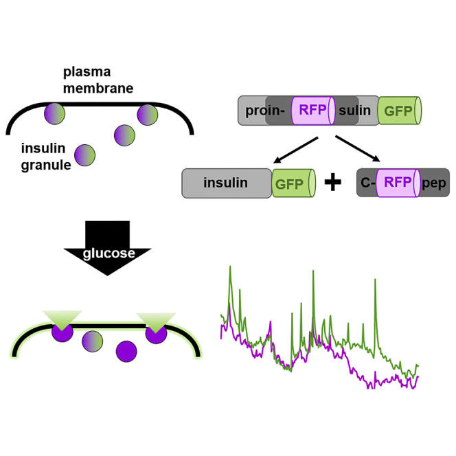
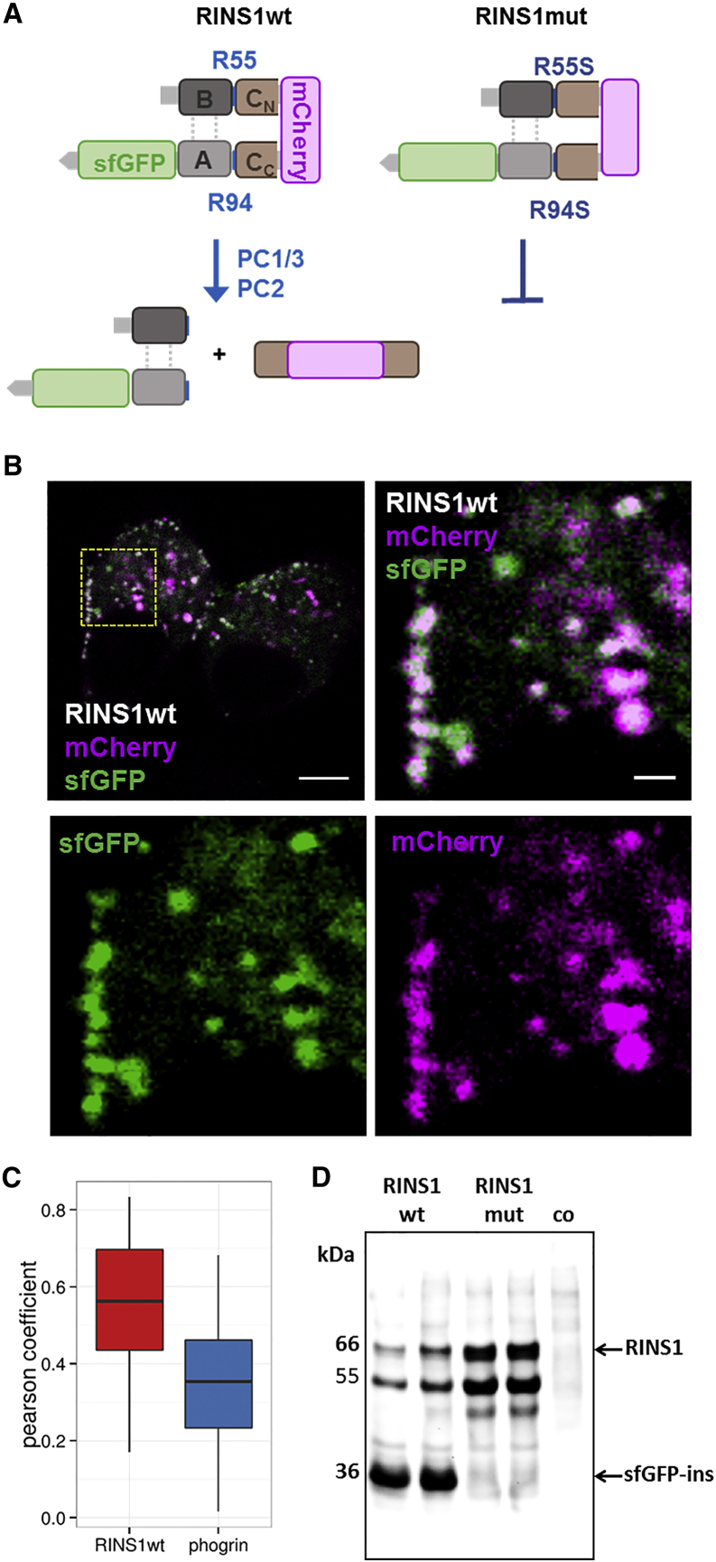
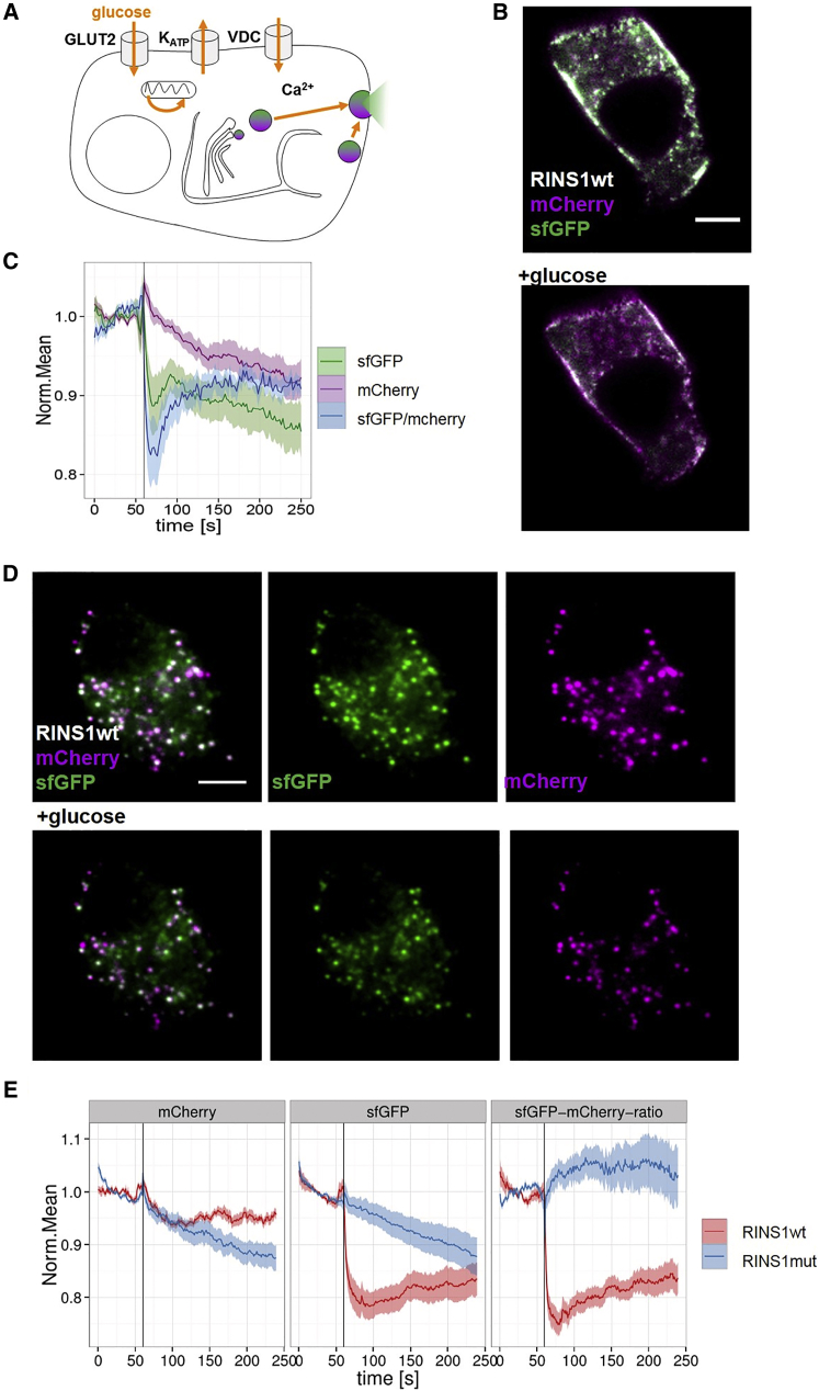
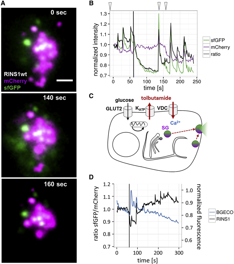
Despite the urgent need for assays to visualize insulin secretion there is to date no reliable method available for measuring insulin release from single cells. To address this need, we developed a genetically encoded reporter termed RINS1 based on proinsulin superfolder GFP (sfGFP) and mCherry fusions for monitoring insulin secretion. RINS1 expression in MIN6 β cells resulted in proper processing yielding single-labeled insulin species. Unexpectedly, glucose or drug stimulation of insulin secretion in β cells led to the preferential release of the insulin-sfGFP construct, while the mCherry-fused C-peptide remained trapped in exocytic granules. This physical separation was used to monitor glucose-stimulated insulin secretion ratiometrically by total internal reflection fluorescence microscopy in single MIN6 and primary mouse β cells. Further, RINS1 enabled parallel monitoring of pulsatile insulin release in tolbutamide-treated β cells, demonstrating the potential of RINS1 for investigations of antidiabetic drug candidates at the single-cell level.
Keywords: TIRF; biosensor; calcium; fluorescence; glucose; granule; insulin; mCherry; oscillation; potassium channel; superfolder GFP; tolbutamide.
Copyright © 2017 The Authors. Published by Elsevier Ltd.. All rights reserved.
Figures
Similar articles
-
Kinetics of insulin secretion from MIN6 pseudoislets after encapsulation in a prototype device of a bioartificial pancreas.Horm Metab Res. 2009 Jan;41(1):5-9. doi: 10.1055/s-0028-1087185. Epub 2008 Oct 14. Horm Metab Res. 2009. PMID: 18855306
-
Effect of 17beta-estradiol on insulin secretion and cytosolic calcium in Min6 mouse insulinoma cells and human islets of Langerhans.Pancreas. 2005 May;30(4):307-13. doi: 10.1097/01.mpa.0000161886.17492.22. Pancreas. 2005. PMID: 15841038
-
Glucose but not KCl diminishes submembrane granule turnover in mouse beta-cells.J Mol Endocrinol. 2017 Oct;59(3):311-324. doi: 10.1530/JME-17-0063. Epub 2017 Aug 1. J Mol Endocrinol. 2017. PMID: 28765259
-
Imaging glucose-regulated insulin secretion and gene expression in single islet beta-cells: control by AMP-activated protein kinase.Cell Biochem Biophys. 2004;40(3 Suppl):179-90. doi: 10.1385/cbb:40:3:179. Cell Biochem Biophys. 2004. PMID: 15289653 Review.
-
β-Cell Pathophysiology: A Review of Advanced Optical Microscopy Applications.Int J Mol Sci. 2021 Nov 26;22(23):12820. doi: 10.3390/ijms222312820. Int J Mol Sci. 2021. PMID: 34884624 Free PMC article. Review.
Cited by
-
Preparation of Whole-mount Mouse Islets on Vascular Extracellular Matrix for Live Islet Cell Microscopy.Bio Protoc. 2023 Nov 5;13(21):e4868. doi: 10.21769/BioProtoc.4868. eCollection 2023 Nov 5. Bio Protoc. 2023. PMID: 37969764 Free PMC article.
-
Genetically Encoded Fluorescent Biosensors Illuminate the Spatiotemporal Regulation of Signaling Networks.Chem Rev. 2018 Dec 26;118(24):11707-11794. doi: 10.1021/acs.chemrev.8b00333. Epub 2018 Dec 14. Chem Rev. 2018. PMID: 30550275 Free PMC article. Review.
-
Systematic Comparison of Vesicular Targeting Signals Leads to the Development of Genetically Encoded Vesicular Fluorescent Zn2+ and pH Sensors.ACS Sens. 2020 Dec 24;5(12):3879-3891. doi: 10.1021/acssensors.0c01231. Epub 2020 Dec 11. ACS Sens. 2020. PMID: 33305939 Free PMC article.
-
Liquid-liquid phase separation facilitates the biogenesis of secretory storage granules.J Cell Biol. 2022 Dec 5;221(12):e202206132. doi: 10.1083/jcb.202206132. Epub 2022 Sep 29. J Cell Biol. 2022. PMID: 36173346 Free PMC article.
-
The Pancreatic ß-cell Response to Secretory Demands and Adaption to Stress.Endocrinology. 2021 Nov 1;162(11):bqab173. doi: 10.1210/endocr/bqab173. Endocrinology. 2021. PMID: 34407177 Free PMC article. Review.
References
-
- Ammala C., Larsson O., Berggren P.O., Bokvist K., Juntti-Berggren L., Kindmark H., Rorsman P. Inositol trisphosphate-dependent periodic activation of a Ca(2+)-activated K+ conductance in glucose-stimulated pancreatic beta-cells. Nature. 1991;353:849–852. - PubMed
-
- Ammala C., Ashcroft F.M., Rorsman P. Calcium-independent potentiation of insulin release by cyclic AMP in single beta-cells. Nature. 1993;363:356–358. - PubMed
-
- Burns S.M., Vetere A., Walpita D., Dancik V., Khodier C., Perez J., Clemons P.A., Wagner B.K., Altshuler D. High-throughput luminescent reporter of insulin secretion for discovering regulators of pancreatic Beta-cell function. Cell Metab. 2015;21:126–137. - PubMed
MeSH terms
Substances
LinkOut - more resources
Full Text Sources
Other Literature Sources
Medical
Research Materials
Miscellaneous
