. 2017 Jun 26;91(14):e02310-16.
doi: 10.1128/JVI.02310-16. Print 2017 Jul 15.

Pathogenic Events in a Nonhuman Primate Model of Oral Poliovirus Infection Leading to Paralytic Poliomyelitis

Affiliations

Pathogenic Events in a Nonhuman Primate Model of Oral Poliovirus Infection Leading to Paralytic Poliomyelitis

Ling Shen et al. J Virol. .

Abstract

Despite a great deal of prior research, the early pathogenic events in natural oral poliovirus infection remain poorly defined. To establish a model for study, we infected 39 macaques by feeding them single high doses of the virulent Mahoney strain of wild type 1 poliovirus. Doses ranging from 107 to 109 50% tissue culture infective doses (TCID50) consistently infected all the animals, and many monkeys receiving 108 or 109 TCID50 developed paralysis. There was no apparent difference in the susceptibilities of the three macaque species (rhesus, cynomolgus, and bonnet) used. Virus excretion in stool and nasopharynges was consistently observed, with occasional viremia, and virus was isolated from tonsils, gut mucosa, and draining lymph nodes. Viral replication proteins were detected in both epithelial and lymphoid cell populations expressing CD155 in the tonsil and intestine, as well as in spinal cord neurons. Necrosis was observed in these three cell types, and viral replication in the tonsil/gut was associated with histopathologic destruction and inflammation. The sustained response of neutralizing antibody correlated temporally with resolution of viremia and termination of virus shedding in oropharynges and feces. For the first time, this model demonstrates that early in the infectious process, poliovirus replication occurs in both epithelial cells (explaining virus shedding in the gastrointestinal tract) and lymphoid/monocytic cells in tonsils and Peyer's patches (explaining viremia), extending previous studies of poliovirus pathogenesis in humans. Because the model recapitulates human poliovirus infection and poliomyelitis, it can be used to study polio pathogenesis and to assess the efficacy of candidate antiviral drugs and new vaccines.IMPORTANCE Early pathogenic events of poliovirus infection remain largely undefined, and there is a lack of animal models mimicking natural oral human infection leading to paralytic poliomyelitis. All 39 macaques fed with single high doses ranging from 107 to 109 TCID50 Mahoney type 1 virus were infected, and many of the monkeys developed paralysis. Virus excretion in stool and nasopharynges was consistently observed, with occasional viremia; tonsil, mesentery lymph nodes, and intestinal mucosa served as major target sites of viral replication. For the first time, this model demonstrates that early in the infectious process, poliovirus replication occurs in both epithelial cells (explaining virus shedding in the gastrointestinal tract) and lymphoid/monocytic cells in tonsils and Peyer's patches (explaining viremia), thereby supplementing historical reconstructions of poliovirus pathogenesis. Because the model recapitulates human poliovirus infection and poliomyelitis, it can be used to study polio pathogenesis, candidate antiviral drugs, and the efficacy of new vaccines.

Keywords: animal model; macaques; oral poliovirus infection; poliomyelitis.


Figures

FIG 1
FIG 1
Validation studies of the cloned type 1 poliovirus Mahoney, plaque assay, and CD155 expression in macaques. (a) Representative confocal microscopy images showing that the Mahoney poliovirus reproducibly infected CD155+ HeLa cells, as detected by confocal staining of CD155 and type 1 poliovirus protein 1 (VP1). Note intracellular staining of PV1 in green in the infected cells (right), but not the uninfected control (left), after overnight infection with cloned virus (MOI = 100:1) in cultures. There was no staining for isotype controls (data not shown). (b) Representative photograph of PFU assay results. The indicated TCID50 titers of cloned type 1 poliovirus Mahoney were mixed with 50 ml feces from uninfected macaques and then subjected to analysis of PFU counts. Note that approximately 10 TCID50 of cloned poliovirus in feces could form detectable PFU, which served as a detection limit, as well. (c) Representative flow histograms showing that the poliovirus receptor CD155 can be expressed in subsets of CD14+ monocytes and CD20+ B cells but rarely in CD3+ T cells from 3 macaque species (cynomolgus, bonnet, and rhesus). (d) Representative immunohistochemistry analysis of CD155 expression on intestinal mucosal cells of macaques. (Left) Image (magnification, ×200) of CD155 expression on surfaces of intestinal epithelial cells in a tissue section from the small intestine of a representative rhesus macaque. Similar images were seen in the gut sections from other rhesus, cynomolgus, and bonnet macaques. (Right) Image (×400) of CD155 expression on gut epithelial cells (orange arrow) and cells with macrophage/DC morphology (light-green arrows) in a lymphoid follicle of Peyer's patch in a section from the small intestine of a representative rhesus macaque.
FIG 2
FIG 2
Poliovirus in feces of macaques following oral infection with type 1 poliovirus Mahoney. (a) Mean poliovirus PFU (± standard errors of the mean [SEM]) per 100 ml in feces from three different species of macaques following oral infection with 109, 108, or 107 TCID50 Mahoney type 1 poliovirus. The “pass through” virus titers measured at day 1 ranged from 106 to 107, depending upon the feeding dose. P values of <0.05 for comparisons between preinfection and days 3 through 13, except day 11, for doses of 109 or 108 TCID50 in rhesus/cynomolgus macaques and except days 11 and 13 for a dose of 107 TCID50. n = 21 for rhesus but decreased to 13 from days 8 through 22 due to euthanasia for paralysis; n = 15 for cynomolgus but decreased to 12 from days 10 through 22; n = 3 for bonnet but decreased to 2 after day 11. (b) Mean (±SEM) poliovirus PFU (left) and poliovirus mRNA (right) per 100 ml in feces from NHP fed with 109 TCID50 (n = 13), 108 TCID50 (n = 16), and 107 TCID50 (n = 10) PFU. The data were pooled, since there were no significant differences in patterns between NHP species (panel a). Arrow, infection time; *, P < 0.05; **, P < 0.01 (between groups).
FIG 3
FIG 3
Poliovirus in gastrointestinal tissues of macaques following oral infection with type 1 poliovirus Mahoney. Live poliovirus titers (left) and poliovirus mRNA (copies per milliliter) (right) in homogenates from tonsils, small (SI) and large (LI) intestines, and mesentery lymph nodes (MLN) collected from 8 paralyzed macaques at days 9 to 12 after oral infection with type 1 poliovirus. Boxes and whiskers: the top and bottom hinges represent 90th and 10th percentile values, respectively; +, mean value; horizontal lines in boxes, medians.
FIG 4
FIG 4
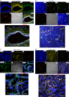
Viral replication proteins were detected in both epithelial and lymphoid cell populations in the tonsil and intestinal mucosa of poliovirus-infected macaques. (a) Poliovirus polymerase 3D was detected in CD155+ stratified squamous epithelia lining the tonsil crypt. The merged image is enlarged at the bottom. Note that CD155/epithelial markers overwhelm, but 3D (red) can be judged by referring to the corresponding positions in the multicolor images at the top. DIC, differential interference contrast. (b) 3D was detected in lymphoid cells (CD155+ CD11+ macrophages/DC) in the tonsil lymphoid follicle. The arrows point to detectable 3D in target cells in the enlarged image. (c) 3D was detected in CD155+ epithelial cells in intestinal villi. The arrows point to clustered areas of detectable 3D in epithelia in the enlarged image. Note that epithelia express weaker CD155 than macrophages (center; also see the CD11C+ macrophages in panel d). (d) 3D was detected in CD155+ CD11+ macrophages/DC in the lamina propria of the small intestine. (e) Representative images showing that 3D was detected in CD11C+ macrophages/DC in the intestinal mucosa containing the Peyer's patch-like area (left), as well as in CD155+ epithelial cells (right, arrows). (f) Representative images showing that control IgG isotypes did not give rise to any staining and anti-3D MAb does not stain tissue sections of tonsil and gut from a healthy macaque not infected with poliovirus. Sections for control isotypes are from intestinal mucosae collected from macaques at days 10 to 12 after oral poliovirus infection, whereas tonsil and small intestine sections for control 3D MAb staining were collected from uninfected macaque RH8335. Shown above are representative images in the sections from the tonsil and intestinal mucosa harboring high titers of replicating poliovirus (Fig. 3). The representative tissues with replicating virus were collected at day 8 or 10 after oral infection of rhesus macaques 8747 and 8745. Poliovirus antigens were detected in tissue sections from five paralyzed rhesus macaques. Fluorescence-labeling Abs and nuclear staining are indicated.
FIG 4
FIG 4
Viral replication proteins were detected in both epithelial and lymphoid cell populations in the tonsil and intestinal mucosa of poliovirus-infected macaques. (a) Poliovirus polymerase 3D was detected in CD155+ stratified squamous epithelia lining the tonsil crypt. The merged image is enlarged at the bottom. Note that CD155/epithelial markers overwhelm, but 3D (red) can be judged by referring to the corresponding positions in the multicolor images at the top. DIC, differential interference contrast. (b) 3D was detected in lymphoid cells (CD155+ CD11+ macrophages/DC) in the tonsil lymphoid follicle. The arrows point to detectable 3D in target cells in the enlarged image. (c) 3D was detected in CD155+ epithelial cells in intestinal villi. The arrows point to clustered areas of detectable 3D in epithelia in the enlarged image. Note that epithelia express weaker CD155 than macrophages (center; also see the CD11C+ macrophages in panel d). (d) 3D was detected in CD155+ CD11+ macrophages/DC in the lamina propria of the small intestine. (e) Representative images showing that 3D was detected in CD11C+ macrophages/DC in the intestinal mucosa containing the Peyer's patch-like area (left), as well as in CD155+ epithelial cells (right, arrows). (f) Representative images showing that control IgG isotypes did not give rise to any staining and anti-3D MAb does not stain tissue sections of tonsil and gut from a healthy macaque not infected with poliovirus. Sections for control isotypes are from intestinal mucosae collected from macaques at days 10 to 12 after oral poliovirus infection, whereas tonsil and small intestine sections for control 3D MAb staining were collected from uninfected macaque RH8335. Shown above are representative images in the sections from the tonsil and intestinal mucosa harboring high titers of replicating poliovirus (Fig. 3). The representative tissues with replicating virus were collected at day 8 or 10 after oral infection of rhesus macaques 8747 and 8745. Poliovirus antigens were detected in tissue sections from five paralyzed rhesus macaques. Fluorescence-labeling Abs and nuclear staining are indicated.
FIG 5
FIG 5
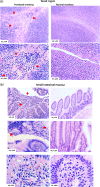
Poliovirus infection in the tonsil and intestinal mucosa coincided with detectable histopathologic lesions and inflammation. (a) (Top left) Representative image showing the hemorrhage and infiltration of numerous necrotic and inflammatory cells in the stratified squamous epithelia in the tonsil crypt (arrows). (Bottom left) Image showing the destruction and disappearance of the tonsil epithelial covering (red arrows) in the tonsil crypt; the tonsil crypt lumen was filled with hemorrhage and exudates (red arrows) comprised of inflammatory and necrotic cells, including macrophages, neutrophils, and lymphocytes. Of note, new or normal epithelia did not emerge underneath the destroyed tissue (under healthy conditions, new epithelia should be seen to outgrow or replace any potential aging or dying cells). (Right) Histology of undamaged tonsil crypts from control macaques not infected with Mahoney poliovirus. Note the intact structures of the stratified squamous epithelia and clean lumen without inflammatory exudates and cells or hemorrhage. (b) (Top left) Large area of necrotic epithelial cells and tissue (arrows) in the surface of the small intestinal mucosa. (Middle left) Necrotic or destroyed epithelial cells/tissue, together with mucus exudates in the gut lumen (arrows). (Bottom left) Infiltration of numerous inflammatory and necrotic cells (arrows) in the lamina propria in small intestine mucosa. (Right) Intact histology of the small intestine mucosa from control macaques not infected with poliovirus Mahoney. The representative tissue sections were collected from macaques 8745 and 8747, who exhibited high-titer poliovirus and viral replication proteins in the tonsil and intestinal mucosae. Similar histopathologic lesions in the tonsil/intestine could be seen in other macaques infected with poliovirus Mahoney.
FIG 6
FIG 6
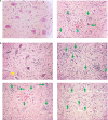
Paralytic poliomyelitis induced by oral poliovirus Mahoney infection was consistent with typical histopathology of poliomyelitis in NHP. (a) (Left) HE-stained section in the left anterior horn of the lumbosacral enlargement from an uninfected healthy control macaque. The section contained spinal motor neurons, astrocytes, and oligodendrocytes. Note the typical appearance of a motor neurons, with a prominent nucleolus in the nucleus and the basophilic Nissl bodies (blue in H&E stain) in the cytoplasm. (Right) Representative H&E-stained section in the lumbosacral enlargement from a paralyzed macaque (RH8203). Note necrotic motor neurons (arrows) and hemorrhage (arrowhead) in the left anterior. (b) (Top left) Section in the left anterior horn of the lumbosacral enlargement from a cynomolgus macaque (CN7821) with paralysis of the right leg. Note that most of the motor neurons in the left anterior horn of the lumbosacral enlargement exhibited “normal” images and only a few degenerative neurons (arrow), with a lack of obvious inflammation and destroyed neurons. (Top right) Section in the right anterior horn of the lumbosacral enlargement from the same cynomolgus macaque (CN7821). Note the completely destroyed neurons, with many neuronophagia (arrows) and infiltration of inflammatory cells. (Bottom left and right) Sections in the left and right anterior horns, respectively, of the lumbosacral enlargement from a rhesus macaque (RH8204) who developed paralysis involving both legs. Note that almost all the neurons in both the left and right anterior horns were destroyed (arrows). RH8203 and the other 2 rhesus macaques with paralysis involving multiple limbs also showed polio lesions in the cerebral cortex and brain stem and the cervical enlargement, respectively (data available upon request).
FIG 7
FIG 7
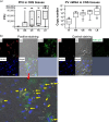
Paralytic poliomyelitis after neuronal spread correlated with high titers of poliovirus in the spinal cord and replicating poliovirus in CD155+ neurons. (a) Poliovirus titers (left) and viral mRNA copies (right) per milliliter in homogenates from CNS tissues from 11 paralyzed macaques sacrificed 24 to 48 h after onset of paralysis. The data were pooled, since there were no significant differences in titers between NHP species or between groups fed with different virus doses. The whiskers for the box plots are the same as in Fig. 3. B, brain; BS, brain stem; CE, cervical enlargement of spinal cord; LE, lumbosacral enlargement; TS, thoracic segment. (b) Representative confocal microscopy images showing detection of poliovirus polymerase 3D in CD155+ neurons in anterior-horn sections of the spinal cords of paralyzed macaques. (Top left) Representative confocal split images of multicolor staining for poliovirus polymerase 3D (red), CD155 (green), and nucleus (blue) on tissue sections prepared from anterior horns in the lumbosacral enlargement of spinal cord from the rhesus macaque RH8204, who became paralyzed at day 11. (Bottom) Enlarged, merged image showing positively stained poliovirus polymerase 3D in CD155+ neurons (arrows) in the anterior-horn sections. Note that the cells harboring 3D are consistent with the sizes of motor neurons, based on the size scale shown. Similar results were seen in other paralyzed macaques. (Top right) There was no staining for isotype IgG controls in spinal cord sections from the paralyzed macaque.
FIG 8
FIG 8
Detectable response of neutralizing Ab coincided temporally with resolution of viremia and virus shedding in oropharynges/stool. (a) Poliovirus-specific neutralization titers (log2) after oral poliovirus feeding. Shown are mean dilution titers of plasma with SEM. In geometric scales, log 1 denotes 0. n = 30. (b) Poliovirus mRNA copies in blood (top) and throat swab eluate (bottom) from orally infected macaques. The data are mean mRNA copies with standard deviations (SD) in 106 PBMC and in 1 ml of throat swab eluate, respectively. Poliovirus mRNA copies in PBMC were pooled from eight macaques who had detectable poliovirus RNA after infection with 108 and 109 TCID50. None of the macaques infected with 107 TCID50 had detectable viral RNA in blood. The data from throat swabs were pooled from 23 macaques. There were no significant differences in frequencies or titers in swabs between species or between the 3 groups infected with 107, 108, and 109 TCID50.

Similar articles

Cited by

References

    1. Nathanson N, Kew OM. 2010. From emergence to eradication: the epidemiology of poliomyelitis deconstructed. Am J Epidemiol 172:1213–1229. doi:10.1093/aje/kwq320. - DOI - PMC - PubMed
    1. Roberts L. 2016. Infectious disease. New polio cases in Nigeria spur massive response. Science 353:738. - PubMed
    1. Hagan JE, Wassilak SG, Craig AS, Tangermann RH, Diop OM, Burns CC, Quddus A, Centers for Disease and Prevention. 2015. Progress toward polio eradication—worldwide, 2014-2015. MMWR Morb Mortal Wkly Rep 64:527–531. - PMC - PubMed
    1. Nathanson N. 2008. The pathogenesis of poliomyelitis: what we don't know. Adv Virus Res 71:1–50. doi:10.1016/S0065-3527(08)00001-8. - DOI - PubMed
    1. Koike S, Horie H, Sato Y, Ise I, Taya C, Nomura T, Yoshioka I, Yonekawa H, Nomoto A. 1993. Poliovirus-sensitive transgenic mice as a new animal model. Dev Biol Stand 78:101–107. - PubMed

Publication types