. 2017 Mar 23;2(6):e89206.
doi: 10.1172/jci.insight.89206.

Physiologically activated mammary fibroblasts promote postpartum mammary cancer

Affiliations

Physiologically activated mammary fibroblasts promote postpartum mammary cancer

Qiuchen Guo et al. JCI Insight. .

Abstract

Women diagnosed with breast cancer within 5 years of childbirth have poorer prognosis than nulliparous or pregnant women. Weaning-induced breast involution is implicated, as the collagen-rich, immunosuppressive microenvironment of the involuting mammary gland is tumor promotional in mice. To investigate the role of mammary fibroblasts, isolated mammary PDGFRα+ cells from nulliparous and postweaning mice were assessed for activation phenotype and protumorigenic function. Fibroblast activation during involution was evident by increased expression of fibrillar collagens, lysyl oxidase, Tgfb1, and Cxcl12 genes. The ability of mammary tumors to grow in an isogenic, orthotopic transplant model was increased when tumor cells were coinjected with involution-derived compared with nulliparous-derived mammary fibroblasts. Mammary tumors in the involution-fibroblast group had increased Ly6C+ monocytes at the tumor border, and decreased CD8+ T cell infiltration and tumor cell death. Ibuprofen treatment suppressed involution-fibroblast activation and tumor promotional capacity, concurrent with decreases in tumor Ly6C+ monocytes, and increases in intratumoral CD8+ T cell infiltration, granzyme levels, and tumor cell death. In total, our data identify a COX/prostaglandin E2 (PGE2)-dependent activated mammary fibroblast within the involuting mammary gland that displays protumorigenic, immunosuppressive activity, identifying fibroblasts as potential targets for the prevention and treatment of postpartum breast cancer.


Conflict of interest statement

Conflict of interest: The authors have declared that no conflict of interest exists.

Figures
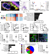
Figure 1
Figure 1. Mammary fibroblasts are activated during weaning-induced gland involution.
(A) Mammary gland thin sections from the Col1a1-GFP reporter mouse immunofluorescence stained for GFP+ collagen 1–expressing cells (red), fibroblast PDGFRα (green), and myoepithelial cell calponin (purple), show colocalization of GFP and PDGFRα (yellow), but not calponin. Nuclei stained with DAPI (blue). White scale bar: 50 μm. Yellow scale bar: 10 μm. Arrows show PDGFRα+GFP+calponin cells. (B) Flow gate for isolating murine mammary PDGFRα+ cells. (C) RT-qPCR on FACS-isolated PDGFRα+ cells for fibroblast markers (Col1a1, Col3a1, Col5a1, and PDGFRα) and markers of potential contaminating cell populations: Cd45 (lymphocyte common antigen), F4/80 (mature macrophage), Cd31 (endothelial cell), Adipoq (adipocyte), and Ecad (epithelial cell). Sorted PDGFRα-negative cells were used as positive controls for Cd45, F4/80, Cd31, Adipoq, and mammary epithelial EpH4 cells were used as the Ecad-positive control. n = 3–4 per group. (D) RNA-Seq heatmap illustrates 870 significantly differentially expressed genes (>1.3-fold changes) between fibroblasts isolated from nulliparous and involution day 6 (InvD6) mammary glands. Red: relative high expression. Blue: relative low expression. (E) Pathway analysis of RNA-Seq data set showing extracellular matrix (ECM) regulation–related pathways that are upregulated in InvD6-fibroblasts. (F) PDGFRα+ fibroblasts reported as number per gram of mammary tissue observed in nulliparous and 4-, 6-, and 8-day postweaning mice; n = 3–10 per group. (G) Fibrillar collagens and Lox gene expression analysis by RT-qPCR in sorted PDGFRα+ mammary fibroblasts from nulliparous and 4-, 6-, and 8-day postweaning mice; n = 3–10 per group. (H) Mmp2, Mmp3, and Tgfb1 gene expression analysis by RT-qPCR in nulliparous and InvD6 fibroblasts, n = 6–9 per group. (I) Immunofluorescence staining shows non-colocalization of α-smooth muscle actin (red) and PDGFRα (green) in mammary glands of nulliparous and InvD6 mice. Boxes show images at high magnification. Arrow: PDGFRα+ cells. Arrowhead: αSMA+ cells. White scale bar: 50 μm. Yellow scale bar: 10 μm. (J) Pathway analysis of RNA-Seq data described in D showing immune regulation–related pathways that are upregulated in InvD6 fibroblasts. (K) RNA-Seq data show chemokine expression pattern of fibroblasts isolated from nulliparous mammary glands. (L) RT-qPCR gene expression for Cxcl12 in sorted PDGFRα+ fibroblasts from nulliparous and InvD6 mammary glands, n = 3–5 per group. Gene expression data are normalized to Actb and are from 2 to 6 independent studies. RNA-Seq data are archived at the NCBI Gene Expression Omnibus (GEO GSE94761). *P < 0.5, **P < 0.01, ***P < 0.001, #P < 0.0001 by unpaired 2-tailed t test. Data represent mean ± SEM.
Figure 2
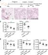
Figure 2. Systemic in vivo ibuprofen treatment during weaning-induced mammary gland involution decreases mammary fibroblast activation.
(A) Schematic shows breeding and weaning-induced mammary gland involution timeline with 0 mg/kg (control), 150 mg/kg (150IBU), and 300 mg/kg (300IBU) ibuprofen provided in chow for 6 days following weaning, with mammary fibroblasts FACS-isolated on involution day 6 (InvD6). (B) Representative H&E images of nulliparous, InvD6, and 300IBU mammary glands depicting normal nulliparous and InvD6 gland morphology in the absence and presence of ibuprofen treatment. Scale bar: 200 μm. (C) Left: RT-qPCR of Col1a1 gene expression in isolated fibroblasts, n = 4–7 per group. Right: type I collagen IHC quantification around InvD5 mammary lobules (InvD5T) with and without 300IBU treatment for 5 days, n = 5–6 per group. (D) RT-qPCR analysis of targeted genes in isolated fibroblasts, n = 4–7 per group. Gene expression data are normalized to Actb. Ibuprofen intervention data are from 2 independent breeding studies. *P < 0.05 by 1-way ANOVA with Tukey correction and unpaired 2-tailed t test. Data represent mean ± SEM.
Figure 3
Figure 3. In vitro mammary fibroblast activation by TGF-β1 is directly inhibited by ibuprofen.
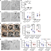
Treatment conditions are: 0.5 TGF-β1 = 0.5 ng/ml TGF-β1, 5 TGF-β1 = 5 ng/ml TGF-β1, Low IBU =10 μg/ml ibuprofen, High IBU = 30 μg/ml ibuprofen (HIBU). (A) Mammary fibroblast morphology when cultured on plastic (left panel) or within floating collagen pads (right panel). Dashed lines show the outline of the cells. (B) α-Smooth muscle actin (αSMA) gene expression in primary mammary fibroblasts cultured on plastic, within floating collagen pads, or from freshly sorted mammary PDGFRα+ fibroblasts, n = 6–10 per condition. (C) Morphologic evidence of TGF-β1–induced fibroblast activation in the floating collagen pad culture model. (D) Increased Col1a1 and Cox2 gene expression in fibroblasts treated with TGF-β1, n = 4–7 per condition. (E) The ability of TGF-β1–treated fibroblasts to contract collagen pads (top) is suppressed by ibuprofen (bottom) and (F) data quantification, n = 5 per condition. (G) Fibroblast morphology with TGF-β1 treatment in the presence or absence of ibuprofen. (H) Col1a1 and Cox2 gene expression in fibroblasts with TGF-β1 treatment in the presence or absence of ibuprofen, n = 5 per condition. Scale bars: 100 μm (A, C, and G) and 1 cm (E). All gene expression data are normalized to Actb and then normalized to the control groups in each experiment. TGF-β1 and ibuprofen combination treatment studies were repeated 5 times. *P < 0.05, **P < 0.01, ***P < 0.001, #P < 0.0001 by 1-way ANOVA with Tukey correction. For data normalized to control, statistics were performed using matched 1-way ANOVA with Tukey correction on the raw data that are not normalized to control. Data represent mean ± SEM. NS, not significant.
Figure 4
Figure 4. Prostaglandin E2 (PGE2) directly stimulates fibroblast collagen and cytokine expression but not cell elongation.
(A) Col1a1, Mmp3, and Cxcl12 gene expression induced by 1 ng/ml PGE2 (1 PGE2) or 5 ng/ml PGE2 (5 PGE2) treatment, n = 4–8 per group. All gene expression data are normalized to Actb and then normalized to the control groups in each experiment. Gene expression data obtained from 4 to 8 independent experiments. (B) Mammary fibroblast morphology when cultured within floating collagen pads in the absence (right) and presence of 5 ng/ml PGE2. Scale bar: 100 μm. (C) Model of fibroblast-induced monocyte-derived cell migration assay setup. (D) Quantification of the number of monocyte-derived cells that migrated into collagen pads in Control (1 μg/ml mouse IgG1 isotype control), 1 PGE2 (1 ng/ml PGE2 with 1 μg/ml mouse IgG1 isotype control), and +anti-CXCL12 (1 ng/ml PGE2 with 1 μg/ml CXCL12-neutralizing antibody) conditions, with data normalized to control, n = 5/condition. Monocyte-derived cell migration assay was repeated 5 times. (E) Representative images of migrated monocyte-derived cells labeled with CFSE (green) fluorescent dye in Control, 1 PGE2, and 1 PGE2+anti-CXCL12 conditions as described in D. Scale bar: 200 μm. *P < 0.5, **P < 0.01 by matched 1-way ANOVA with Tukey correction on the raw data that are not normalized to control. Data represent mean ± SEM. NS, not significant.
Figure 5
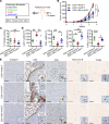
Figure 5. Mammary involution-fibroblasts are tumor promotional via a COX2-dependent mechanism that suggests immune modulation.
(A) Schematic design for mammary fibroblast treatment, isolation, and use in an orthotopic mammary tumor model. FACS-isolated mammary fibroblasts (20,000 cells) from nulliparous, involution day 6 (InvD6), or InvD6 hosts treated during involution with 300 mg/kg ibuprofen in chow (IBU InvD6 F), were mixed with D2A1 tumor cells at a 1:1 ratio, injected into mammary fat pads of nulliparous BALB/c mice, and tumor tissue collected 3.5 weeks after tumor cell injection. (B) Tumor growth curve of D2A1 cells coinjected with sorted fibroblasts from nulliparous (D2A1+Nullip F), InvD6 (D2A1+InvD6 F) and InvD6 with ibuprofen treatment (D2A1+IBU InvD6 F) mice, n = 7–10 tumors per group. (C) IHC quantification of intratumoral type I collagen, n = 5–8 tumors per group. (D) IHC quantification of tumor border Ly6C, intratumoral CD8, granzyme B (Gzmb), and TUNEL; n = 5–10 tumors per group. (E) Representative IHC images of markers quantified in D. From left to right: intratumoral collagen I, tumor border Ly6C (dashed lines show tumor border), intratumoral CD8, granzyme B, and TUNEL. Black scale bars: 100 μm. Red scale bars: 50 μm. Green scale bar: 10 μm. Counterstaining (blue) is not shown in granzyme B– and TUNEL-stained images to enhance visualization of the positive cells. All tumor data were obtained from 2 independent breeding studies. *P < 0.05, **P < 0.01, ***P < 0.001, #P < 0.0001 by 1-way ANOVA with Tukey correction. Data represent mean ± SEM. NS, not significant.
Figure 6
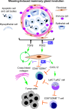
Figure 6. Schematic overview of the potential tumor promotional contributions of mammary involution-fibroblasts.
Activation of involution-fibroblasts by TGF-β and prostaglandin E2 (PGE2) in the involution microenvironment leads to increased fibrillar collagen expression, which is tumor promotional. Involution-fibroblasts also have increased CXCL12 expression, which recruits Ly6C+Ly6G monocytes, blocking CD8+ T cell tumor infiltration and tumor cell death. Involution-fibroblasts also block CD45+GZMA+ cell infiltration into tumors by an unknown mechanism (dashed block arrows). These tumor promotional attributes of the involution-fibroblasts can be blocked by ibuprofen (IBU). GZMA and GZMB, granzymes A and B.

Similar articles

Cited by

References

    1. Stensheim H, Møller B, van Dijk T, Fosså SD. Cause-specific survival for women diagnosed with cancer during pregnancy or lactation: a registry-based cohort study. J Clin Oncol. 2009;27(1):45–51. doi: 10.1200/JCO.2008.17.4110. - DOI - PubMed
    1. Johansson AL, Andersson TM, Hsieh CC, Cnattingius S, Lambe M. Increased mortality in women with breast cancer detected during pregnancy and different periods postpartum. Cancer Epidemiol Biomarkers Prev. 2011;20(9):1865–1872. doi: 10.1158/1055-9965.EPI-11-0515. - DOI - PubMed
    1. Callihan EB, et al. Postpartum diagnosis demonstrates a high risk for metastasis and merits an expanded definition of pregnancy-associated breast cancer. Breast Cancer Res Treat. 2013;138(2):549–559. doi: 10.1007/s10549-013-2437-x. - DOI - PMC - PubMed
    1. Amant F, et al. Prognosis of women with primary breast cancer diagnosed during pregnancy: results from an international collaborative study. J Clin Oncol. 2013;31(20):2532–2539. doi: 10.1200/JCO.2012.45.6335. - DOI - PubMed
    1. Lyons TR, et al. Postpartum mammary gland involution drives progression of ductal carcinoma in situ through collagen and COX-2. Nat Med. 2011;17(9):1109–1115. doi: 10.1038/nm.2416. - DOI - PMC - PubMed

Publication types

MeSH terms

Substances