

An investigation on cytotoxic effect of bioactive AgNPs synthesized using Cassia fistula flower extract on breast cancer cell MCF-7
- PMID: 28352579
- PMCID: PMC4980703
- DOI: 10.1016/j.btre.2015.10.004
An investigation on cytotoxic effect of bioactive AgNPs synthesized using Cassia fistula flower extract on breast cancer cell MCF-7
Abstract
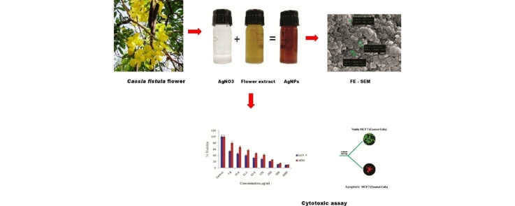
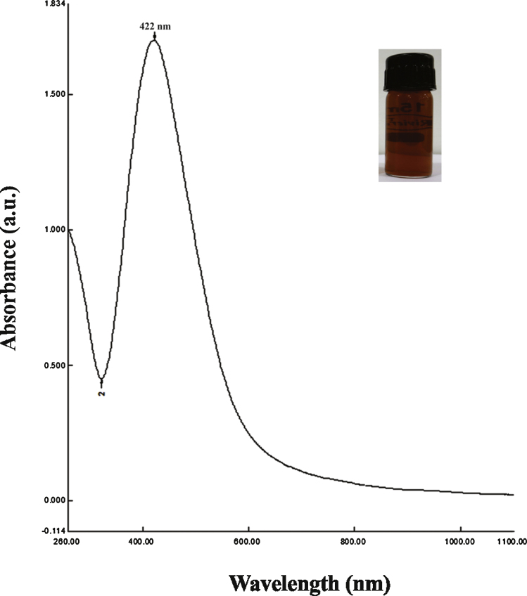
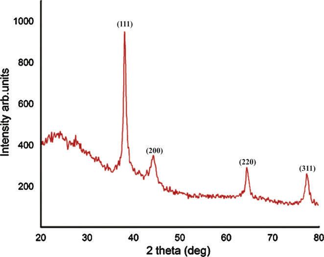
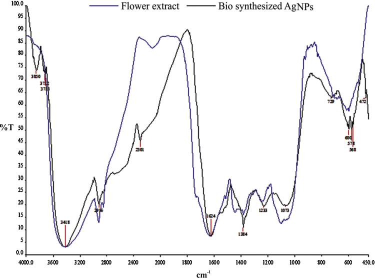
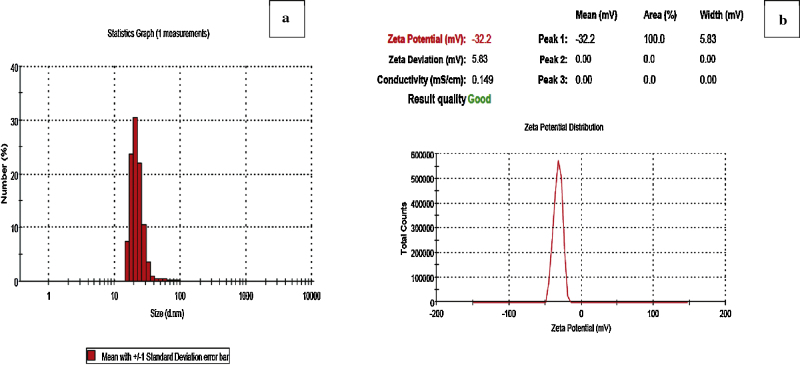
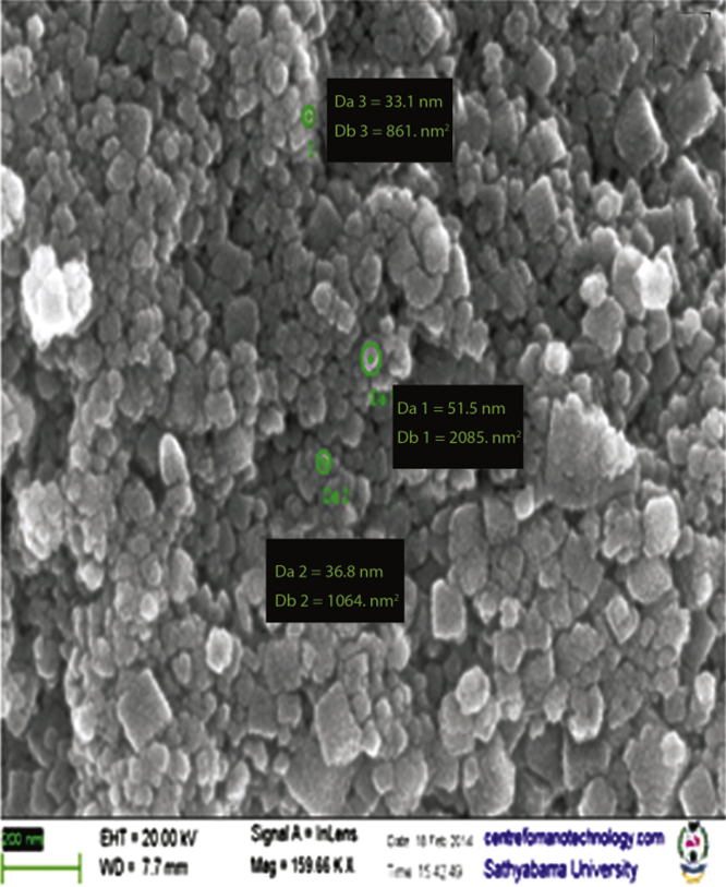
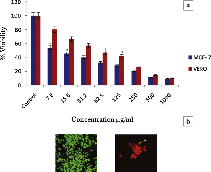
A single step protocol to produce biofunctionalized silver nanoparticles (AgNPs) using the aqueous extract of Cassia fistula flower as "natural factory" was investigated. The reaction between silver ions and aqueous flower extract after the bioreduction process has resulted in the formation of reddish brown color colloidal solution. XRD pattern showed the face centered cubic crystalline structure of AgNPs and exhibited spherical morphology as characterized by FE-SEM. FTIR studies identified different functional groups involved in effective capping of AgNPs. The zeta potential affirmed the phytoreduced AgNPs possess good stability and the size of the particle was measured by DLS. The synthesized AgNPs displayed effective cytotoxic potential against MCF7 and the inhibitory concentration (IC50) was recorded at 7.19 μg/mL. The apoptotic effects of the AgNPs were also confirmed by AO/EB staining. The investigation presents preliminary evidence that biosynthesized AgNPs can be used in the development of novel anticancer drugs.
Keywords: Cassia fistula; Cytotoxicity; Silver nanoparticles.
Figures
Similar articles
-
Biosynthesis and Characterization of Silver Nanoparticles from Methanol Leaf Extract of Cassia didymobotyra and Assessment of Their Antioxidant and Antibacterial Activities.J Nanosci Nanotechnol. 2015 Dec;15(12):9818-23. doi: 10.1166/jnn.2015.10966. J Nanosci Nanotechnol. 2015. PMID: 26682418
-
Characterization, Antibacterial and Antioxidant Properties of Silver Nanoparticles Synthesized from Aqueous Extracts of Allium sativum, Zingiber officinale, and Capsicum frutescens.Pharmacogn Mag. 2017 Jul;13(Suppl 2):S201-S208. doi: 10.4103/pm.pm_430_16. Epub 2017 Jul 11. Pharmacogn Mag. 2017. PMID: 28808381 Free PMC article.
-
Biomimetic synthesis of silver nanoparticles using Matricaria chamomilla extract and their potential anticancer activity against human lung cancer cells.Mater Sci Eng C Mater Biol Appl. 2018 Nov 1;92:902-912. doi: 10.1016/j.msec.2018.07.053. Epub 2018 Jul 21. Mater Sci Eng C Mater Biol Appl. 2018. PMID: 30184820
-
Antioxidant and anticancer activities of green synthesized silver nanoparticles using aqueous extract of tubers of Pueraria tuberosa.Artif Cells Nanomed Biotechnol. 2018;46(sup3):S71-S85. doi: 10.1080/21691401.2018.1489265. Epub 2018 Jul 25. Artif Cells Nanomed Biotechnol. 2018. PMID: 30043665
-
Green engineered biomolecule-capped silver and copper nanohybrids using Prosopis cineraria leaf extract: Enhanced antibacterial activity against microbial pathogens of public health relevance and cytotoxicity on human breast cancer cells (MCF-7).Microb Pathog. 2017 Apr;105:86-95. doi: 10.1016/j.micpath.2017.02.019. Epub 2017 Feb 16. Microb Pathog. 2017. PMID: 28214590
Cited by
-
Alginate-based hydrogel platform embedding silver nanoparticles and cisplatin: characterization of the synergistic effect on a breast cancer cell line.Front Mol Biosci. 2023 Oct 23;10:1242838. doi: 10.3389/fmolb.2023.1242838. eCollection 2023. Front Mol Biosci. 2023. PMID: 37936720 Free PMC article.
-
Cytotoxicity of Plant-Mediated Synthesis of Metallic Nanoparticles: A Systematic Review.Int J Mol Sci. 2018 Jun 11;19(6):1725. doi: 10.3390/ijms19061725. Int J Mol Sci. 2018. PMID: 29891772 Free PMC article. Review.
-
Phycobiliprotein-mediated synthesis of biogenic silver nanoparticles, characterization, in vitro and in vivo assessment of anticancer activities.Sci Rep. 2018 Jun 12;8(1):8925. doi: 10.1038/s41598-018-27276-6. Sci Rep. 2018. PMID: 29895869 Free PMC article.
-
Antibacterial applications of biologically synthesized Pichia pastoris silver nanoparticles.Heliyon. 2024 Feb 6;10(4):e25664. doi: 10.1016/j.heliyon.2024.e25664. eCollection 2024 Feb 29. Heliyon. 2024. PMID: 38375309 Free PMC article.
-
Green-Synthesized Silver Nanoparticles Induced Apoptotic Cell Death in MCF-7 Breast Cancer Cells by Generating Reactive Oxygen Species and Activating Caspase 3 and 9 Enzyme Activities.Oxid Med Cell Longev. 2020 Oct 5;2020:1215395. doi: 10.1155/2020/1215395. eCollection 2020. Oxid Med Cell Longev. 2020. PMID: 33082906 Free PMC article.
References
-
- Zhang L., Gu F.X., Chan J.M., Wang A.Z., Langer R.S., Farokhzad O.C. Nanoparticles in medicine: therapeutic applications and developments. Clin. Pharmacol. Ther. 2008;83:761–769. - PubMed
-
- Kawasaki E.S., Player A. Nanomedicine, and the development of new, effective therapies for cancer. Nanomed. NBM. 2005;1:101–109. - PubMed
-
- Kumar V., Yadav S.K. Plant-mediated synthesis of silver and gold nanoparticles and their applications. J. Chem. Technol. Biotechnol. 2009;84:151–157.
-
- Zargar M., Shameli K., Najafi G.R., Farahani F. Plant mediated green biosynthesis of silver nanoparticles using Vitex negundo L. extract. J. Ind. Eng. Chem. 2014;20:4169–4175.
-
- Wiley B.J., Im S.H., McLellan J., Siekkinen A., Xia Y. Maneuvering the surface plasmon resonance of silver nanostructures through shape-controlled synthesis. J. Phys. Chem. B. 2006;110:15666–15675. - PubMed
LinkOut - more resources
Full Text Sources
Other Literature Sources